

 PDF
PDF ePub
ePub Citation
 Print
Print


INTRODUCTION
Autophagy, responsible for a variety of cellular homeostasis functions, is necessary for the degradation of long-lasting proteins and nonfunctional organelles [1,2]. It is activated to promote cellular survival by degrading unnecessary proteins and organelles [3]. When over-activated, however, the process of autophagy may deplete essential molecules and organelles responsible for cellular survival [4,5]. It has been shown that lifelong calorie restriction by 40% increases the expression of autophagic markers in the heart [6]. This finding implies that up-regulation of autophagy by calorie restriction may have a protective role on the cardiomyocytes by reducing levels of oxidative damage due to aging and cardiovascular diseases.
One study has shown that a lack of Atg5, a cardiac-specific protein that is required for the activation of autophagy, leads to cardiac hypertrophy, left ventricular dilatation, and contractile dysfunction, along with increased levels of ubiquitination [7]. This study also demonstrated that a reduced expression level of Atg7, a protein necessary for the formation of autophagosome, is responsible for reduced cellular viability and remarkable features of cardiomyocyte hypertrophy [7]. In addition, lysosomal-associated membrane protein 2 is thought to be involved in the pathogenesis of cardiomyopathy, as its deficiency has been shown to accumulate autophagic vacuoles and impair the autophagic degradation of dysfunctional proteins [8].
Overall, these findings unanimously support that cardiomyocyte autophagy is essential for controlling protein quality, and maintaining cellular function and survival [7]. However, it should be noted that increased or excessive autophagy may be detrimental under some circumstances, including pressure overload-induced heart failure [9].
UNDER ENERGY DEPRIVATION CONDITIONS
Autophagy is important for the preservation of energy status in response to energy deprivation. Glucose deprivation, a condition similar to myocardial ischemia, induces autophagy, while pharmacological inhibition of autophagy has been shown to reduce cellular survival in cultured cardiomyocytes [10]. Such findings collectively support the notion of the cardioprotective roles of autophagy. It may serve to maintain energy production in response to energy deprivation, including acute ischemia, and also may be responsible for the clearance of long-lived proteins and dysfunctional organelles during chronic ischemia or reperfusion.
A state of nutrient deprivation, such as myocardial ischemia, induces macroautophagy activation. Macroautophagy degrades proteins and organelles, thereby generating fatty acids and amino acids that are all used for mitochondrial adenosine triphosphate (ATP) production, which promotes cardiomyocyte survival [10,11].
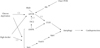
Adenosine monophosphate (AMP)-activated protein kinase (AMPK) is an energy sensing kinase, taking action when the cellular AMP-to-ATP ratio increases. AMPK activates autophagy by activating ULK1, a serine/threonine-protein kinase, and by relieving the mammalian target of rapamycin (mTOR)-mediated inhibition of macroautophagy [12]. mTOR tightly regulates autophagy by inhibiting the ULK1 kinase complex and accordingly prevents autophagy activation, along with phosphorylation of the tuberous sclerosis complex (TSC). This signaling pathway is accordingly called AMPK-mTOR, and is considered critical in regulating the activation of autophagy under circumstances such as energy stress and glucose starvation (Fig. 1). The TSC-mTOR pathway can function in diabetic hearts. In diabetic hearts, phosphorylation of raptor at both Ser722 and Ser792 is decreased, and phosphorylation of mTOR at both Ser2448 and Thr2446 is increased. Also, 4 E binding protein 1 and p70 ribosomal protein S6 kinase 1, downstream effectors of mTOR, are increased [13]. Studies collectively suggest that mTOR complex 1 (mTORC1) activation may be detrimental under cardiac energy deprivation, while mTORC1 inhibition is protective because of energy preservation [14]. In addition, it has been revealed that mTORC1 activation may be responsible for cell growth.
Rheb, a Ras homolog guanosine triphosphate-binding protein, is inhibited in response to energy deprivation for autophagy activation. In addition, suppression of autophagy activation by inhibiting Beclin-1 counteracts the protective action of Rheb protein for energy deprivation. The action of Rheb-regulated autophagy has been shown to be protective against nutrient starvation and ischemia in cardiomyocytes through the preservation of ATP content and the reduction of misfolded protein accumulation [14].
Rheb controls the activation of autophagy partly through Atg7, where Atg7 overexpression induces autophagy and suppresses Rheb-induced cell death in response to glucose deprivation. In addition, mTORC1 may be involved in regulating autophagy through ULK1/2 regulation [15], though its exact function remains to be elucidated.
Under a starvation state, such as in myocardial ischemia, AMPK acts as a checkpoint by suppressing cellular growth and promoting autophagy activation in cardiomyocytes. Therefore, the AMPK-mTOR pathway is certainly a crucial regulator of autophagy in such circumstances, as inhibition of AMPK reduces autophagy and increases cell death in cardiomyocytes [10]. This phenomenon was also observed in transgenic mice with cardiac-specific expression of a dominant negative AMPK. This setting has shown reduced autophagy induction in states of fasting in vivo [10], which suggests that AMPK-induced autophagy may be controlled by inhibiting the expression of mTOR in response to ischemia. One study has shown that glycogen synthase kinase (GSK)-3, an enzyme involved in gene transcription regulation, protein translation, and apoptosis, as well as hexose metabolism, may be a regulator of the mTOR pathway in cardiomyocytes [16]. In addition, inhibition of GSK-3β has been reported to be cardioprotective [17-20] by inhibiting mTOR signaling and thus activating autophagy via phosphorylation of TSC2 [21,22]. The function of GSK-3β also includes the regulation of mTOR during both myocardial ischemia and reperfusion [20].
In response to glucose deprivation, cardiomyocytes initiate the nuclear translocation of FoxO1 and FoxO3 to the nucleus where the transcription of genes responsible for autophagy are activated [23,24]. FoxO3 overexpression in the heart is associated with increased autophagy, which may be related to the development of cardiac atrophy [25], while genetic deletion of FoxO3 resulted in the development of cardiac hypertrophy [26]. Under a starvation state, Sirtuin 1 (Sirt1), a NAD-dependent deacetylase, is up-regulated [27,28]. Sirt1 mediates the deacetylation of FoxO1 and upregulation of Rab7, which functions as the center for mediating increased autophagic flux in response to starvation, which in turn maintains left ventricular function during these events [23].
Atg13 binds to Atg1 and Atg17 in response to glucose deprivation, promoting the induction of autophagy at the phagophore assembly site. This complex can be found in yeast; the mammalian counterpart shows slight differences [29]. During starvation, mTOR dissociates and promotes the activation of ULK1, which up-regulates autophagy by increasing the phosphorylation of mATG13 and focal adhesion kinase interacting protein of 200 kD (FIP200). A class III phosphoinositide 3-kinase (PI3K) complex is then recruited to the assembly site and Vps34 lipid kinase protein binds to the phagophore via Vps15. This complex contains Beclin1/ATG6 and ATG14, which control the induction of Vps34 lipid kinase protein. It is believed that this lipid kinase is an essential protein for recruiting additional ATG proteins, where they complete the autophagosome formation [30].
UNDER NUTRIENT-RICH CONDITIONS
Under nutrient-rich conditions, including obesity, the Akt signaling pathway is activated, and Akt phosphorylates and activates mTOR kinase and the FoxO family. It has been shown that inhibition of the TOR pathway is responsible for cardioprotection against cardiac dysfunction induced by a high fat diet [31]. Activated mTOR interacts with ULK1, the mammalian ATG13, and FIP200 complex. This leads to the phosphorylation of ULK1, which suppresses autophagy (Fig. 1).
High fat diet-induced obesity activates the Rheb/mTORC1 pathway and reduces the activation of autophagy in the heart of mice [14]. Increased myocardial injury in these mice in response to prolonged ischemia suggests that an increased level of mTORC1 activity may be associated with an increased susceptibility. It has been demonstrated that reactivation of autophagy is a critical mechanism underlying the beneficial effects of mTORC1 inhibition in high fat diet-induced obesity, as shown in the failure of pharmacological mTORC1 inhibition to reduce ischemic injury by inhibiting Beclin-1. Physiological inhibition of the Rheb-mTORC1 signaling pathway during myocardial ischemia can be impaired in such conditions as obesity and metabolic syndrome, which consequently exacerbates myocardial injury. This Rheb-dependent mTORC1 pathway seems to be critical in regulating the activation of autophagy during ischemia in cardiomyocytes and its dysfunction is thereby associated with human diseases.
Nonobese mice with fructose-induced insulin resistance have shown activation of myocardial autophagy [32], while Ossabaw swine with excessive nutrition have shown inhibition of cardiac autophagy, which may be responsible for myocardial injury [33]. The expression of conjugated Atg12-Atg5 is increased in obesity and metabolic syndrome, as well as in defective hepatic autophagy of obese individuals [34], while unc-51-like kinase-1, Beclin-1, and LC3 conversion, which are indicative of autophagic activity, all decrease in metabolic syndrome, indicating the suppressed formation of nascent and mature autophagosomes. In addition, insulin resistance impairs the action of autophagy as highlighted by the up-regulation of the autophagy inhibitor mTOR in metabolic syndrome. On the other hand, Sirt1 is responsible for activating autophagy by inhibiting the expression mTOR [35]; its down-regulation may be associated with the inhibition of autophagy in metabolic syndrome. Inhibition of autophagy, in turn, leads to accumulation of dysfunctional cellular organelles and proteins that thereby result in apoptosis or cell death [36,37].
CONCLUSIONS
Increased numbers of autophagosomes have been observed in patients with left ventricular hypertrophy [41], hibernating myocardium [42], aortic valve stenosis [43], and heart failure [44]. Unfortunately, the exact roles of autophagy activation in cardiac diseases have not been fully elucidated. It is still ambiguous whether autophagy is activated to promote cell death in these conditions, or to prevent it. The increased expression of autophagic proteins and occurrence of autophagic vacuoles in chronic myocardial ischemia suggest that autophagy may show cardioprotective effects [45]. This cardioprotective role of autophagy has also been implicated in the autophagic degradation of dysfunctional organelles, misfolded proteins, and the importance of autophagy in nutrient supply and preservation of energy in times of limitation, such as ischemia. Some studies have suggested that a transition from obesity to metabolic syndrome may involve progressive changes in myocardial inflammation, mitochondrial dysfunction, fibrosis, apoptosis, and myocardial autophagy [33].
It is still unclear whether autophagy activation promotes cell survival or cell death. Further research on autophagy is warranted to clarify whether autophagy plays a beneficial role or has a deleterious effect, and to elucidate the exact role of autophagy in obesity-associated cardiac dysfunction.
 XML Download