

 PDF
PDF ePub
ePub Citation
 Print
Print


INTRODUCTION
Type 2 diabetes mellitus (T2DM) is a public health problem in the world with a high prevalence which is the most noticeable disease in developing countries [1]. T2DM is characterized formally by insulin resistance or insulin deficiency in patients with high blood glucose [2]. T2DM is a multifactorial disease such that both environmental and genetic factors lead to its pathogenesis [3]. Hence, it is essential to recognize the population with genetic inclination and protect them from exposure to environmental risks.
Sex hormones may act as an important role in patients with diabetes mellitus. Different studies showed that estrogen can inhibit the deduction of insulin dependent diabetes, modulates insulin secretion, regulates calcium signals through plasma membrane receptors, and regulates K-ATP channel activity [4,5]. Estrogen shows its physiological effect via the estrogen receptors (ERs) which might prevent osteoporosis, menopause syndrome, diabetes, arteriosclerosis, etc. [6-8]. There are two main forms of ER including ERα and ERβ. ERα gene is located on chromosome 6q25.1 whereas ERβ gene is located on chromosome 14q22-24 [9]. ERα gene encompasses 140 kb of DNA including eight exons, encodes a protein of 595 amino acids and its molecular weight is about 66 kDa. The first intron of the gene, like a promoter, usually involves a larger number of regulatory sequences than other introns do. Several single nucleotide polymorphism (SNPs) or sequence variations have been identified in the ERα gene and its association with increased or decreased frequencies of various diseases has been shown in several cases. Estrogen-related receptor has been proposed to modulate estrogen signaling, and the proposal has recently been reviewed from a metabolic perspective in this journal [10]. Thus, the gene encoding ERα gene is a potential candidate gene for susceptibility to T2DM. The PvuII and XbaI restriction fragment length polymorphisms (RFLPs) are common markers for genetic analysis of the ERα that are located in intron 1 of the ERα [7,9].
An association has been found among the PvuII and XbaI with different pathological conditions, including prostate and breast cancer, cardiovascular disorders, severe preeclampsia, and osteoporosis [11-15]. PvuII and XbaI may affect the alteration of protein expression that results from splicing of ER mRNA. Results are still conflicting and the transcriptional regulation of ERα is as yet unclear. Nevertheless, only a small number of regulatory regions have been characterized as well [16]. Possible practical mechanisms referred to PvuII and XbaI polymorphisms include a change of ERα gene expression by influencing on alternative splicing of ERα gene and altering binding of transcription factors. In the last few years, ERα polymorphisms attracted huge interest and the PvuII and XbaI polymorphisms are the most widely investigated issues. Nevertheless, it is still unknown how the polymorphisms of ERα gene may act as genetic markers of diabetes. Since the evidence regarding this issue is scarce, we performed the present study to find the association between PvuII and XbaI polymorphisms and T2DM in the inpatient population of a hospital in southern Iran.
METHODS
Study design
The target subjects of present based descriptive cross-sectional survey consisted of women and men aged 35 to 65 years residing in Jahrom, Iran between the spring of 2010 and the fall of 2011. Of them, 174 adults with T2DM were compared with 174 nondiabetic ones as controls. Control subjects were frequency matched to patients by sex, age, and body mass index (BMI). All of case subjects were selected from patients who admitted to clinical centers in Jahrom, and control subjects were randomly selected from the healthy patients of our parent study [17]. We included the healthy subjects in the analyses of present study who were all normal fasting glucose and normal glucose tolerance. Randomization was done by a computer-based random digit generator. None of the patients and control subjects received hormone replacement therapy, had history of a sex hormone dependent disease, and had significant renal dysfunction and liver damage. All of the participants consented to donate biological specimens for present study. Patients were diagnosed as T2DM according to the World Health Organization criteria 1999 which is adopted by the American Diabetes Association 1997 standard. They were treated by drugs based on National Institute for Health and Clinical Excellence guideline 87. The study protocol was approved by the Ethics Committee of Jahrom University of Medical Sciences and all of the participants gave their written informed consent.
Biochemical analysis
Body height and weight were measured in participants, then the measurements were used to calculate BMI. After 12-hour fasting, blood samples were selected from the patients. For diagnosis of T2DM, a standard oral glucose tolerance test was performed (fasting blood glucose [FBG] ≥7.0 mmol/L). Individuals whose FBG was lower than 5.6 mmol/L (less than 7.8 mmol/L 2 hours after the test) were put in the controls. Others with 7.0 mmol/L>FBG≥5.6 mmol/L (7.9 to 11 mmol/L 2 hours after the test) were excluded. Serum levels of high density lipoprotein cholesterol (HDL-C), total cholesterol (TC), and triglyceride (TG) were determined by using standard methods of commercial kits (Pars Azmon, Tehran, Iran). Low density lipoprotein cholesterol (LDL-C) was calculated based on to the Friedewald formula. The lipid and glucose data of patients were not analyzed in present study, because most of patients were in treatment by drugs.
Genotyping
All of the participants were genotyped for the PvuII and XbaI polymorphisms which are also known as T/C, rs2234693, and A/G, rs9340799, respectively. Polymerase chain reaction (PCR)-based RFLP assays were used to analyze the presence of PvuII and XbaI polymorphisms within ERα gene. A 1.37 kb DNA fragment which contains the two polymorphic sites was amplified by using forward, 5'-CTG CCA CCC TAT CTG TAT CTT TTC CTA TTC TCC-3', and reverse, 5'-TCT TTC TCT GCC ACC CTG GCG TCG ATT ATC TGA-3' primers. PCR amplification was carried out in a 20 µL reaction mixture with each primer as the following steps: an initial denaturation step at 95℃ for 5 minutes; followed by 40 cycles of 95℃ for 1 minute, 62℃ for 1 minute, and 72℃ for 1 minute; and finally elongation at 72℃ for 10 minutes. The PCR products were completely digested with the restriction endonucleases PvuII and XbaI separately for 16 hours at 37℃. Digested products electrophoresed in a 2% agarose gel which was stained with ethidium bromide. Genotype of heterozugous Pp and Xx exhibited fragments 1,300,850, and 450 bp lengths, while the XbaI is approximately 50 bp away from PvuII polymorphism site. Capital P or X and lower-case p or x represent the absence of restriction site and the presence of the restriction site, respectively.
Statistical analysis
Results are reported as mean±standard deviation (SD) or median for quantitative variables, and percentages for categorical variables. A 2-sided of P values of 0.05 or less was considered statistically significant. All the statistical analyses were performed using SPSS version 14.0 for Windows (SPSS Inc., Chicago, IL, USA). The groups were compared using the Student t-test and the chi-square test (or Fisher exact test if required) for continuous and categorical variables, respectively. Allele frequencies were calculated for each genotype by allele counting. With the observed number of events/nonevents or cases/noncases and the assumption of 2-sided values of P values of 0.05 or less, we calculated the hazard ratio/odds ratio (OR) which could be detected in present studies at 90% power. Logistic regression analysis was done to indentify the association of risk factors including age and sex with genotypes differences in the studied groups.
RESULTS
One hundred and seventy-four patients with T2DM and 174 controls without diabetes were genotyped for the PvuII and XbaI gene polymorphisms in ERα. All of participants were aged 35 to 65 years. Although most of women were between the ages of 40 and 65, but all of them were in premenopause. No significant differences between demographic characteristics of control and patients groups were observed. Seventy-nine men (45.40%) had T2DM and 82 men (47.12%) were in control group. The average age of the participants in the present study was 55.82±10.6. The clinical data and baseline characteristics of participants are shown in Table 1.
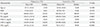
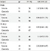
PvuII and XbaI polymorphisms were detected in the study. The frequencies of PP, Pp, pp, XX, Xx, and xx genotypes and alleles in control subjects and the patients were investigated. Both frequencies of p and x alleles were higher than P and X ones. Allele frequencies of both PvuII and XbaI polymorphisms were significantly different between patients and control subjects (P=0.014 vs. P=0.002, respectively), so Table 2 showed that PvuII and XbaI variants were associated to the T2DM. Logistic regression analysis of genotype distribution of PvuII (pp vs. Pp+PP) in both sexes revealed that there was no significantly association in men (P=0.89) and women (P=0.12). When the sex factor was controlled by logistic regression, analysis of genotype distribution of PvuII (pp vs. Pp+PP) found no significant differences in 35 to 44 years (OR, 0.95; 95% confidence interval [CI], 0.48 to 1.85; P=0.88), 45 to 54 years (OR, 1.1; 95% CI, 0.52 to 2.32; P=0.81), and 55 to 65 years age group (OR, 2.32; 95% CI, 0.88 to 6.07; P=0.08), but OR of gene polymorphism ascend with aging (Table 3).
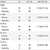
Logistic regressions showed that there was no significant association between men and women in genotype distribution of XbaI (P=0.32 vs. P=0.39). Moreover, after adjusting the sex factor by logistic regression analysis, data showed no significant differences in 35 to 44 years (OR, 1.17; 95% CI, 0.60 to 2.25; P=0.63), 45 to 54 years (OR, 1.33; 95% CI, 0.63 to 2.81; P=0.45), and 55 to 65 years age group (OR, 1.45; 95% CI, 0.55 to 3.80; P=0.44) in genotype distribution of XbaI (Table 4).
Analysis of the association between gene polymorphisms and clinical characteristics in healthy group showed that PvuII and XbaI genotypes were significantly related to high level of FBS (PvuII: OR, 2.7, 95% CI, 1.05 to 6.95, P=0.03; XbaI: OR, 2.51, 95% CI, 1.03 to 6.15, P=0.04) in women and (PvuII: OR, 2.99, 95% CI, 1.31 to 6.80, P=0.008; XbaI: OR, 2.66, 95% CI, 1.20 to 5.90, P=0.01) in 55 to 65 years age group with PP, Pp, XX, and Xx genotypes than in those with pp and xx genotypes. After adjusting age or sex by logistic regression analysis in control group, data showed that PvuII genotype (PP vs. Pp) was significantly related to high level of TC and TG (TC: OR, 3.11, 95% CI, 1.37 to 7.07, P=0.005; TG: OR, 3.07, 95% CI, 1.34 to 7.05, P=0.007) in women and (TC: OR, 2.91, 95% CI, 1.35 to 6.25, P=0.005; TG: OR, 3.32, 95% CI, 1.51 to 7.31, P=0.002) in 55 to 65 years age group (Table 5). Moreover, logistic regression analysis showed that XbaI genotype (XX vs. Xx) was significantly related to high level of TC and TG (TC: OR, 3.07, 95% CI, 1.44 to 6.54, P=0.003; TG: OR, 2.61, 95% CI, 1.25 to 5.45, P=0.009) in women (Table 6).
DISCUSSION
T2DM, which is caused by both acquired and genetic abnormalities, is a multifactorial disease that affects insulin secretion and insulin sensitivity. Hallmarks of T2DM are insulin resistance in liver, skeletal muscle and fat, combined with insulin insufficiency owing to a slump in β-cell function [18]. Recognition of the susceptibility genes for T2DM may come to initial prevention of disease. Genetic changes have an additive and partial effect on T2DM. Moreover, environmental factors play a fundamental role in facilitating or reprieving the expression of the disease.
The distribution of PvuII and XbaI polymorphisms in our study showed an increased ratio of Pp to pp and that of Xx to xx genotypes and a reduced frequency of PP and XX genotypes of patients in comparison to that of Asian and Caucasian populations of European ancestry, even though African population showed a lower frequency of pp and xx genotypes [11,18-21]. As a matter of fact, linkage disequilibrium differential degree among different racial populations may explain previous contradiction among ERα gene polymorphism studies [22]. These frequencies of genotypes may be due to incomplete disequilibrium and may result from multiple mutations and recombination that have occurred at or between polymorphic sites.
Sex steroids apparently have a considerable role in insulin resistance risk. Role of estradiol in adjusting energy metabolism has been reported by recent studies, which revealed new evidences about the important role of ERs [23]. ERs seem to play a role in the occurrence or prevention of T2DM. ERα is a member of the nuclear hormone receptor superfamily and regulates the transcription of target genes in response to estrogen [24]. In the present study, there was an association between PvuII and XbaI polymorphisms in ERα and T2DM. There are few studies on the relationship of PvuII and XbaI polymorphisms with diabetes. Our study reports a significant association between both ERα gene polymorphisms (PvuII and XbaI) and T2DM in both men and women subjects of the inpatient population. A gender specific association has been reported from Hungrian and Chinease populations [5,23]. A study of 100 Iranian with T2DM and control group showed no significant association between PvuII and XbaI polymorphisms with diabetes in women [8]. In a study of Swedish population, no significant association was found between PvuII and XbaI polymorphisms and T2DM [25]. Our finding did not correlate with these studies, while a study in China supports our study in which Huang et al. [20] reported that PvuII gene polymorphism in ERα is associated with T2DM. However, in contrast to our data, they did not find any association between XbaI polymorphisms and T2DM. The exact reasons behind these discrepancies are not entirely clear; recruitment procedures and differences in environmental background and/or genetics may have played a role.
PvuII and XbaI genes polymorphisms are possible markers for several diseases. These genotypic variations have been implicated in development of multitudinous diseases, among which are osteoporosis, cancers, cardiovascular diseases, neurodegenerative diseases, and lupus erythematosus [11,12,26]. Unfortunately, how these genetic polymorphisms influence receptor activity is still unknown. It has been suggested that a binding site produced by C allele of PvuII is for B-myb transcription factor. Estrogen induces B-myb's expression which can accrue transcription the construct 10-fold of a downstream reporter [27]. It is concluded that C allele might produce ERα isoforms or amplify the ERα transcription. A to G transition of XbaI may also have an effect on expression and alteration of the ERα.
Huang et al. [20] confirmed that PvuII and XbaI polymorphisms were more frequent in female with T2DM, but our data showed no significant differences of PvuII and XbaI polymorphisms distribution between female and male. The reason for this discrepancy can be ascribed to several factors such as sample size, the study design, gene-environment interactions, and population heterogeneity. After logistic regression analysis, OR showed that PvuII and XbaI polymorphisms in ERα increased with the aging; although it could be because small sample size in our study rendered this association statistically insignificant. FBS is an important factor to judge the T2DM, so data from healthy group suggest the possibility that both PvuII and XbaI polymorphisms in ERα is significantly related to the development of female with T2DM, and improved with increasing age. It suggests that PvuII and XbaI polymorphisms in ERα might be a risk factor for T2DM in women. Nevertheless, because the data is from the control group, the results can be associated with the metabolic syndrome, not with T2DM, thus it is a logic jump to assert that the polymorphisms are related to T2DM. On the contrary, these finding shows that difference in the locations of the two SNPs does not influence in the roles. However, further studies are needed to confirm these finding.
An association between obesity, hypertriglyceridemia, and T2DM has been detected since the early 1960s [28,29]. It is clear that estrogen affects the lipoprotein metabolism in many potential beneficial ways. Estrogen increases the rates of all lipoprotein fractions formation so it leads to an increase in HDL-C, a decrease in LDL-C, and an increase in the mRNA for LDL receptor [6]. Our data revealed that PvuII polymorphisms are related to high serum lipids of TC and TG, that is high serum lipid can increase the risk of T2DM in healthy group. Several other studies including a study of Italians indicating that PvuII polymorphisms are strongly associated with familial hypercholesterolemia, confirmed our finding [30-33].
The present study had some advantages and limitations. The first advantage of this study is that our studied population was collection of a homogeneous sample well characterized controls and cases that increases the sensitivity of detecting the associations. The second advantage of this study is that we do not dichotomize continuous variables data which gives an additional impact on exactness. We acknowledge that the number of sample size might be small in the population and this doesn't allow drawing any definitive conclusions. Therefore, a larger population is required to establish a definitive role for these variations in the inpatient population.
In conclusion, findings of present study suggest the possibility that PvuII and XbaI polymorphisms in ERα are related with T2DM in inpatient men and women population. It would be invaluable to conduct studies in other heterogeneous populations in order to check the replication validity of present findings. It is suggested that future studies focus on the role of ERα in the progression and development of different diseases which may help to identify therapeutic or diagnostic markers.


 XML Download