

 PDF
PDF ePub
ePub Citation
 Print
Print


INTRODUCTION
Gestational diabetes mellitus (GDM) is a subtype of diabetes that occurs during pregnancy, and its prevalence in females who have been pregnant is 3% to 8% worldwide. A Korean survey showed that the prevalence of GDM is 2.2% [1]. Given that the number of obese and older pregnant women is increasing, the number of cases of gestational diabetes is continuously increasing as well [2]. Women who have been diagnosed with gestational diabetes have a high probability of developing type 2 diabetes after giving birth, and the incidences of atherosclerosis and cardiovascular disease risk factors are also higher than those in normal women [3,4].
Recently, a study to predict or diagnose cardiovascular disease was performed by measuring carotid intimal-medial thickness (CIMT). Studies have shown that CIMT tends to increase with age, and that CIMT values of smokers and patients with dyslipidemia, hypertension, and type 2 diabetes are increased compared with those of control groups [5-10]. In addition, in individuals with thicker CIMTs, the risk of stroke and coronary artery disease is known to be higher [11-13].
The association between GDM and CIMT has not yet been clearly established. However, females who have a history of GDM have been reported to have increased CIMTs compared to those of females with normal pregnancies [14,15]. On the other hand, in a comparison of carotid and femoral artery intimal- medial thickness (IMT) in females who had insulin resistance associated with pre-eclampsia, Blaauw et al. [16] reported increased femoral artery IMTs in these females compared to those of the normal pregnancy group, and that there was no significant difference between the two groups in CIMT value.
In this study, the CIMTs of Korean women with a history of gestational diabetes were measured using a high-resolution B-mode ultrasound. Results were examined to determine if there was a difference between the CIMT of women with a history of GDM and that of women who had normal pregnancies and if there is a connection among CIMT, cardiovascular risk factors and glucose intolerance in women with history of GDM.
METHODS
Target group
This study was performed between January 1999 and December 2002 with females who were diagnosed with gestational diabetes between the 24th and 28th week of pregnancy at the Department of Endocrinology and Metabolism Internal Medicine, Cheil General Hospital. Among the diagnosed patients, 101 agreed to participate in this study. The gestational diabetes diagnostic criteria used in this study are those recommended at the Third International Workshop-Conference on GDM [17]. The control group (normal pregnancy, NP) was comprised of women who had a negative result on gestational diabetes screening tests performed during a previous pregnancy. A total of 19 females were enrolled who had no family history of diabetes. There were no statistical differences in age or body mass index (BMI) between the two study groups.
This study was approved by the Institutional Review Board of Seoul National University Hospital. The study participants received a summary of the study purpose and method and submitted written consent prior to participation in the study.
Study method
One year after women with a history of GDM gave birth, and one to two years after women in the control group gave birth, CIMT and cardiovascular risk indicators were measured. Body measurements, including height, weight and waist and hip circumference, were collected from all subjects. The area of abdominal fat was measured using a CT scan (Somatom Sensation 16; Siemens, Erlangen, Germany), an image was taken at the navel with 10 mm thickness and the areas between -250 to -50 Hounsfield units were measured. The images were edited to maintain only the portion containing the innermost abdominal muscles, while the rest was deleted, and the internal fat was used for measurements.
A 75 g oral glucose tolerance test was performed after an eight hour fast, and fasting plasma glucose, insulin, glycated hemoglobin and plasma lipid concentrations were measured. The plasma glucose levels were measured using the oxidase method YSI 2300 STAT (Yellow Springs Instrument Co., Yellow Springs, OH, USA), glycated hemoglobin was measured using the principles of a variant of ion-exchange high performance liquid chromatography (Bio-Rad Laboratories, Hercules, CA, USA) and plasma insulin concentration was measured using a human-specific radioimmunoassay kit (Linco Research, St. Charles, MO, USA). Total cholesterol, triglycerides, and high density lipoprotein/low density lipoprotein cholesterol (HDL-C/LDL-C) levels were measured using a Hitachi 747 autoanalyzer (Hitachi Ltd., Tokyo, Japan), and lipoprotein concentration was calculated using the Friedewald formula: ([total cholesterol-HDL-C-triglyceride]/5) [18]. The homeostasis model assessment of insulin resistance (HOMA-IR) was used as an indicator of insulin resistance and was calculated using the following formula: fasting plasma glucose (mmol/L)×fasting plasma insulin (mU/L)/22.5 [19].
Carotid IMT measurement
One technician measured CIMTs of all subjects using a high-resolution B-mode ultrasound on each side of the carotid artery. Starting 1 cm from the proximal end of the left and right carotid arteries, the CIMT was measured at ten 1 mm intervals, for a total of 20 locations on the distal walls, using a Digmatic calipus (CD-15B; Mitutoyo, Tokyo, Japan) caliper, and the mean value was calculated (coefficient of variation value, 9%).
Data analysis
The statistical analysis of the data was conducted using SPSS version 12.0 (SPSS Inc., Chicago, IL, USA). Data that followed a normal distribution are expressed as mean±standard deviation, and variables that did not follow a normal distribution were logarithmically transformed prior to statistical analysis. Results are presented as medians (interquartile range). The characteristics of the GDM group and the NP control group were compared using an independent t-test. A linear regression analysis was used for women with a history of GDM in order to analyze the associations between CIMT and cardiovascular risk factors, and the association between CIMT and glucose intolerance was verified using ANOVA.
RESULTS
Clinical characteristics of women with a history of GDM versus those of the NP control group
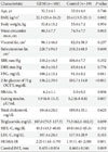
In this study, according to the physical measurements taken one year after delivery, the waist circumference of the females with a history of gestational diabetes group was thicker than that of the control group, but there was no difference in BMI or internal fat mass. In the results of the 75 g oral glucose tolerance test, the fasting plasma glucose levels were 100.2±15.1 mg/dL in the GDM group vs. 91.0±8.1 mg/dL in the NP control group (P=0.011). Two hours after the glucose tolerance test, blood glucose levels were 136.2±39.9 mg/dL in the GDM group vs. 105.7±14.0 mg/dL in the NP group (P<0.001). The results from the GDM group were statistically and significantly higher. The glycated hemoglobin levels were higher in the GDM group (6.3±1.1% in GDM vs. 5.9±0.5% in NP, P=0.096), but this difference was not statistically significant. The fasting plasma insulin concentration was 9.9 (6.8 to 15.3) µU/mL in the GDM group vs. 8.2 (6.8 to 8.8) µU/mL in the NP group (P=0.007), and the HOMA-IR results in women with a history of GDM appeared to be significantly higher: 2.25 (1.61 to 3.79) in the GDM group vs. 1.91 (1.41 to 2.00) in the NP group (P=0.001). Total cholesterol, triglycerides, HDL-C and LDL-C concentrations were similar in both groups. When CIMT results from the GDM group were compared with those of the NP group, there did not appear to be a statistically significant difference: 0.435±0.054 mm in GDM vs. 0.460±0.046 mm in NP, respectively (P=0.069) (Table 1).
Cardiovascular risk factors that affect the CIMT of females with a history of gestational diabetes
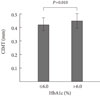
In order to determine the factors affecting CIMT in women with a history of GDM, a linear regression analysis that adjusted for age was performed. Waist circumference, blood pressure, fasting glucose, fasting insulin, and lipids did not affect CIMT. However, as glycated hemoglobin increased, CIMT increased as well (P=0.011) (Table 2). When the glycated hemoglobin group was separated into two groups, a control group (glycated hemoglobin ≤6.0%) and a high glycated hemoglobin group (glycated hemoglobin >6.0%), and compared, the CIMT of the high glycated hemoglobin group was significantly thicker: 0.420±0.047 mm vs. 0.448±0.057 mm, respectively (P=0.011) (Fig. 1).
Differences in CIMT based on glucose tolerance
Among the 101 females who had histories of GDM, 94 received a 75 g oral glucose tolerance test six weeks and again one year after delivery. When the glucose tolerance state was judged based on the test results after six weeks, 49 patients (52.1%) had normal glucose tolerance (NGT), 11 patients (11.7%) had impaired fasting glucose (IFG), 14 patients (14.9%) had impaired glucose tolerance (IGT), 16 patients (17.0%) had both impaired fasting glucose and impaired glucose tolerance (IFG/IGT) and four patients (4.3%) were diagnosed with diabetes (DM). After one year had passed, 31 patients (33.0%) had NGT, 16 patients (17.0%) had IFG, 16 patients (17.0%) had IGT, 22 patients (23.4%) had IFG/IGT and nine patients (9.6%) were diagnosed with DM.
No statistically significant difference was seen at six weeks or one year when the differences in IMT were compared among all five groups (Table 3). In addition, the results were the same whether the IFG group, IGT group, and the IFG/IGT group were analyzed together or separated into three groups (Table 4). There was also no difference observed when the NGT and DM groups were compared (data not shown).
DISCUSSION
Placental growth hormone and placental lactogen are hormones produced by the placenta during pregnancy. During the late stages of pregnancy, the concentrations of insulin antagonizing hormones, such as prolactin, cortisol and glucagon, and cytokines such as tumor necrosis factor-alpha (TNF-α) increase, and insulin resistance is lower [20-22]. During normal pregnancies, the compensatory insulin secretion from the pancreas increases. However, when adequate amounts of insulin are not secreted to overcome the compensatory insulin resistance, gestational diabetes occurs [23]. Compared with normal pregnancies, women who previously had GDM have a higher risk for developing type 2 diabetes, and as a result, future risk of cardiovascular complications have been known to increase [24,25].
In this study, the clinical characteristics of females who had gestational diabetes and of females with a normal pregnancy were compared. The results showed that, compared to normal pregnancies, women who had gestational diabetes had higher concentrations of insulin and had higher insulin resistance. As mentioned earlier, during pregnancy, the effects of several physiological hormones increase insulin resistance. Particularly in patients with gestational diabetes who suffer from chronic insulin resistance, as physiological insulin resistance increases, they develop a higher level of insulin resistance compared to that in women with normal pregnancy [26-28], as was shown in the present study.
Considering that the patients with a history of gestational diabetes are at future risk for cardiovascular disease, we measured the CIMT in such patients, and the resulting correlations with cardiovascular risk factors were analyzed. When a multiple linear regression analysis was performed, none of waist circumference, blood pressure, internal fat, HDL- and LDL-C or triglycerides showed a significant correlation with CIMT. However, as glycated hemoglobin increased, the CIMT significantly increased as well. In previous studies performed on people with impaired glucose tolerance, CIMT was reported to increase as glycated hemoglobin increased [29], and studies targeting type 2 diabetes patients reported that there was a positive correlation between CIMT and glycated hemoglobin levels [30,31]. In the present study, a statistical significance in the increase in glycated hemoglobin levels and CIMT was observed in women with a history of GDM.
In this study, we observed no difference in CIMT between the two study groups. When CIMT results of the GDM and control groups in Western countries were analyzed in a previous study, the results showed that women with histories of GDM were reported to have thicker CIMT measurements compared to those of normal healthy females [14]. In that study, the group that had a history of gestational diabetes had a mean CIMT of 0.582±0.066 mm, while that of the control group was 0.543±0.049 mm, showing that the group with a history of GDM had significantly thicker CIMT [14]. However, in our study, the mean CIMT of women with history of GDM was 0.435±0.054 mm, and the mean CIMT of the control group was 0.460±0.046 mm, which was significantly lower than those in the Western cases. Ethnic background and culturally-based obesity have been suggested as causes to explain this difference. One major factor that influences CIMT is obesity [32]. However, compared to Western populations, obesity is much less common in Koreans, in whom the average overall BMI is also much lower. The BMI of the GDM group and the NP group in this study fell within the normal range. By way of comparison, in the Western study [14], the mean BMI of the GDM group was 28.65±4.75 kg/m2, and that of the control group was 27.17±2.90 kg/m2, which both fall in the overweight range. The differences in the weight ranges are believed to influence the CIMT measurements. In addition, the average BMI of the women who had a history of GDM in our study was low compared to the average BMIs of study groups in other countries. Therefore, the inclusion of a high number of very lean GDM patients is considered to be a limitation which may have affected the study results.
In addition, one thing that must be considered when interpreting the results for CIMT measurements is the age of our study population. The mean CIMTs of Korean females in their 30s and 40s are 0.57±0.10 mm and 0.60±0.10 mm, respectively, which seems to increase gradually with age [33]. However, in the present study, the mean CIMT values of patients in their 30s were significantly lower than those of patients in their 40s. The mean age of the GDM group in this study was 32.2±4.1 years, and the mean age of the NP group was 32.6±4.6 years. In addition, some patients were still in their 20s. Due to their young ages, their results were not considered to contribute to the determination of significant changes in blood vessels due to arteriosclerosis. Finally, the size of the study groups could have influenced the results. There were 101 patients in the GDM group, but the NP group consisted of only 19 patients.
In this study, despite the similarities in CIMT results between the GDM groups and the NP group, the insulin resistance of the GDM group was greater than that of the NP group, and we confirmed that increases in glycated hemoglobin occurred with increases in CIMT in the GDM group. Considering the young age of the study group, it is difficult to observe a difference between the CIMT measurements of young women with history of GDM and women without, and there was no visible difference or correlation with existing known cardiovascular risk factors. Therefore, it was difficult to use CIMT as a measure to predict future risk of atherosclerosis. In addition, as seen in previous studies, one year after giving birth, the group that transitioned to diabetes was limited by its small size. When the significant correlation of CIMT and glycated hemoglobin in females with history of gestational diabetes who have increasing insulin resistance and who are considered at high risk for cardiovascular disease is considered, those women can be expected to experience significant changes in CIMT and cardiovascular risk factors. Therefore, in the examinations performed shortly after giving birth in Korean women with history of gestational diabetes, although CIMTs in the normal range and normal glucose tolerances were seen, as time passes and the rate of those who transition to diabetes increases, and when the accompaniment of hyperlipidemia and cardiovascular risk factors are considered, follow-up visits would be helpful. This study has been performing prospectively, the difference in CIMT between the transitioned to diabetes group and the non-transitioned to diabetes group will be determined sooner or later. These findings should be considered in women with GDM.


 XML Download