

 PDF
PDF ePub
ePub Citation
 Print
Print


INTRODUCTION
It is well known that the renin-angiotensin-aldosterone (RAA) system is an important pathway of progression in cardiovascular disease, diabetic nephropathy, and chronic renal disease through a mechanism of inflammation, fibrosis, and necrosis [1,2]. For this reason, angiotensin converting enzyme inhibitor (ACEI) and angiotensin receptor blocker (ARB) are effective in the treatment of chronic heart failure and diabetic nephropathy [3-6]. However, long term use of RAA system blockade therapies have been shown to result in increased plasma aldosterone levels in 40% of patients with diabetic nephropathy and 20% of patients with chronic renal failure. This condition is referred to as the aldosterone escape phenomenon [7]. The proteinuria-reducing effects of ACEI and ARB are also decreased in these patients [8]. It is expected that a reduction in aldosterone levels alone could be a crucial treatment target in diabetic nephropathy.
Recent data suggests that an aldosterone receptor blocker could reduce proteinuria by decreasing various growth factors [9,10]. It has also been reported that aldosterone receptor blocker treatment can reduce proteinuria in patients with chronic renal disease who did not respond to ACEI therapy [11].
Vascular endothelial growth factor (VEGF), a strong angiogenic factor, is known to play a major role in the neovascularization of atherosclerotic plaques and solid cancers [12,13], and in the progression of diabetic nephropathy [14]. Although it is reported that ARB treatment protects against diabetic nephropathy by reducing VEGF [15,16], there is no data demonstrating the effect of aldosterone receptor blocker therapy on renal VEGF expression.
In this study, we investigated the effects of losartan, spironolactone, and a combination of these two drugs on albuminuria and renal VEGF expression in a type 2 diabetic rat model.
METHODS
Experimental rats
Thirty-three male Otsuka-Long-Evans-Tokushima-Fatty rats (OLETF rats; Otsuka Pharmaceutical, Tokushima, Japan) were divided into four groups and received different medication regimens: spironolactone (50 mg/kg/day), losartan (20 mg/kg/day), or a combination of the two from weeks 25 to 50. The first group consisted of untreated diabetic OLETF controls (CO; n=5), the second group was spironolactone-treated diabetic rats (SPR; n=10), the third group was losartan-treated diabetic rats (LO; n=9) and the fourth group was spironolactone and losartan combination-treated diabetic rats (COM; n=10). All groups had access to standard rat chow and drinking water, ad libitum. This research protocol was approved by the animal ethics committee of the Yonsei University Wonju College of Medicine (Wonju, Korea).
Basic parameters
Body weights and blood glucose levels (Surestep; Lifescan Inc., Milpitas, CA, USA) were recorded at weeks 15, 30, and 50. Blood pressure was measured using tail-cuff plethysmography at weeks 30 and 50. Twenty-four hour urines were collected at weeks 15, 30, and 50 for measuring and urine protein levels (Roche Molecular Biochemicals, Indianapolis, IN, USA) and albumin-creatinine ratio (ACR) values (by ELISA; Shibayagi, Shibukawa, Japan) were determined.
Kidney extraction
At week 50, all experimental rats were sacrificed under anesthesia by intraperitoneal injection of Zoletil® (30 mg/kg). One kidney was rapidly fixed in 4% paraformaldehyde for 24 hours and paraffin-embedded for immunohistochemical study and histologic examination. The other kidney was flash frozen in liquid nitrogen for subsequent protein and RNA extraction. Frozen kidneys were stored at -70℃ until analyzed by Western blot and real time reverse transcription-polymerase chain reaction (RT-PCR) for VEGF, transforming growth factor (TGF)-β, and collagen type IV.
Histologic examination of kidney
Paraffin-embedded kidney tissues were cut into 7 µm sections and stained with periodic acid-Schiff (PAS). A glomerular matrix index (GMI) score was measured and the degree of sclerosis for each glomerulus was graded from 0 to 4 as follows [17]: grade 0: normal; grade 1: mild sclerosis (less than 25% of glomerulus); grade 2: moderate sclerosis (25 to 50% of glomerulus); grade 3: moderate-severe sclerosis (50 to 75% of glomerulus); grade 4: severe sclerosis (75 to 100% of glomerulus). Ten glomeruli were scored in each kidney section.
Immunohistochemical staining of VEGF
Tissue sections were affixed onto slides and deparaffinized for immunohistochemical staining. The slides were then transferred to a 10-mmol/L citrate buffer solution (pH 6.0), washed with distilled water and blocked with 0.05% H2O2-methanol for 15 minutes. A 1:1,000 dilution of anti-VEGF monoclonal antibody (Santa Cruz Biotechnology Inc., Santa Cruz, CA, USA) was applied and slides were incubated at room temperature. A biotinylated secondary antibody from a rat ABC staining kit (Santa Cruz Biotechnology Inc.) was used followed by incubation in a horseradish peroxidase Avidin-Biotin Complex solution (ABC reagent). The slides were then incubated in a peroxidase substrate containing 0.05% 3,3'-diaminobenzidine tetrahydrochloride (DAB). Stained sections were viewed with a light microscope equipped with a charge-coupled device camera (Pulnix America Inc., Sunnyvale, CA, USA) and glomerular VEGF optical densities were measured by image analysis.
Real time RT-PCR
Total RNA was extracted from frozen kidney tissues using TRIzol® LS (GIBCO-BRL, Grand Island, NY, USA) and then reverse transcribed into cDNA using oligo-(dT) primers (Promega, Madison, WI, USA). Real time RT-PCR was performed using a SYBR Green RT-PCR kit (Qiagen, Valencia, CA, USA) and analyzed with a Rotor-Gene RG-3000 cycler (Corbett Research, Mortlake, NSW, Australia). Primer oligonucleotide sequences for VEGF, TGF-β, collagen type IV and GAPDH are as follows:
VEGF forward: 5'-GTATATCTTCAAGCCGTCCTGTGTG-3', VEGF reverse: 5'-GATCCGCATGATCTGCATAGTGAC-3', TGF-β forward: 5'-TGAGTGGCTGTCTTTTGACG-3', TGF-β reverse: 5'-TGGGACTGATCCCATTGATT-3', Collagen type IV forward: 5'-CCAGGATTCCAAGGTCAGAA-3', Collagen type IV reverse: 5'-CCCTGGTTCTCCTTTGATGA-3', GAPDH forward: 5'-TCAGGTCATCACTATCGGCAATG-3', GAPDH reverse: 5'-GGAATTGAATGTAGTTTCATGGATGC-3'
Real-time RT-PCR amplification was performed with one cycle at 95℃ for 10 minutes followed by 40 cycles that consisted of denaturation for 15 seconds at 94℃, annealing for 30 seconds at 58℃ and extension for 30 seconds at 72℃. After rinse and melting processes, fluorescent products were detected during the 92℃ extension cycle. The cycle threshold (ΔCt=Ct VEGF-Ct β-actin) of each sample was calculated and the relative change ratio determined using an mRNA ratio of VEGF/β-actin.
Western blot analysis
The cortex of each kidney was homogenized in RIPA buffer, incubated on ice for 20 minutes and centrifuged at 15,000 rpm to remove cellular debris. 10 µg samples of protein lysate were electrophoresed on 10% SDS-PAGE gels at 100 V and transferred onto polyvinylidene fluoride (PVDF) membranes for 1 hour at 280 mA in a Tris-based buffer. Non-specific binding sites were blocked with a 5% non-fat dried milk solution for 1 hour. Membranes were incubated overnight with anti-rat β-actin antibody (1:2,000 dilution; Cell Signaling Technology Inc., Beverly, MA, USA) or VEGF antibody (1:1,000 dilution; R&D System, Minneapolis, MN, USA) followed by incubation with anti-rabbit IgG or anti-goat IgG HRP antibodies. Signal was visualized using the ELC Western Blotting Analysis System (Amersham Biosciences, Buckinghamshire, UK).
Monocyte chemotactic protein-1 (MCP-1) and malondialdehyde (MDA)
Twenty-four hour urine MCP-1 levels were measured by quantitative sandwich ELISA (Biosource Inc., Camarillo, CA, USA) and used as an inflammatory marker. Urine MDA levels were measured by a rapid and sensitive fluorometric HPLC method and used as an oxidative stress marker [18].
Statistical analyses
All data are presented as mean±standard deviation. The data were analyzed statistically using one-way ANOVA and the Tukey test (multiple comparisons). All analyses were performed using a Windows-based, SPSS statistical package version 12.0 (SPSS Inc., Chicago, IL, USA). P<0.05 was considered to be statistically significant.
RESULTS
Basic parameters of experimental rats
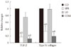
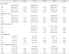
There were no significant differences in body weights and plasma glucose levels at each time point. However, blood pressure levels of the COM group at week 50 were significantly lower than that of the CO group (Table 1). At weeks 15 and 30, there were no differences in 24-hour urine protein levels and ACR among all groups. At week 50, the LO and COM groups showed significant decreases in 24-hour urine protein levels and ACR, but the SPR group did not show reduced proteinuria and ACR when compared to the CO group (Table 1).
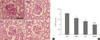
PAS staining of glomeruli and GMI score
Glomerular mesangial expansion was observed in the CO group compared to the other groups in PAS staining of glomeruli (Fig. 1A). The GMI scores were significantly decreased in all medicated groups compared to the CO group. Also, the COM group showed a marked decrease in GMI scores compared to the SPR and LO groups (Fig. 1B).
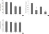
Analysis of renal VEGF expression
Glomerular VEGF immunohistology revealed darker staining in the CO group compared to all of the medication treated groups (data not shown). The optical density of immunohistochemical staining for VEGF in the LO and COM groups was significantly decreased compared to the CO group. The SPR group showed no appreciable difference when compared with the CO group (Fig. 2A). Real time RT-PCR analysis revealed that VEGF mRNA expression was 0.35-fold in the SPR group, 0.67-fold in the LO group, and 0.22-fold in the COM group when compared to the CO group (Fig. 2B). Western blot analysis, however, did not show any significant differences between the groups (Fig. 2C).
DISCUSSION
Our data shows that losartan (ARB) alone or a combination treatment consisting of losartan and spironolactone (aldosterone receptor blocker) can reduce proteinuria by reducing renal VEGF expression in a type 2 diabetic rat model.
Diabetic nephropathy is the most common cause of end-stage renal disease (ESRD). Since diabetic ESRD patients are more prone to cardiovascular mortality than other ESRD patients, early identification of diabetic nephropathy and prompt renoprotective treatment are critical for the prevention of end organ damage from diabetic nephropathy [19].
It is well known that therapies with ACEI and ARB can retard the progression of diabetic nephropathy, chronic heart failure and chronic renal failure by reducing blood pressure and by decreasing inflammatory and sclerosing effects [3-6]. Recent data indicates that spironolactone, an aldosterone receptor blocker, can reduce proteinuria by providing anti-inflammatory protection and by decreasing oxidative stress [10,20]. In our study, losartan- and the combination regimen-treated rat groups showed significant decreases in 24-hour urine protein levels and ACR while the spironolactone monotherapy group did not show these significant reductions. This discrepancy may be explained by the differences in treatment initiation time periods. Recent studies reported that treatment with aldosterone receptor blockers in the early stages of nephropathy may reduce proteinuria but has no beneficial effects at later stages of the disease [21,22]. Since we administered spironolactone treatment as late as week 25, its protective actions against diabetic nephropathy could have been inadequate compared to other studies that initiated treatment at earlier stages [10]. Another possible explanation is the different effects of spironolactone on hemodynamics and the fibrosis process. We observed decreased mesangial expansion and sclerosis in both the SPR and COM groups, which is consistent with results from previous studies [23,24]. However, there were no differences in blood pressure between the groups. It could be explained that spironolactone can act on diabetic nephropathy by non-hemodynamic mechanisms [25].
In diabetic nephropathy, VEGF expression may be increased by various growth factors including platelet derived growth factor and the accumulation of hyperglycemia-induced, advanced glycation end products [26,27]. Recent studies revealed that ACEI and ARB can decrease proteinuria by reducing VEGF expression in type 2 diabetic rats [15,28]. Our data also suggests that a combination therapy with losartan and spironolactone may affect diabetic nephropathy by reducing VEGF expression despite incidental, disparate results between VEGF mRNA expression, immunohistochemical staining, and Western blot analysis. The discrepancies between mRNA expression and protein levels may be due to the interpretation of our data, which was in part based on inconsistent tissue sampling. While we could analyze individual glomeruli by immunohistochemical staining, we could not isolate glomeruli from tubules and interstitial tissue when extracting total RNA and protein for real time RT-PCR and Western blot analysis.
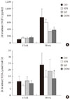
Activation of the renin-angiotensin-aldosterone (RAA) system causes increased production of reactive oxygen species through activation of the NADPH oxidase enzymatic complex in numerous tissues, including the kidney [29,30]. Angiotensin and other cytokines can also cause diabetic nephropathy through inflammatory mechanisms [29,30]. Blockade of the RAA system may have protective effects against diabetic nephropathy through anti-oxidative and anti-inflammatory mechanisms [20]. In our study, only a combination therapy of losartan and spironolactone reduced MDA levels. MCP-1 levels showed a slight decreasing trend in the combination regimen treated group but this was not statistically significant. These results indirectly explain that spironolactone and losartan combination therapy may be an effective treatment regimen for diabetic nephropathy by reducing oxidative stress.
In conclusion, we suggest that ARB therapy in combination with ARB and aldosterone receptor blocker therapies may have protective effects against diabetic nephropathy by reducing VEGF expression. We also propose that combination treatments could reduce proteinuria by anti-oxidative mechanisms and by decreasing TGF-β and type IV collagen expression.
 XML Download