Abstract
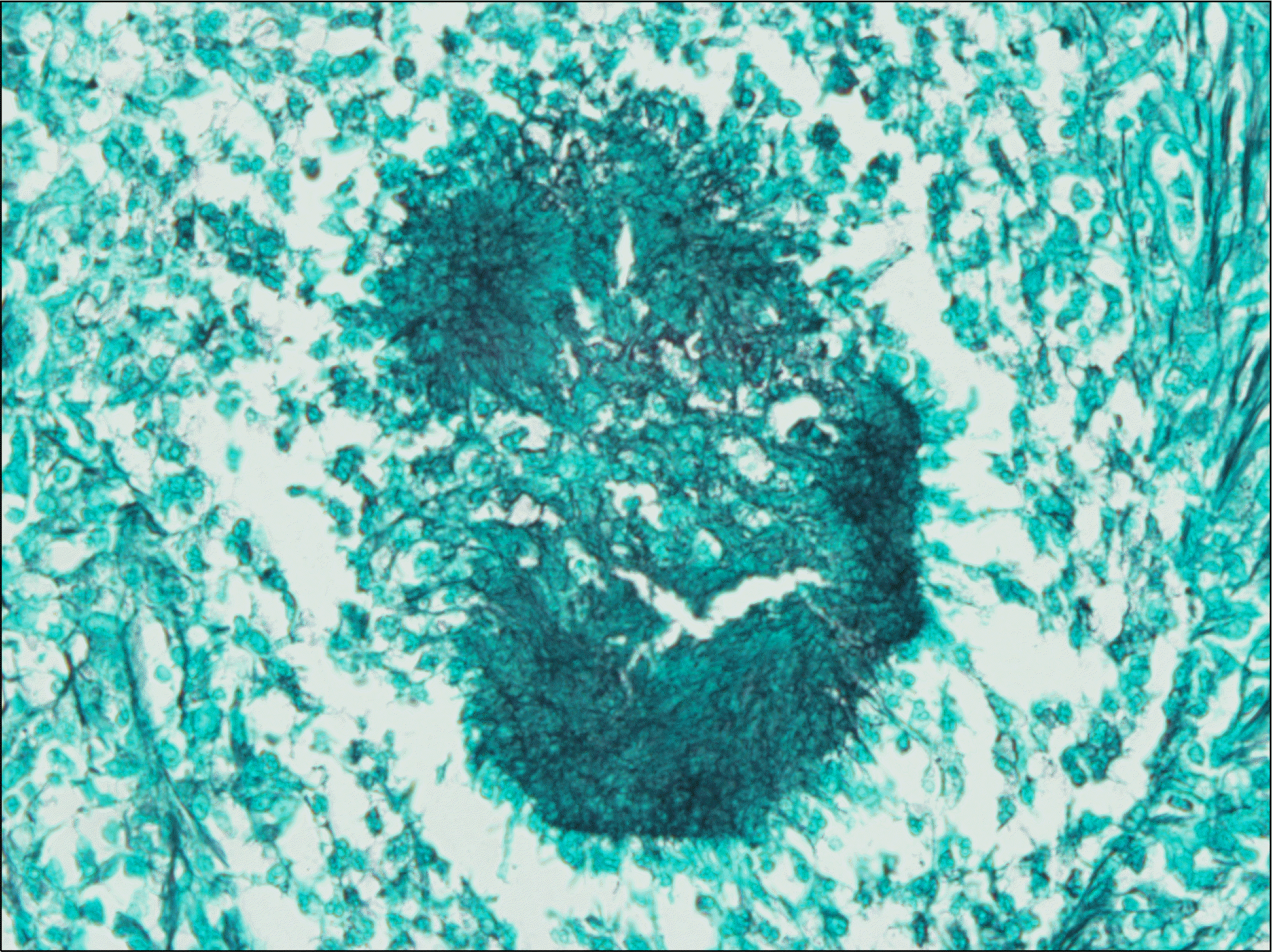
This case report describes an uncommon case of renal actinomycosis in a 63-year-old man. The patient underwent radical nephrectomy for suspicious renal cell carcinoma with renal vein thrombosis and spinal metastasis. The postoperative diagnosis of renal and spinal actinomycosis was established in accordance with the results from histological examination. Three years after surgery, the patient did not show any symptoms of recurrence.
REFERENCES
1.Mueller MC., Ihrler S., Degenhart C., Bogner JR. Abdominal actinomycosis. Infection. 2008. 36:191.

2.Das N., Lee J., Madden M., Elliot CS., Bateson P., Gilliland R. A rare case of abdominal actinomycosis presenting as an inflammatory pseudotumour. Int J Colorectal Dis. 2006. 21:483–4.

3.Lee IJ., Ha HK., Park CM., Kim JK., Kim JH., Kim TK, et al. Abdominopelvic actinomycosis involving the gastrointestinal tract: CT features. Radiology. 2001. 220:76–80.

4.Wagenlehner FM., Mohren B., Naber KG., Mannl HF. Abdominal actinomycosis. Clin Microbiol Infect. 2003. 9:881–5.

5.Chang DS., Jang WI., Jung JY., Chung S., Choi DE., Na KR, et al. Renal actinomycosis with concomitant renal vein thrombosis. Clin Nephrol. 2012. 77:156–60.

6.Hayashi M., Asakuma M., Tsunemi S., Inoue Y., Shimizu T., Komeda K, et al. Surgical treatment for abdominal actinomycosis: a report of two cases. World J Gastrointest Surg. 2010. 2:405–8.

Fig. 1.
An abdomino-pelvic computed tomography scan showed two exophytic renal masses—1.5 cm and 3.7 cm in size (A, B)—with renal vein thrombosis (C, arrow). The masses showed unclear margins with possible direct invasion to the ascending colon (arrows of A, B). (D) A magnetic resonance imaging scan showed a 2-cm soft-tissue mass on the anterior portion of the L2 vertebral body with cortical disruption (arrow).


 PDF
PDF ePub
ePub Citation
 Print
Print


 XML Download