

 PDF
PDF ePub
ePub Citation
 Print
Print


Abstract
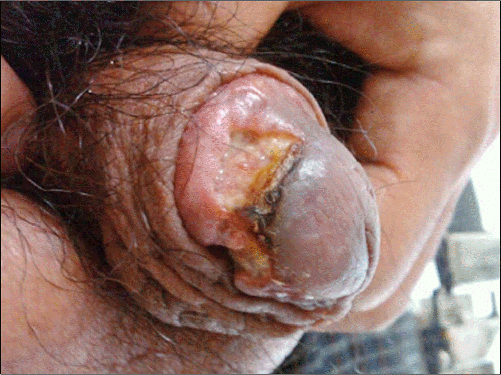
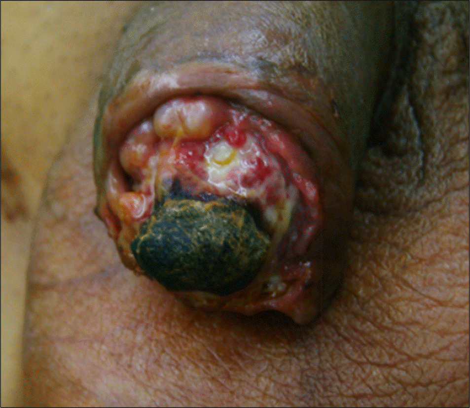
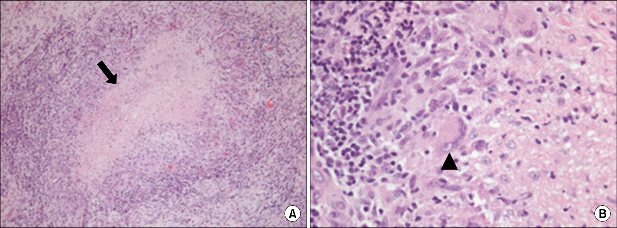
Tuberculosis of the glans penis is rare. The clinical features of tuberculosis of the glans penis include the appearance of a superficial ulcer of the glans. We recently experienced a case of tuberculosis of the glans penis presenting as an ulcer, which progressed to glans necrosis. A 46-year-old man presented at our institution with painful ulcerative penile lesions. Initial pathological findings of the ulcers showed non-granulomatous inflammatory changes. He was treated with antibiotics and anti-inflammatory drugs for 6 months, and during that time, his glans ulcers progressed to glans gangrene. A partial glansectomy showed multiple epithelioid granulomas with central caseous necrosis, which was compatible with the findings of tuberculosis. The patient received anti-tuberculosis chemotherapy. The current case is reported to alert physicians to consider the possibility of tuberculosis when evaluating unusual penile glans lesions.
References
1. Lee JY, Park HY, Park SY, Lee SW, Moon HS, Kim YT, et al. Clinical characteristics of genitourinary tuberculosis during a recent 10-year period in our center. Korean J Urol. 2011; 52:200–5.
2. Jacob JT, Nguyen TM, Ray SM. Male genital tuberculosis. Lancet Infect Dis. 2008; 8:335–42.

3. Yonemura S, Fujikawa S, Su JS, Ohnishi T, Arima K, Sugimura Y. Tuberculid of the penis with a scab on the nodule. Int J Urol. 2004; 11:931–3.
4. Jaisankar TJ, Garg BR, Reddy BS, Riba B, Ramarao AP. Penile lupus vulgaris. Int J Dermatol. 1994; 33:272–4.
5. Kar JK, Kar M. Primary tuberculosis of the glans penis. J Assoc Physicians India. 2012; 60:52–3.
6. Sah SP, AshokRaj G, Joshi A. Primary tuberculosis of the glans penis. Australas J Dermatol. 1999; 40:106–7.
7. Richard BO, William DJ, Timothy GB. Disease of the skin. Philadelphia: WB Saunders;2000.
8. Yanagihara H. Risk of tuberculosis infection among care workers during an outbreak of tuberculosis at a care facility for the elderly. Kekkaku. 2014; 89:631–6.
9. Brisson-Noel A, Gicquel B, Lecossier D, Levy-Frebault V, Nassif X, Hance AJ. Rapid diagnosis of tuberculosis by amplification of mycobacterial DNA in clinical samples. Lancet. 1989; 2:1069–71.
 XML Download