Abstract
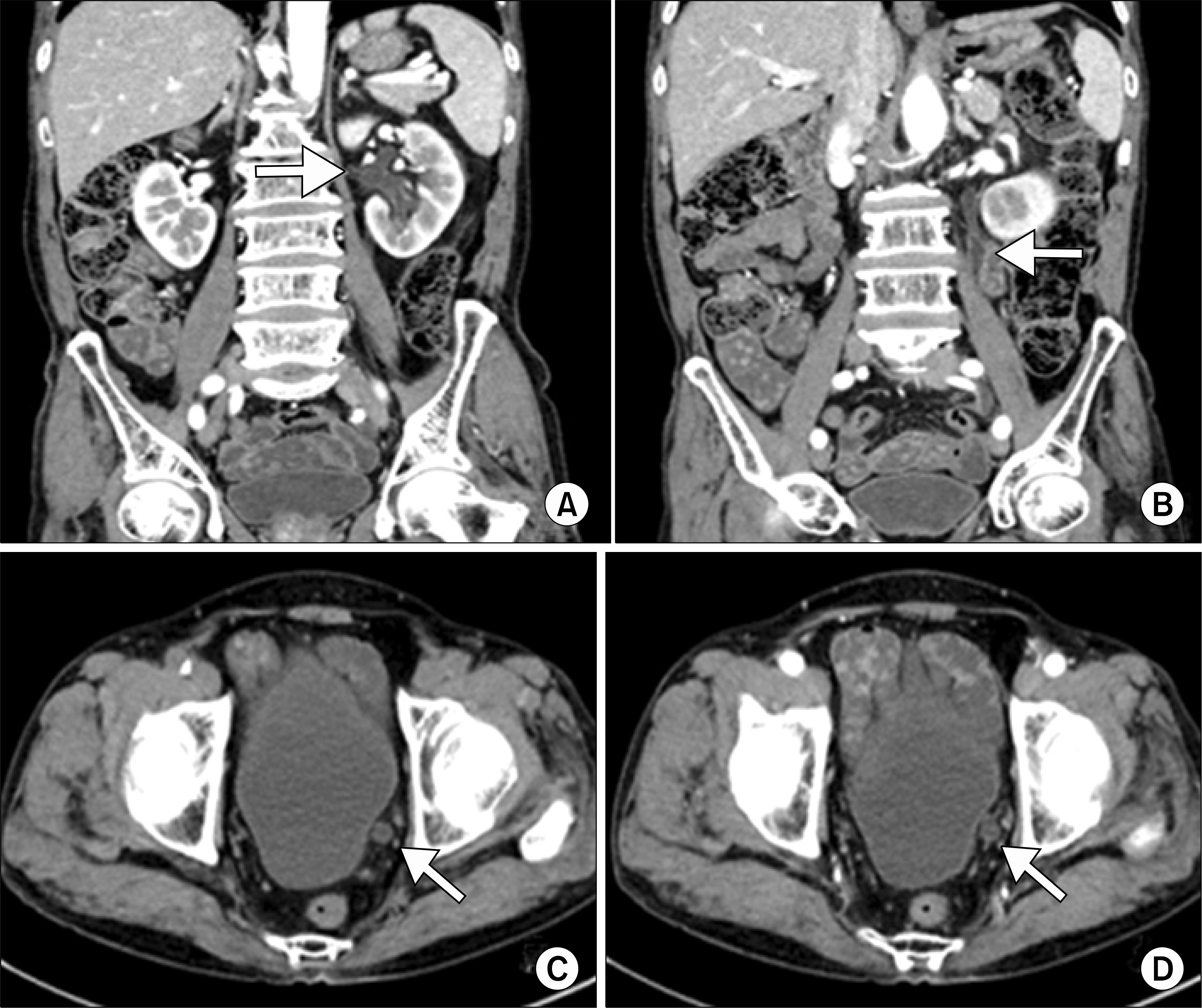
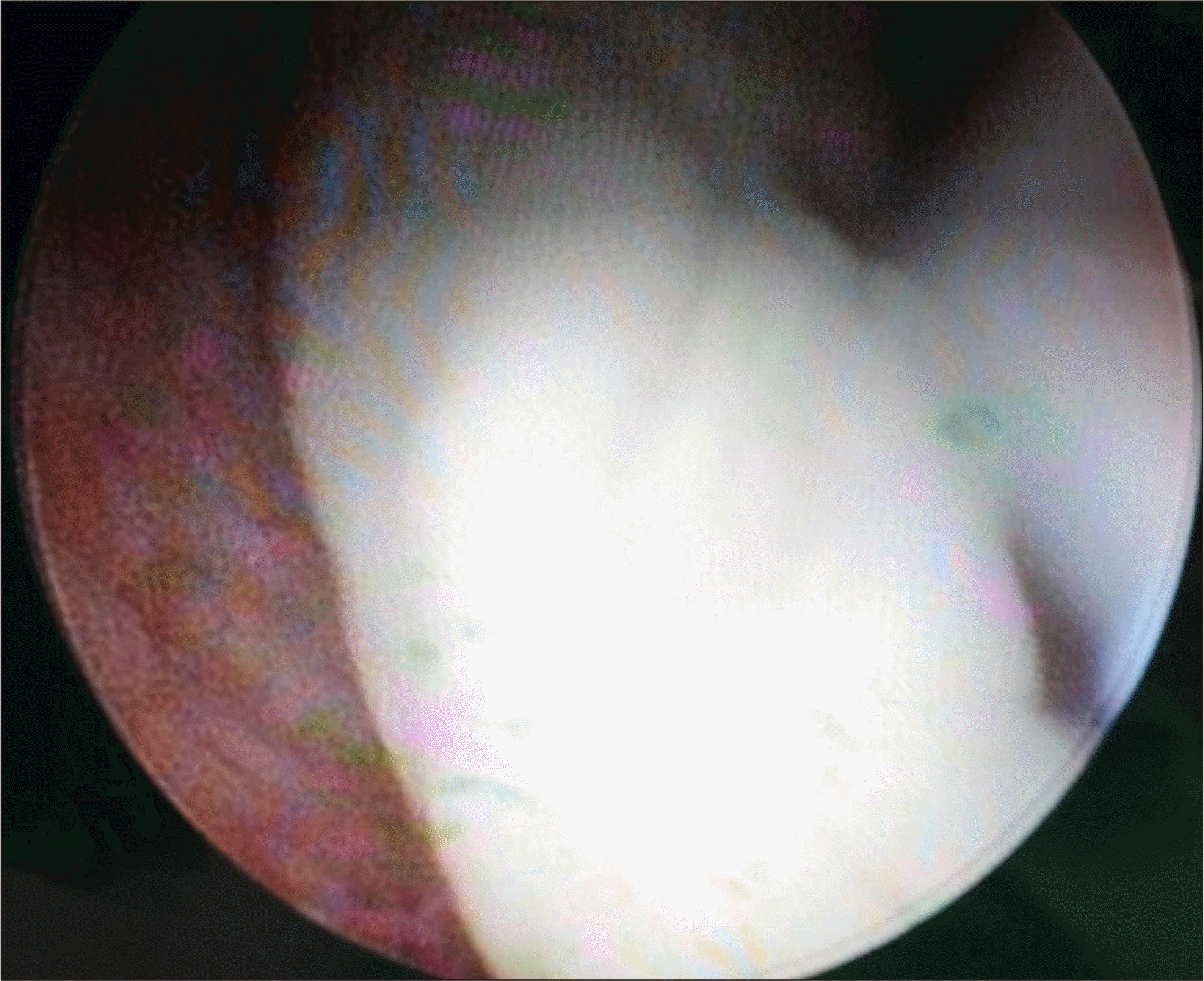
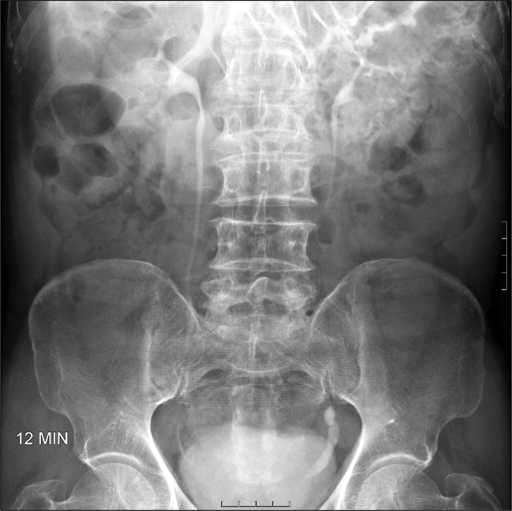
Although rarely, aspergillosis can cause obstructive uropathy. This generally occurs in patients with immunosuppressive conditions. Herein, we report a case of aspergilloma that caused ureteral obstruction in a 79-year-old man with no immunosuppressive conditions. A computed tomography revealed that his left pelvocalyceal system and ureter showed mild dilation, without a definite obstructive lesion. The fungal bezoar was removed using an ureteroscopy. The patient was successfully treated with antifungal medication.
Go to : 
REFERENCES
1.Stevens DA., Kan VL., Judson MA., Morrison VA., Dummer S., Denning DW, et al. Practice guidelines for diseases caused by Aspergillus. Infectious Diseases Society of America. Clin Infect Dis. 2000. 30:696–709.
2.Yoon YK., Kang EH., In KH., Kim MJ. Unilateral ureteral obstruction caused by Aspergillus, subgenus Nidulantes in a patient on steroid therapy: a case report and review of the literature. Med Mycol. 2010. 48:647–52.
3.Rao P. Aspergillus infection in urinary tract post-ureteric stenting. Indian J Med Microbiol. 2015. 33:316–8.

4.Lee SW. An Aspergilloma mistaken for a pelviureteral stone on nonenhanced CT: a fungal bezoar causing ureteral obstruction. Korean J Urol. 2010. 51:216–8.

5.Choi H., Kang IS., Kim HS., Lee YH., Seo IY. Invasive aspergillosis arising from ureteral aspergilloma. Yonsei Med J. 2011. 52:866–8.

6.el Fakir Y., Kabbaj N., Dafiri R., Imani F. Imaging of urinary Candida bezoars. Prog Urol. 1999. 9:513–7.
Go to : 


 PDF
PDF ePub
ePub Citation
 Print
Print


 XML Download