

 PDF
PDF ePub
ePub Citation
 Print
Print


Abstract
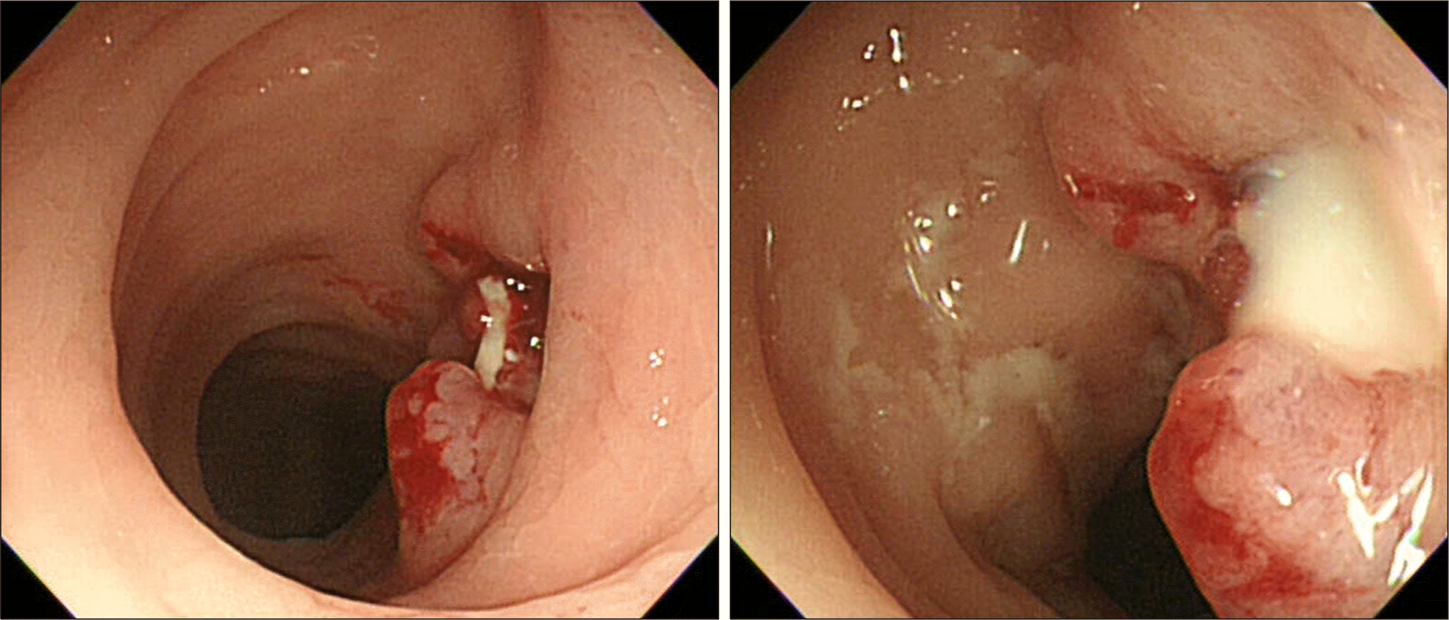
Seminal vesicle abscess is a rare urologic disease. Herein, we report our experience of the first case of a 41-year-old male patient with neurogenic bladder who underwent successful treatment of seminal vesicle abscess with rectal fistula after rectal decompression. Only a simple insertion of the rectal tube with intravenous antibiotics was able to remove the seminal vesicle abscess with rectal fistula without any percutaneous, transvesical, or transurethral drainage of the abscess. Rectal decompression should be considered in advance as a treatment of seminal vesicle abscess with rectal fistula before performing any invasive abscess drainage or fistulectomy.
REFERENCES
1.Eastham JA., Spires KS., Abreo F., Johnson JB., Venable DD. Seminal vesicle abscess due to tuberculosis: role of tissue culture in making the diagnosis. South Med J. 1999. 92:328–9.
2.Frye K., Loughlin K. Successful transurethral drainage of bilateral seminal vesicle abscesses. J Urol. 1988. 139:1323–4.

3.Rajfer J., Eggleston JC., Sanders RC., Walsh PC. Fever and prostatic mass in a young man. J Urol. 1978. 119:555–8.
4.Pandey P., Peters J., Shingleton WB. Seminal vesicle abscess: a case report and review of literature. Scand J Urol Nephrol. 1995. 29:521–4.
5.Sağlam M., Uğurel S., Kilciler M., Taşar M., Somuncu I., Uçöz T. Transrectal ultrasound-guided transperineal and transrectal management of seminal vesicle abscesses. Eur J Radiol. 2004. 52:329–34.
6.Dewani CP., Dewani N., Bhatia D. Case report: tubercular cold abscess of seminal vesicle: minimally invasive endoscopic management. J Endourol. 2006. 20:436–42.
7.Zagoria RJ., Papanicolaou N., Pfister RC., Stafford SA., Young HH 2nd. Seminal vesicle abscess after vasectomy: evaluation by transrectal sonography and CT. AJR Am J Roentgenol. 1987. 149:137–8.
8.Gulanikar A., Clark J., Feliz T. Prostatic abscess: an unusual presentation of metastatic prostate cancer. Br J Urol. 1998. 82:309–10.
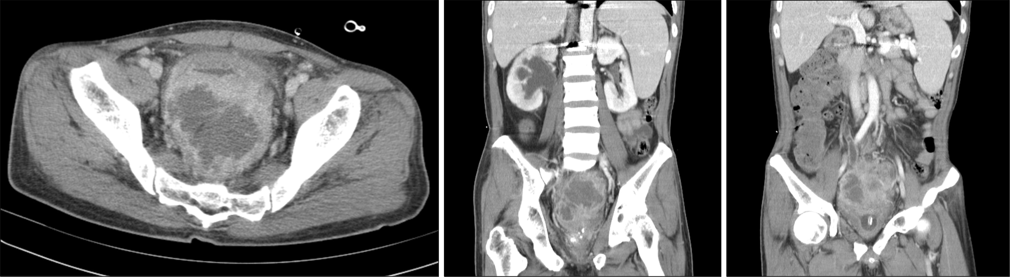
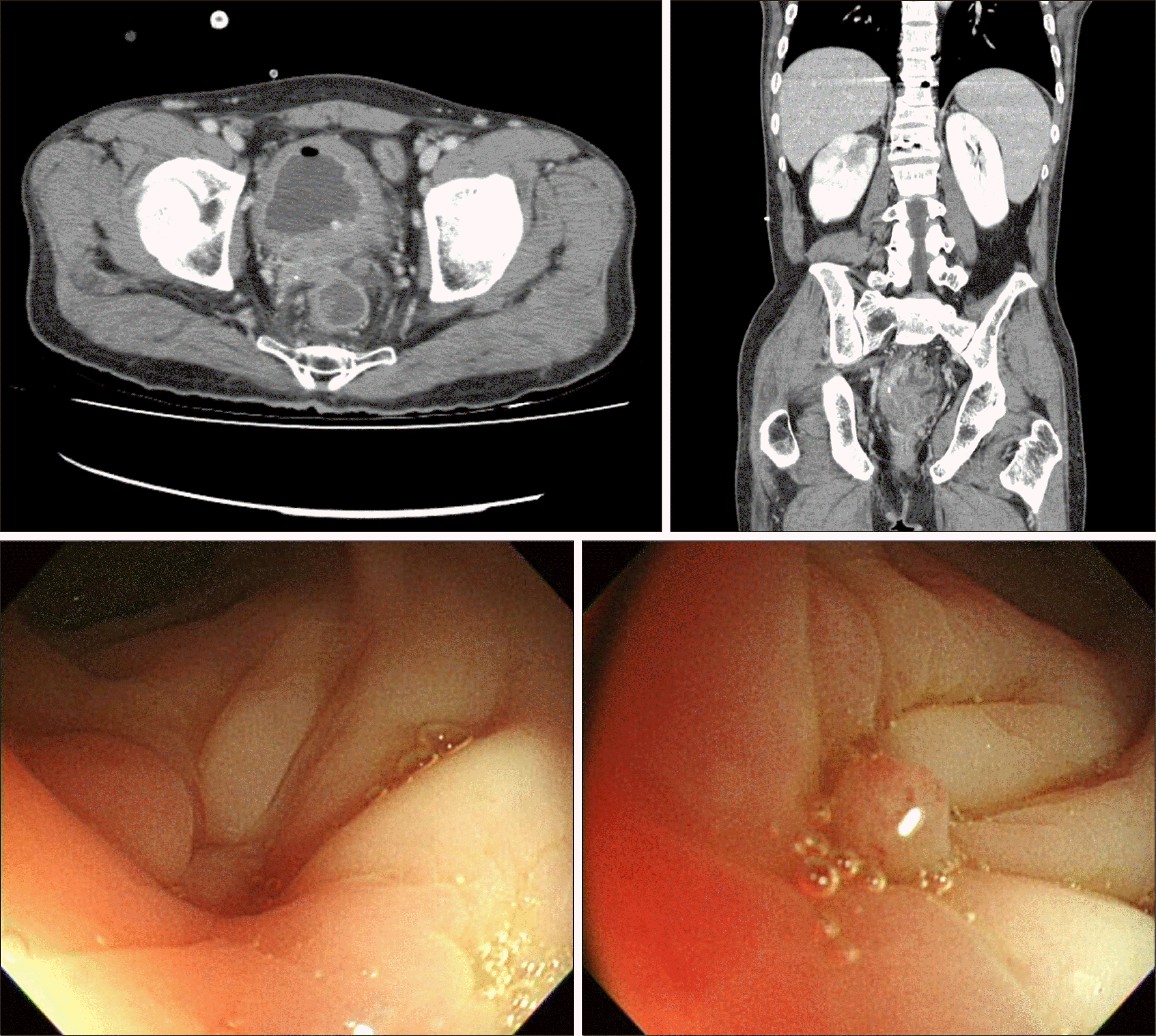
Fig. 1.
Computed tomography revealed 8 cm sized thick-walled pelvic abscess and right hydronephrouretersosis due to pelvic abscess.

 XML Download