Abstract
Brachial radiculoplexitis is characterized by acute onset of shoulder and arm pain followed by weakness and sensory loss. Brachial radiculoplexitis by herpes zoster is a rare disease, which can be diagnosed by careful history, electrodiagnosis and MRI. It has remained uncertain about clinical characteristics, treatment, and prognosis. Better understanding of this disease helps earlier diagnosis and prompt treatment to minimize neurologic sequale. We present two cases of subacute brachial radiculoplexitis preceded by herpes zoster infection. (Korean J Clin Neurophysiol 2015;17:86-90)
Go to : 
REFERENCES
1.Weaver BA. Herpes zoster overview: natural history and incidence. J Am Osteopath Assoc. 2009. 109:2–6.
2.Merchut MP., Gruener G. Segmental zoster paresis of limbs. Electromyogr Clin Neurophysiol. 1996. 36:369–375.
3.Haanpaa M., Hakkinen V., Nurmikko T. Motor involvement in acute herpes zoster. Muscle Nerve. 1997. 20:1443–1448.

4.Tsairis P., Dyck PJ., Mulder DW. Natural history of brachial plexus neuropathy. Report on 99 patients. Arch Neurol. 1972. 27:109–117.
5.Beqhi E., Kurland LT., Mulder DW., Nicolosi A. Brachial plexus neuropathy in the population of Rochester, Minnesota. Ann Nuurol. 1985. 18:320–323.
6.Ayoub T., Raman V., Chowdhury M. Brachial neuritis caused by varicella-zoster diagnosed by changes in brachial plexus on MRI. J Neurol. 2010. 257:1–4.

Go to : 
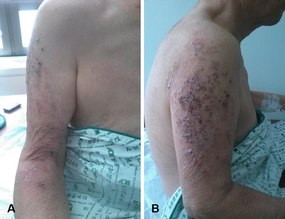
 | Fig. 1.Skin lesions of case 1. The patient had multiple erythematous crusted plaques on the right arm (C5-6 dermatome areas). |
Table 1.
Nerve conduction study, electromyography of case 1 and case 2 at 10 days after the onset of motor weakness
 | Fig. 2.Brachial plexus magnetic resonance imaging (MRI) of case 1 and 2. They were performed at 10 days after the onset of motor weakness. In case 1 (A-D), abnormal diffuse enhancement and thickening involving right brachial plexus, especially C5-6 root are shown on T2 short tau inversion recovery (STIR) coronal image (A), and modified Dixon contrast enhanced image (B). Moreover, abnormal enhancement of right upper trunk is shown on T2 STIR coronal (C) and 3 mm reconstruction coronal image (D). In case 2 (E-H), T2 STIR coronal (E) and 3 mm reconstruction coronal image (F) demonstrate abnormal diffuse enhancement and thickening involving left brachial plexus, especially C5-7 root. In addition, T2 STIR coronal (G) and 5 mm reconstruction coronal image (H) show enhancement and thickening of left upper and middle trunk. |


 PDF
PDF ePub
ePub Citation
 Print
Print


 XML Download