

 PDF
PDF ePub
ePub Citation
 Print
Print


Abstract
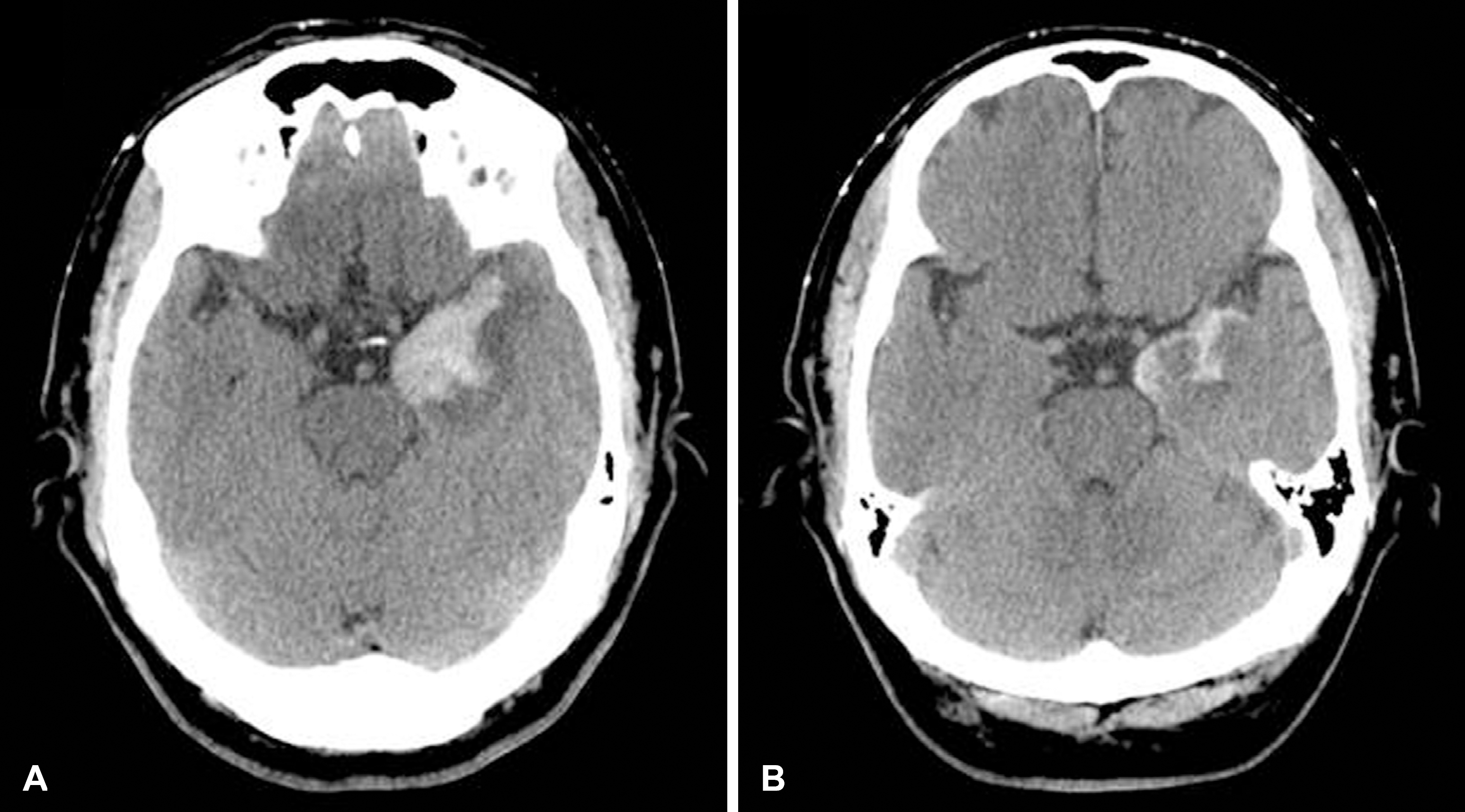
A 59-year old man was admitted for drowsiness and stiff neck. CSF examination showed lymphocytic pleocytosis and PCR for herpes simplex virus (HSV)-1 was positive in CSF. Brain MRI revealed enhanced lesions in left temporal lobe. His symptom improved with acyclovir. Follow-up studies showed red blood cells in CSF and a hematoma in the left temporal lobe. There was no additional symptom related to the hematoma. He was discharged after conservative care. Although rare, hematoma can develop in HSV-1 meningoencephalitis. (Korean J Clin Neurophysiol 2015;17:82-85)
Go to : 
REFERENCES
1.Raschilas F., Wolff M., Delatour F., Chaffaut C., De Broucker T., Chevret S, et al. Outcome of and prognostic factors for herpes simplex encephalitis in adult patients: results of a multicenter study. Clin Infect Dis. 2002. 35:254–260.

2.Rodriguez-Sainz A., Escalza-Cortina I., Guio-Carrion L., Matute-Nieves A., Gomez-Beldarrain M., Carbayo-Lozano G, et al. Intracerebral hematoma complicating herpes simplex encephalitis. Clin Neurol Neurosurg. 2013. 115:2041–2045.
3.Jeong H., Ji K., Chung E., Bae J., Kim E., Kim S. Herpes meningoencephalitis complicated by cerebral hematomas during acyclovir therapy. Korean J Stroke. 2011. 13:144–146.
4.Kang B., Ahn J., Kwon S. Bilateral medial temporal hematomas in HSV-1 encephalitis. J Korean Neurol Assoc. 2008. 26:136–138.
5.Ropper AH., Samuels MA., Klein JP. Adams and Victor's principles of neurology. 9th ed.New York: McGraw Hill;2009. p. 720.
Go to :
 | Fig. 1.Brain magnetic resonance imaging (MRI). Fluid attenuated inversion recovery MRI shows high signal intensity in the left medial temporal lobe area, which is enhanced by gadolinium on T1 weighted imaging (A, B). Gradient echo imaging shows no hemorrhage, and magnetic resonance angiography is normal (C, D). |
 XML Download