Abstract
Foot drop is usually derived from peroneal nerve injury. Traumatic causes of peroneal nerve injury are more common than insidious causes including metabolic syndromes and mass lesions. We present a case with common peroneal neuropathy due to schwannoma, which is extremely rare. Complete excision of the mass lead to a gradual improvement of the symptoms. Schwannoma should be considered as a cause of common peroneal neuropathy. (Korean J Clin Neurophysiol 2014;16:74-76)
REFERENCES
2.Byun JH., Hong JT., Son BC., Lee SW. Schwannoma of the superficial peroneal nerve presenting as sciatica. J Korean Neurosurg Soc. 2005. 38:306–308.
3.Baima J., Krivickas L. Evaluation and treatment of peroneal neuropathy. Curr Rev Musculoskeletal Med. 2008. 1:147–153.

4.Kim DH., Murovic JA., Tiel RL., Kline DG. Management and outcomes in 318 operative common peroneal nerve lesions at the Louisiana State University Health Sciences Center. Neurosurgery. 2004. 54:1421–1428.

5.Laurencin CT., Bain M., Yue JJ., Glick H. Schwannoma of the superficial peroneal nerve presenting as web space pain. J Foot Ankle Surg. 1995. 34:532–533.

6.Sharma RR., Pawar SP., Dey P. An occult schwannoma of the deep peroneal nerve presenting with neuralgia mimicking sciatica: case report and review of the literature. Ann Saudi Med. 2000. 20:57–59.

7.Mahitchi E., Van Linthoudt D. Schwannoma of the deep peroneal nerve. An unusual presentation in rheumatology. Praxis (Bern 1994). 2007. 96:69–72.
8.Shariq O., Radha S., Konan S. Common peroneal nerve schwannoma: an unusual differential for a symptomatic knee lump. BMJ Case Rep 2012 Dec 3.[Epub].DOI: doi: 10.1136/bcr-20 12-007346.
9.Andrychowski J., Czernicki Z., Jasielski P. Schwannoma of the common peroneal nerve. A differential diagnosis versus rare popliteal cyst. Neurol Neurochir Pol. 2012. 46:396–400.
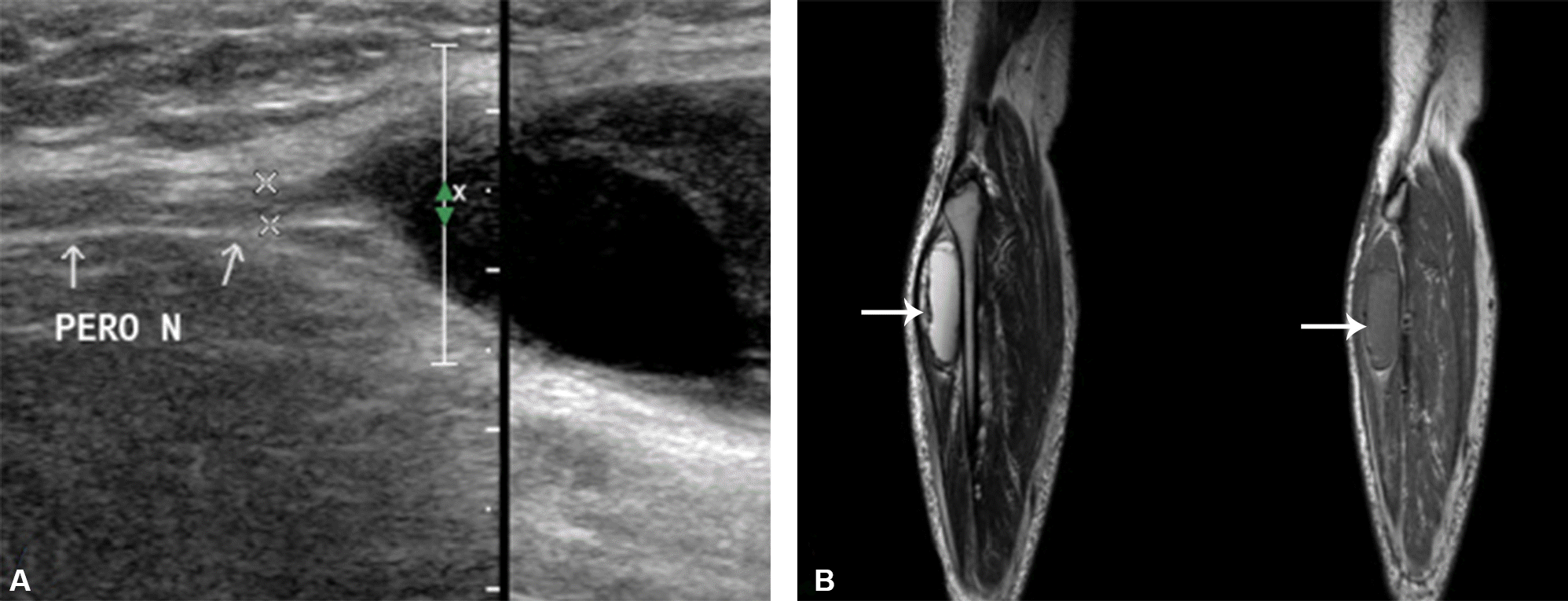
Figure 1.
(A) Ultrasonographic finding. It shows 6.2×3.8×2.8 cm in size mass lesion including anechoic cystic portion and echogenic large cystic portion in anterolateral portion of the right lower leg. (B) T2-(left) and T1-(right) weighted MRI findings. It shows well-defined oval mass with multiloculations just below the bifurcation of right common peroneal nerve (white arrow). PERO; common peroneal nerve.


 PDF
PDF ePub
ePub Citation
 Print
Print


 XML Download