Abstract
Nonconvulsive status epilepticus usually presents with altered mentation without distinct manifestations of seizures. It may be related with various medical disorders. Hashimoto’s encephalopathy is characterized by various neurological manifestations accompanied by high titers of anti-thyroid antibodies. Here, we report a patient with nonconvulsive status epilepticus caused by Hashimoto’s encephalopathy who showed a dramatic response to steroids. (Korean J Clin Neurophysiol 2014;16:70-73)
REFERENCES
1.Niedermeyer E., Khalifeh R. Petit mal status (“spike-wave stupor”): an electro-clinical appraisal. Epilepsia. 1965. 6:250–262.
2.Meierkord H., Holtkamp M. Non-convulsive status epilepticus in adults: clinical forms and treatment. Lancet Neurol. 2007. 6:329–339.

4.Chong JY., Rowland LP., Utiger RD. Hashimoto encephalopathy: syndrome or myth? Arch Neurol. 2003. 60:164–171.
5.Maganti R., Gerber P., Drees C., Chung S. Nonconvulsive status epilepticus. Epilepsy Behav. 2008. 12:572–586.

7.Brain L., Jellinek EH., Ball K. Hashimoto's disease andencepha-lopathy. Lancet. 1966. 2:512–514.
8.Engum A., Bjøro T., Mykletun A., Dahl AA. Thyroid autoimmunity, depression and anxiety; are there any connections? An epidemiological study of a large population. J Psychosom Res. 2005. 59:263–268.

9.Selva-O'Callaghan A., Redondo-Benito A., Trallero-Araguás E., Martínez-Gómez X., Palou E., Vilardell-Tarres M. Clinical significance of thyroid disease in patients with inflammatory myopathy. Medicine (Baltimore). 2007. 86:293–298.
10.Genovesi G., Paolini P., Marcellini L., Vernillo E., Salvati G., Polidori G, et al. Relationship between autoimmune thyroid disease and Alzheimer's disease. Panminerva Med. 1996. 38:61–63.
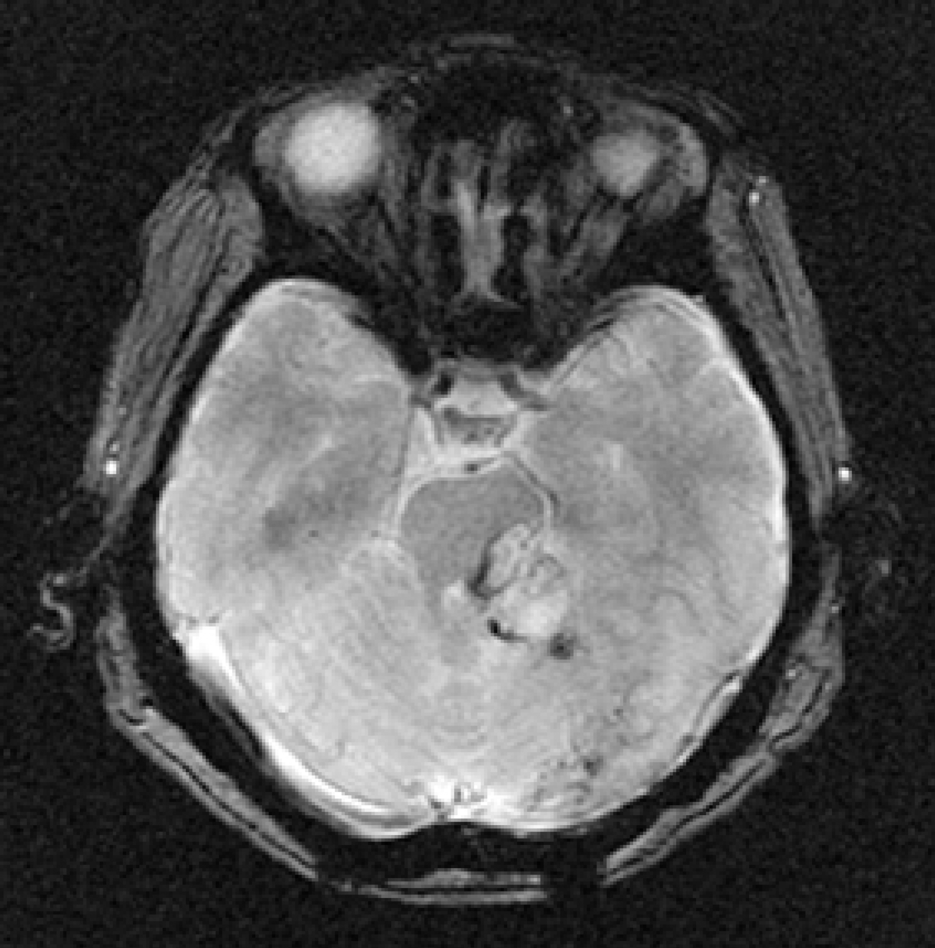
Figure 1.
Diffusion MRI shows no acute lesion other than remnant tumor and minimal hematoma in left pons and cerebellum.

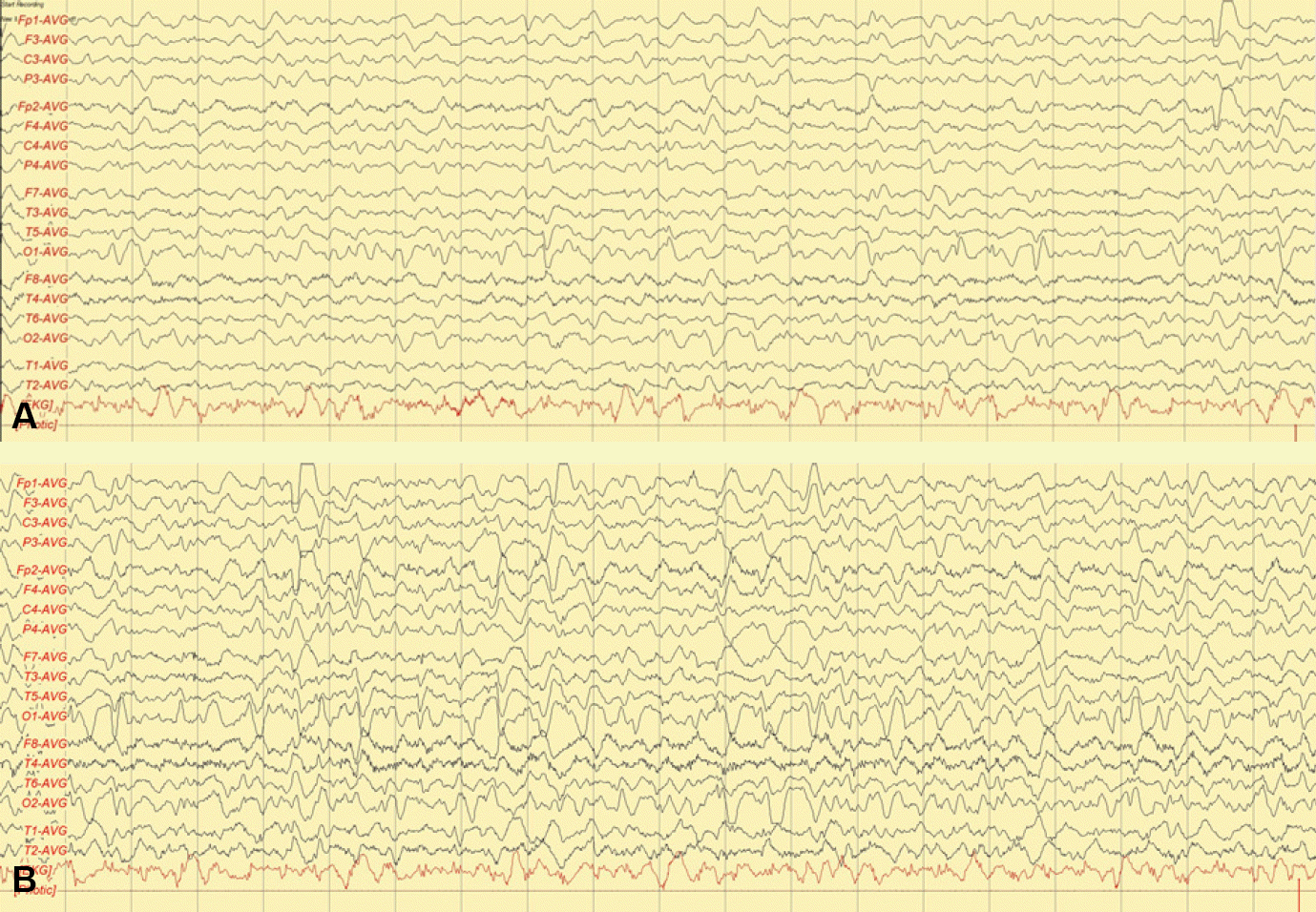
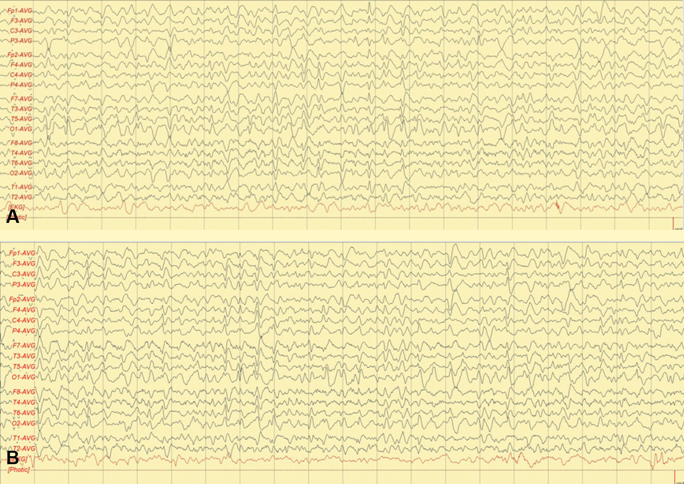
Figure 2.
Frequent sharp-and-slow waves are observed in the left occipital area (A). Intermittent rhythmic delta activity in the left occipital area spreads to the right occipital and bilateral frontotemporal area with evolution to higher amplitude (B).


 PDF
PDF ePub
ePub Citation
 Print
Print


 XML Download