

 PDF
PDF ePub
ePub Citation
 Print
Print


Abstract
Heart rate variability is significantly associated with cardiovascular complications in various neurological disorders with cardiac impairment. Measures of spontaneous heart rate variability might be different from provocating tests of heart rate variability such as deep breathing and Valsava maneuver. Methods for analysis are divided into time domain methods and frequency domain methods. There are standard deviation of NN interval, standard deviation of average NN interval, root mean square of the successive differences, NN50, and pNN50 in time domain methods. Frequency domain bands can be divided into very low, low, and high frequency. Each variables are influenced by sympathetic and/or parasympathetic activity. (Korean J Clin Neurophysiol 2014;16:49-54)
REFERENCES
1.Heart rate variability. Standards of measurement, physiological interpretation, and clinical use. Task Force of the European Society of Cardiology and the North American Society of Pacing and Electrophysiology. Eur Heart J. 1996. 17:354–381.
2.Rajendra Acharya U., Paul Joseph K., Kannathal N., Lim CM., Suri JS. Heart rate variability: a review. Med Biol Eng Comput. 2006. 44:1031–1051.

3.Vaseghi M., Shivkumar K. The role of the autonomic nervous system in sudden cardiac death. Prog Cardiovasc Dis. 2008. 50:404–419.
4.Metelka R. Heart rate variability - current diagnosis of the cardiac autonomic neuropathy. A review. Biomed Pap Med Fac Univ Palacky Olomouc Czech Repub. 2014. 158:327–338.
5.Mancia G., Paleari F., Parati G. Early diagnosis of diabetic autonomic neuropathy: present and future approaches. Diabetolo-gia. 1997. 40:482–484.
6.Hon EH., Lee ST. Electronic Evaluation of the Fetal Heart Rate. Viii. Patterns Preceding Fetal Death, Further Observations. Am J Obstet Gynecol. 1963. 87:814–826.
7.Sayers BM. Analysis of heart rate variability. Ergonomics. 1973. 16:17–32.
8.Ewing DJ., Martyn CN., Young RJ., Clarke BF. The value of cardiovascular autonomic function tests: 10 years experience in diabetes. Diabetes Care. 1985. 8:491–498.
9.Wolf MM., Varigos GA., Hunt D., Sloman JG. Sinus arrhythmia in acute myocardial infarction. Med J Aust. 1978. 2:52–53.
10.Akselrod S., Gordon D., Ubel FA., Shannon DC., Berger AC., Cohen RJ. Power spectrum analysis of heart rate fluctuation: a quantitative probe of beat-to-beat cardiovascular control. Science. 1981. 213:220–222.
11.Bilchick KC., Berger RD. Heart rate variability. J Cardiovasc Electrophysiol. 2006. 17:691–694.
12.Nolan J., Batin PD., Andrews R., Lindsay SJ., Brooksby P., Mullen M, et al. Prospective study of heart rate variability and mortality in chronic heart failure: results of the United Kingdom heart failure evaluation and assessment of risk trial (UK-heart). Circulation. 1998. 98:1510–1516.
13.DeGiorgio CM., Miller P., Meymandi S., Chin A., Epps J., Gordon S, et al. RMSSD, a measure of vagus-mediated heart rate variability, is associated with risk factors for SUDEP: the SUDEP-7 Inventory. Epilepsy Behav. 2010. 19:78–81.
14.Dash S., Chon KH., Lu S., Raeder EA. Automatic real time detection of atrial fibrillation. Ann Biomed Eng. 2009. 37:1701–1709.
15.Ewing DJ., Neilson JM., Shapiro CM., Stewart JA., Reid W. Twenty four hour heart rate variability: effects of posture, sleep, and time of day in healthy controls and comparison with bedside tests of autonomic function in diabetic patients. Br Heart J. 1991. 65:239–244.
16.Campos LA., Pereira VL Jr., Muralikrishna A., Albarwani S., Bras S., Gouveia S. Mathematical biomarkers for the autonomic regulation of cardiovascular system. Front Physiol. 2013. 4:279.
17.Bigger JT., Fleiss JL., Rolnitzky LM., Steinman RC. The ability of several short-term measures of RR variability to predict mortality after myocardial infarction. Circulation. 1993. 88:927–934.
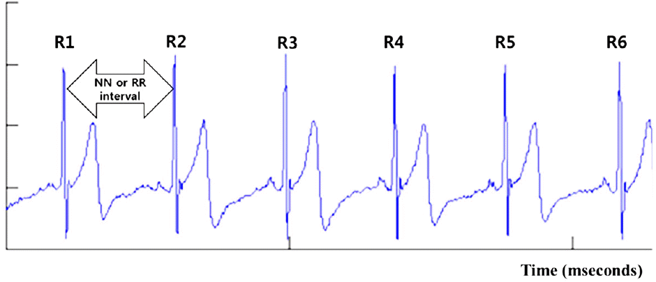
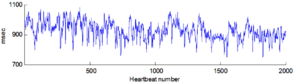
Figure 3.
RR intervals in ECG. R1, R2 and R3 mean 1st R, 2nd R, and 3rd R, respectively. A, B, and C mean RR interval.
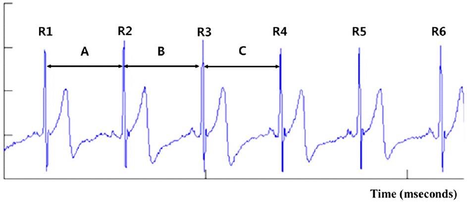
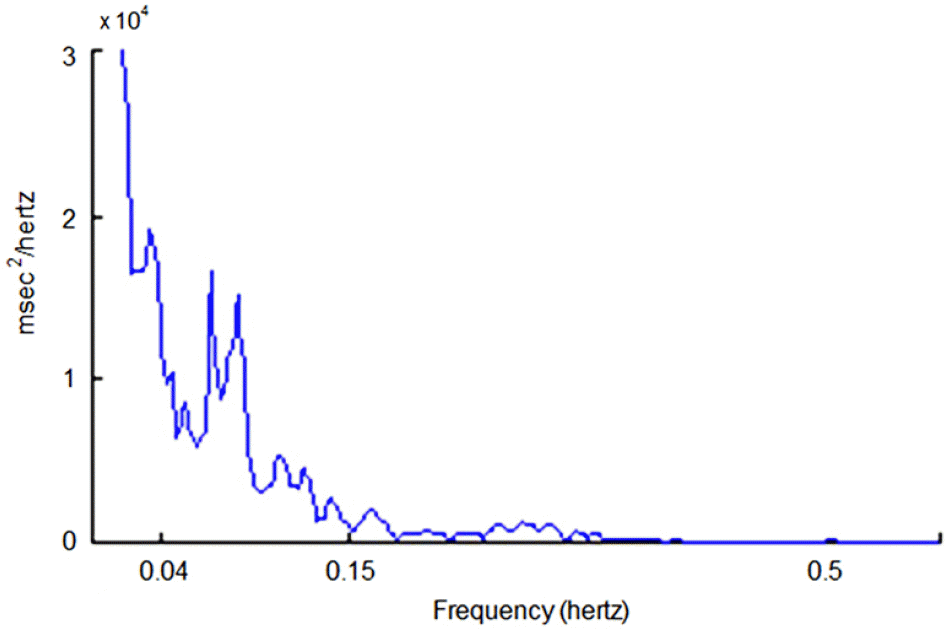
Table 1.
Time domain variables of heart rate variability1
Table 2.
Frequency domain measures of HRV1
 XML Download