

 PDF
PDF ePub
ePub Citation
 Print
Print


Abstract
Hemiplegia cruciata (HC) manifests as paralysis of the ipsilateral arm and contralateral leg. Herein, we report a 64-year-old man with weakness of the right leg and of the left arm after multiple sclerosis (MS). His brain and spine magnetic resonance imaging show a lower medulla lesion, which is extended to posterior part of C1 spine through cervicomedullary junction. HC usually results from stroke or trauma, but it is rare as presenting symptom of MS. (Korean J Clin Neurophysiol 2014;16:39-41)
REFERENCES
1.Wallenberg A. Anatomischer Befund in einem als „acute Bul-bäraffection (Embolie der Art. cerebellar. post. inf. sinistr.?)"1) beschriebenen Falle. Arch Psychiat Nervenkr. 1901. 34:923–959.

2.Dumitru D., Lang JE. Cruciate paralysis. Case report. J Neurosurg. 1986. 65:108–110.
3.Inamasu J., Hori S., Ohsuga F., Aikawa N. Selective paralysis of the upper extremities after odontoid fracture: acute central cord syndrome or cruciate paralysis? Clin Neurol Neurosurg. 2001. 103:238–241.
4.Ciappetta P., Salvati M., Raco A., Artico M. Cruciate hemiplegia: a clinical syndrome, a neuroanatomical controversy. Report of two cases and review of the literature. Surg Neurol. 1990. 34:43–47.
5.Coppola AR. "Cruciate paralysis": a complication of surgery. South Med J. 1973. 66:684 passim.
6.Arseni C., Maretsis M. Cruciate paralysis. Rev Roum Neurol. 1973. 10:359–362.
7.Ladouceur D., Veilleux M., Levesque RY. Cruciate paralysis secondary to C1 on C2 fracture-dislocation. Spine (Phila Pa 1976). 1991. 16:1383–1385.
8.Polman CH., Reingold SC., Banwell B, et al. Diagnostic criteria for multiple sclerosis: 2010 revisions to the McDonald criteria. Ann Neurol. 2011. 69:292–302.
9.Jacques S., Trippi AC., Shelden CH. Alternating Bell's palsy associated with diabetes mellitus. A report of four cases. Bull Los Angeles Neurol Soc. 1976. 41:78–81.
10.Yayama T., Uchida K., Kobayashi S, et al. Cruciate paralysis and hemiplegia cruciata: report of three cases. Spinal Cord. 2006. 44:393–398.
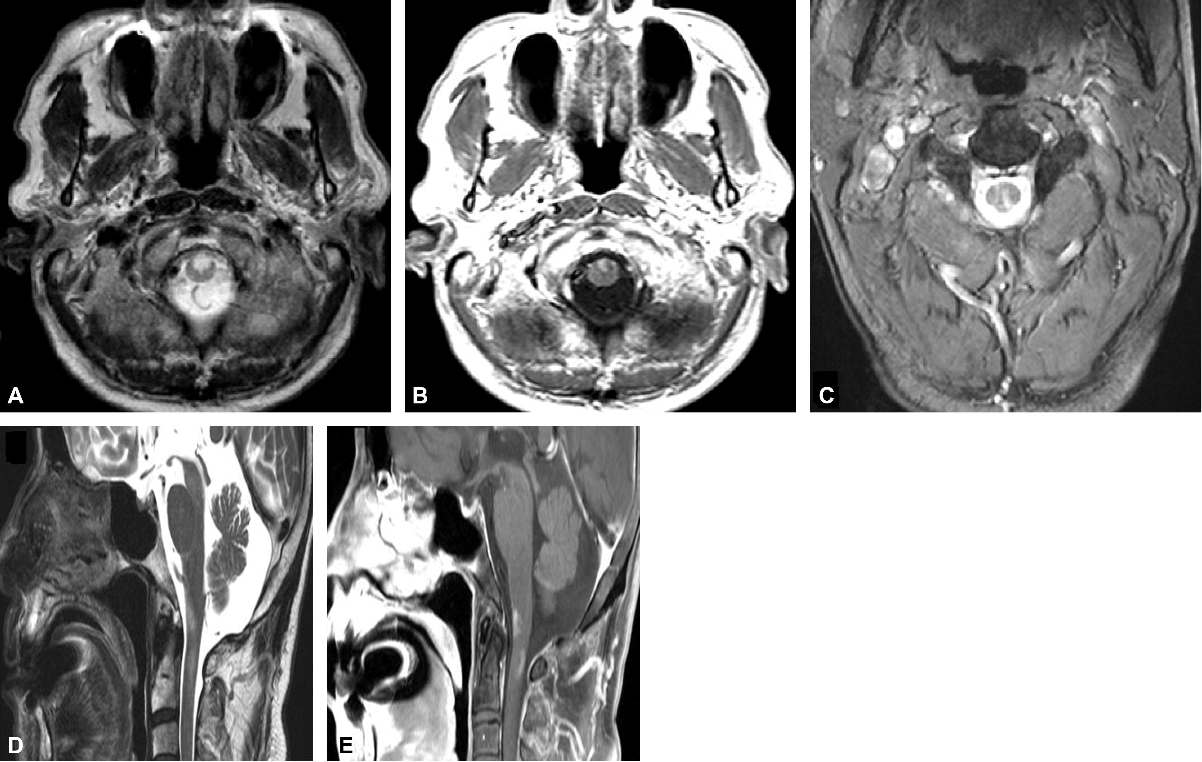
Figure 1.
Brain and cervical MRI. Axial T2-weighted image at the level of lower medullar reveals a hyperintense lesion (A). This lesion shows a gadolinum enhancement on T1 weighted image (B). Cervical MRI shows a hyper-intense lesion around the left posterior column at the level of first cervical vertebra in axial T1-weighted enhanced image (C). Prominent signal changes in T2-weighted (D) and T1-weighted enhanced images (E) are shown between the lower medulla and the upper cervical spinal cord, extending to the first cervical vertebra level.
 XML Download