Abstract
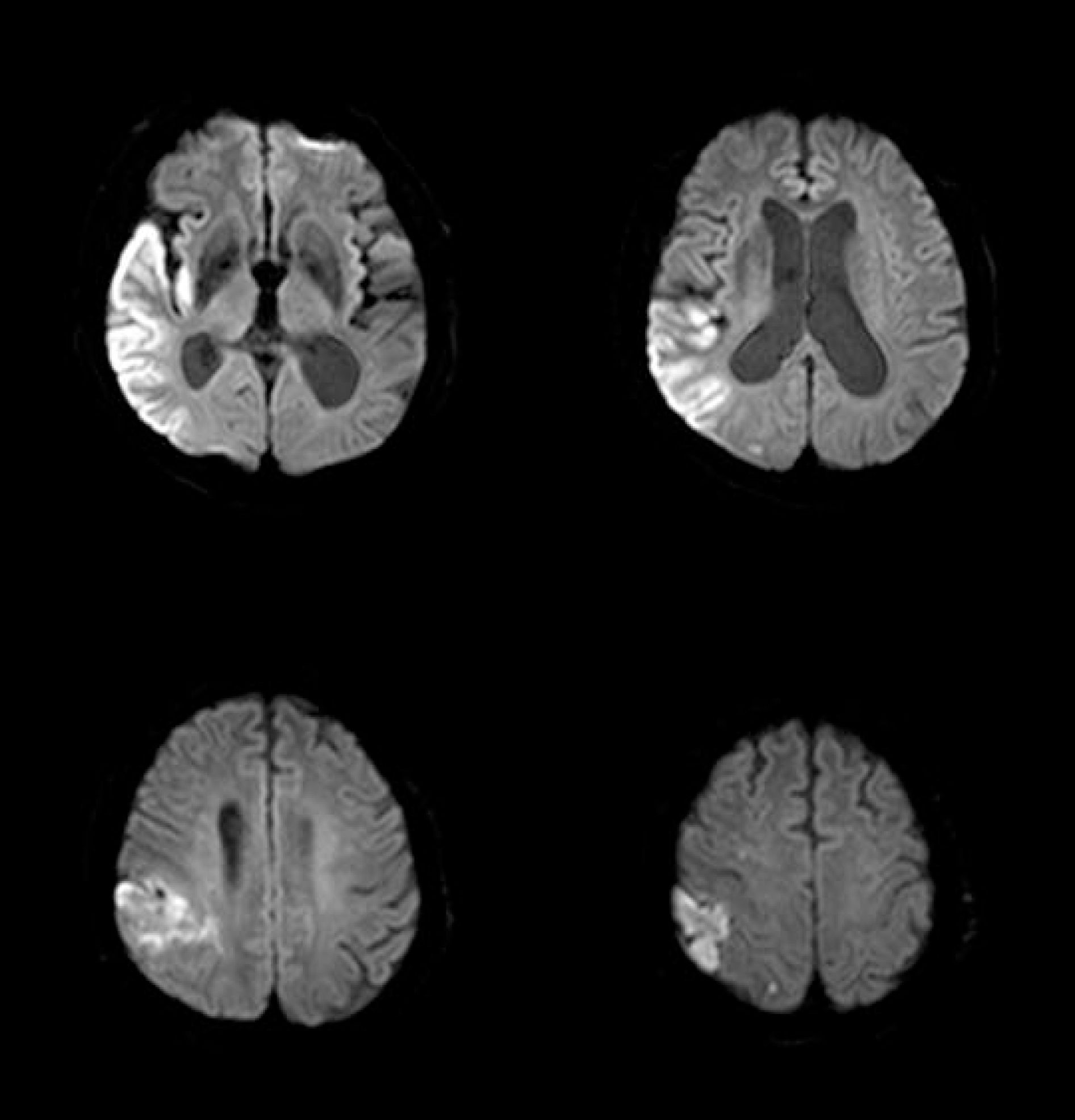
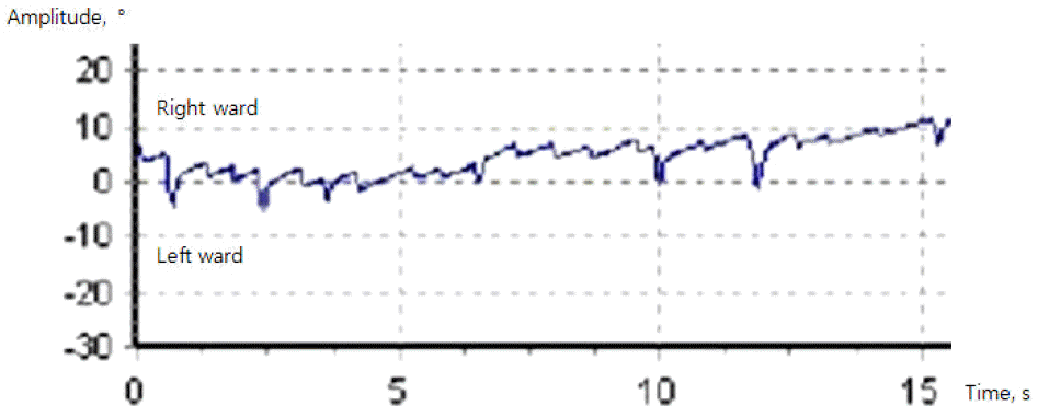
A 77-year-old man developed acute vertigo and unsteady gait. Neurological examination revealed spontaneous left-beating nystagmus in the primary position. He fell to the left when walking without support. Magnetic resonance imaging showed an acute infarction involving the right parieto-temporal lobe. Although the vertigo and unsteady gait are most often associated with vestibular disorders involving the infratentorial structures, those may occur in cerebral infarction of the parieto-temporal lobe. (Korean J Clin Neurophysiol 2014;16:32-34)
Go to : 
REFERENCES
1.Naganuma M., Inatomi Y., Yonehara T., Fujioka S., Hashimoto Y., Hirano T, et al. Rotational vertigo associated with parietal cortical infarction. J NeurolSci. 2006. 246:159–161.

2.Haines DE. Fundamental neuroscience for basic and clinical applications. 4th ed.Philadelphia: Elsevier/Saunders;2013. p. 218–385.
3.Brandt T., Botzel K., Yousry T., Dieterich M., Schulze S. Rotational vertigo in embolic stroke of the vestibular and auditory cortices. Neurology. 1995. 45:42–44.

4.Ahn BY., Bae JW., Kim DH., Choi KD., Kim HJ., Kim EJ. Pseudovestibular neuritis associated with isolated insular stroke. J Neurol. 2010. 257:1570–1572.

5.zu Eulenburg P., Caspers S., Roski C., Eickhoff SB. Meta-analytical definition and functional connectivity of the human vestibular cortex. Neuroimage. 2012. 60:162–169.

Go to : 


 PDF
PDF ePub
ePub Citation
 Print
Print


 XML Download