

 PDF
PDF ePub
ePub Citation
 Print
Print


Abstract
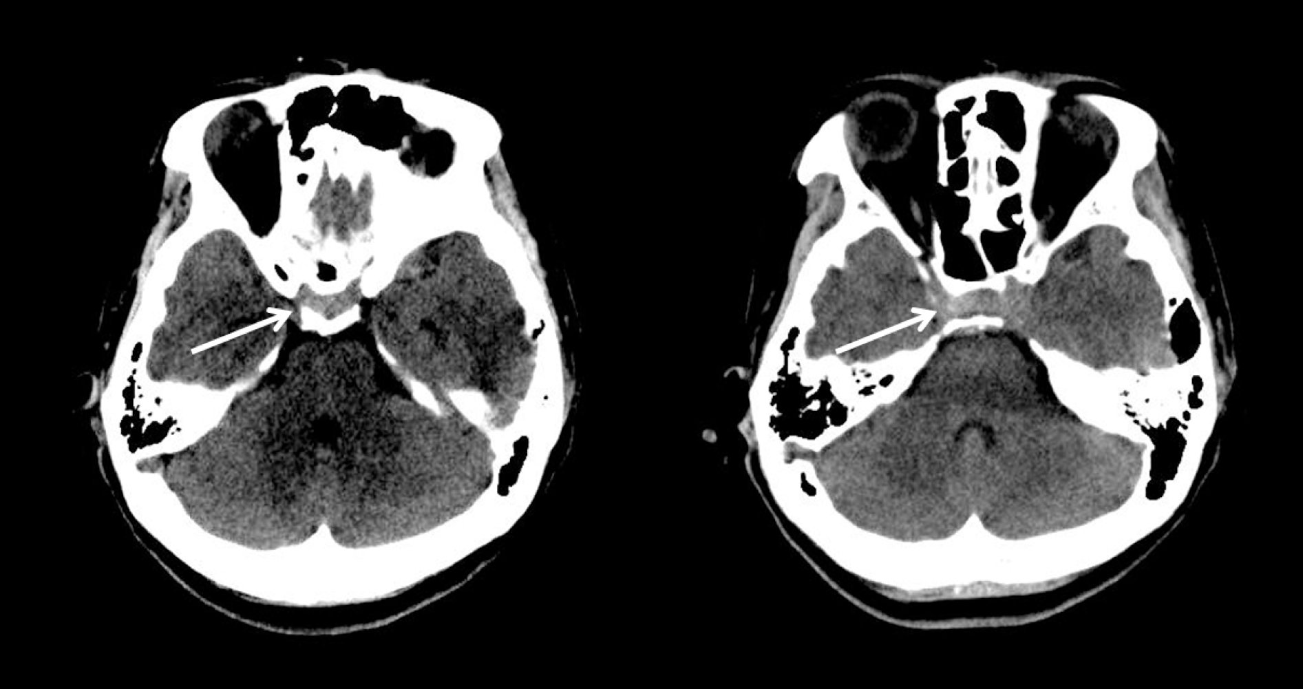
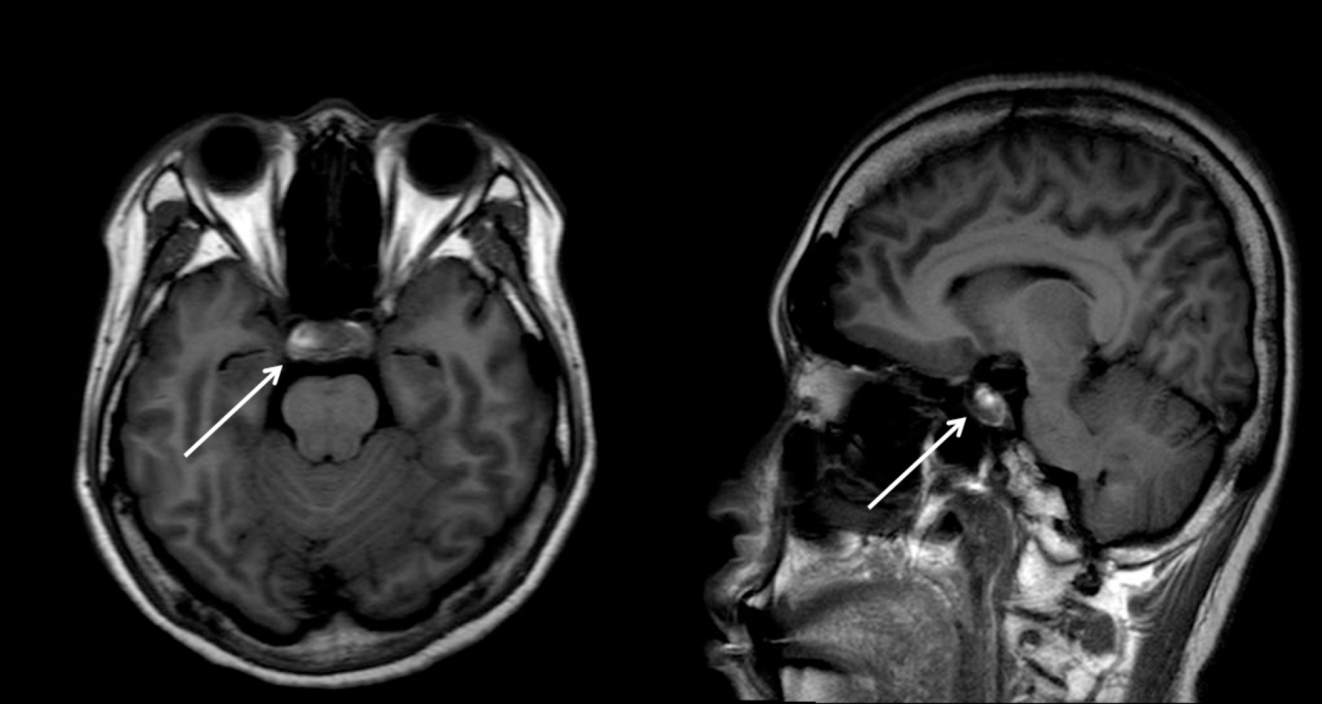
We encountered a case of pituitary apoplexy who presented with isolated headache and vomiting without visual disturbance or ophthalmoplegia. The cerebrospinal fluid examination was compatible with aseptic meningitis. A computed tomography revealed slightly high density in the pituitary fossa and suprasella area, but the signal change was very faint. Our case suggests that clinicians should take into account the possibility of pituitary apoplexy without visual disturbance or ophthalmoplegia, when aseptic meningitis is suspected.
Go to : 
REFERENCES
1.Watson CJ. The problem of hepatic coma. J Med Liban. 1955. 8:205–213.
2.Reutens DC., Edis RH. Pituitary apoplexy presenting as aseptic meningitis without visual loss or ophthalmoplegia. Aust N Z J Med. 1990. 20:590–591.

3.Haviv YS., Goldschmidt N., Safadi R. Pituitary apoplexy manifested by sterile meningitis. Eur J Med Res. 1998. 3:263–264.
4.Valente M., Marroni M., Stagni G., Floridi P., Perriello G., Santeu-sanio F. Acute sterile meningitis as a primary manifestation of pituitary apoplexy. J Endocrinol Invest. 2003. 26:754–757.
5.Chibbaro S., Benvenuti L., Carnesecchi S., Faggionato F., Gagliardi R. An interesting case of a pituitary adenoma apoplexy mimicking an acute meningitis. Case report. J Neurosurg Sci. 2007. 51:65–69. discussion 68-69.
6.Jeon BC., Park YS., Oh HS., Kim YS., Chun BK. Pituitary apoplexy complicated by chemical meningitis and cerebral infarction. J Korean Med Sci. 2007. 22:1085–1089.
7.Winer JB., Plant G. Stuttering pituitary apoplexy resembling meningitis. J Neurol Neurosurg Psychiatry. 1990. 53:440.
8.Murad-Kejbou S., Eggenberger E. Pituitary apoplexy: evaluation, management, and prognosis. Curr Opin Ophthalmol. 2009. 20:456–461.
9.Turgut M., Ozsunar Y., Basak S., Guney E., Kir E., Meteoglu I. Pituitary apoplexy: an overview of 186 cases published during the last century. Acta Neurochir (Wien). 2010. 152:749–761.
10.Chung SE., Lee SJ., Choi KS., Park SH. Comparison of the normal visual fields between the Goldmann and Humphrey kinetic perimetries. J Korean Ophthalmol Soc. 2009. 50:904–910.
Go to :
 XML Download