

 PDF
PDF ePub
ePub Citation
 Print
Print


Introduction
Rotatory deformity of the atlanto-axial joint was termed by Wortzman and Dewar in 1968.17) In 1977, Fielding and Hawkins2) described atlanto-axial rotatory fixation (AARF) as a persistent rotatory deformity of the atlanto-axial joint caused by subluxation or dislocation of the articular surfaces.
The condition is usually associated with an infection or a traumatic event, but it may also arise spontaneously or in association with other conditions. It occurs much more frequently in children than in adult, because of the unique biomechanical features of the atlanto-axial articulation. In children, the joint surface of the lateral mass is shallower and more horizontally oriented.5) In addition, the relative elasticity of the ligaments, the not yet fully developed neck muscles, and the relatively large head might be predisposing factors for AARF.5) We report a rare case of post-traumatic AARF in an adult that was treated by Halter-traction and Halo-vest.
Case Report
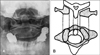
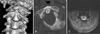
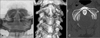
A 50-year-old male patient was transferred to the emergency room after a traffic accident that had occurred the day before. He complained of severe neck pain. Neurological examination was normal but his head was fixed in a position rotated to the right side and tilted to the left side. Initially, the patient underwent intermittent Halter-traction with seven pounds for twenty-four hours at another hospital. Unfortunately, the reduction failed, and the patient was transferred to our unit for further treatment. Cervical spine open mouth radiography showed the left C1 lateral mass to be closer to the odontoid process than the right one (Figure 1). Three-dimensional surface re-constructions of the CT images showed that the atlas was rotated to the right side and the anterior lip of the left superior facet of the C2 vertebra and the left transverse process of the C2 vertebra were fractured. Fortunately, cervical spine MRI revealed no evidence of cord injury (Figure 2). After applying the haltertraction for seven days, successful reduction was gained and neck pain was diminished. After that, neck pain was resolved rapidly. After successful closed reduction, the patient was advised to wear a halo-brace for three months. After three months, follow-up radiograph images confirmed that the atlas and axis has been completely corrected and the patient was relieved of the neck pain without any neurological deficits (Figure 3). Then the patient wore a Philadelphia brace for three months and received rehabilitation therapy care for a year. The patient did not show any range of motion limitations and the neurological examination and the follow-up radiographic examination were normal.
Discussion
The atlanto-axial joint is unique and complex.13) The primary function of the atlanto-axial joints is rotation rather than flexion or extension.3) The transverse ligament is the primary stabilizer of the atlanto-axial joint, and prevents forward subluxation of the atlas on the axis during head flexion.12) The alar ligaments also prevent excessive rotation at the atlanto-axial joints.5) Rotatory fixation may be associated with anterior or, rarely, posterior displacement of the atlas on the axis.3) The common cause of AARF is usually an infection or a traumatic event,3) but other conditions such as ankylosing spondylitis,7) metastatic tumor,16) generalized ligamentous laxity,9) eosinophilic granuloma,9) and following a suboccipital craniotomy and C1-3 laminectomy15) have also been reported.
The pathophysiology of these lesions are not well defined.4) Wortzman and Dewar17) concluded that the rotational fixation is not due to a fracture or ligamentous rupture but to damage of an unknown nature at the atlanto-axial joint itself. At the same time, they noted that the most believable theory is a tear and invagination of capsular ligaments the atlanto-axial synovial joints. Fielding and Hawkins2) concluded that swollen capsular and synovial tissues and muscle spasm prevent reduction in the early stages and that ligament and capsular contractures develop later, causing fixation. The patient suffering from AARF present with persistent torticollis and 'cock robin position', where the head is tilted to one side and rotated to the other side, These are important clinical manifestations of this condition.2,3,14)
Diagnosis is often delayed in this condition. CT scanning is the best method to detect this abnormality, and three dimensional CT reconstruction can demonstrate subluxation.2,3) A CT scan that reveals an atlantoaxial injury would justify performing further imaging with MR studies.11)
Fielding and Hawkins2) described four types of AARF based on the extent of the shift of C1 on C2 and the integrity of transverse ligament, which have been widely accepted. Type I is the most common pattern and the transverse ligament is intact. The fixed atlanto-axial rotation is symmetrical and within the normal range of rotation for atlantoaxial joints. Type II deformity is associated with mild deficiency of the transverse ligament with an atlanto-dental interval (ADI) of 3 to 5 mm. The intact joint acts as the pivot point for unilateral anterior displacement of the opposite side. Type III deformity is greater than 5 mm ADI, with deficiency of both transverse ligament and alar ligament. Type IV lesion is described as a posterior shift of one or both lateral masses of the atlas. this classification provides some guidance as to prognosis and treatment. in 1994, Li and Pang8) attempted to more clearly define this disorder, to establish criteria for its diagnosis, and to provide guidelines for its management.
The usual physiotherapeutic measures (immobilization, traction) will usually help in the early stage of the clinical presentation.12) Generally, early stage of AARF closed reduction can be achived easily. Optimal management of AARF depends on early diagnosis. Roche et al.13) concluded that if the abnormality is detected within 1 month of onset, then traction in combination with muscle relaxants and analgesia is recommended and prognosis for recovery is good. Moreover, if the diagnosis is delayed more than three months, most of the patients need surgical fusion. The delayed diagnosis led to elevated rates of recurrence and failure of non-surgical reduction.10) Therefore, if the diagnosis could be made as soon as possible, good results may be achieved by non-surgical approach. but, in spite of the adequate time interval for diagnosis, the surgical approach-fusion-is needed for cases of AARF with neural involvement and spinal instability, and in cases of failure to maintain reduction by conservative measures.2,13)
Fortunately, the diagnosis was made within a month and the Halter-traction was applied instantly, successfully achieving reduction. Afterwards, although controversial, we judged that a Philadelphia brace would not give enough fixation support, therefore in order to achieve a stronger fixation effect we applied a Halo-vest, external immobilization. The patient was followed up for 6 months and he presented a good outcome.
Conclusion
Misdiagnosis of AARF is more common in adults than in children, because the incidence of AARF in adults is lower than that in children.13) Patients may be treated with non-surgical approach due to early diagnosis. Therefore, when 'cock robbin', the typical posture and neck motion limitation, is found in adults with head trauma, the immediate and accurate diagnosis is essential to prevent surgery.
We reported a case of post traumatic AARF in adults which recovered completely without surgery. Precise diagnosis and an early treatment are the keys to prevent operative treatment.
 XML Download