Abstract
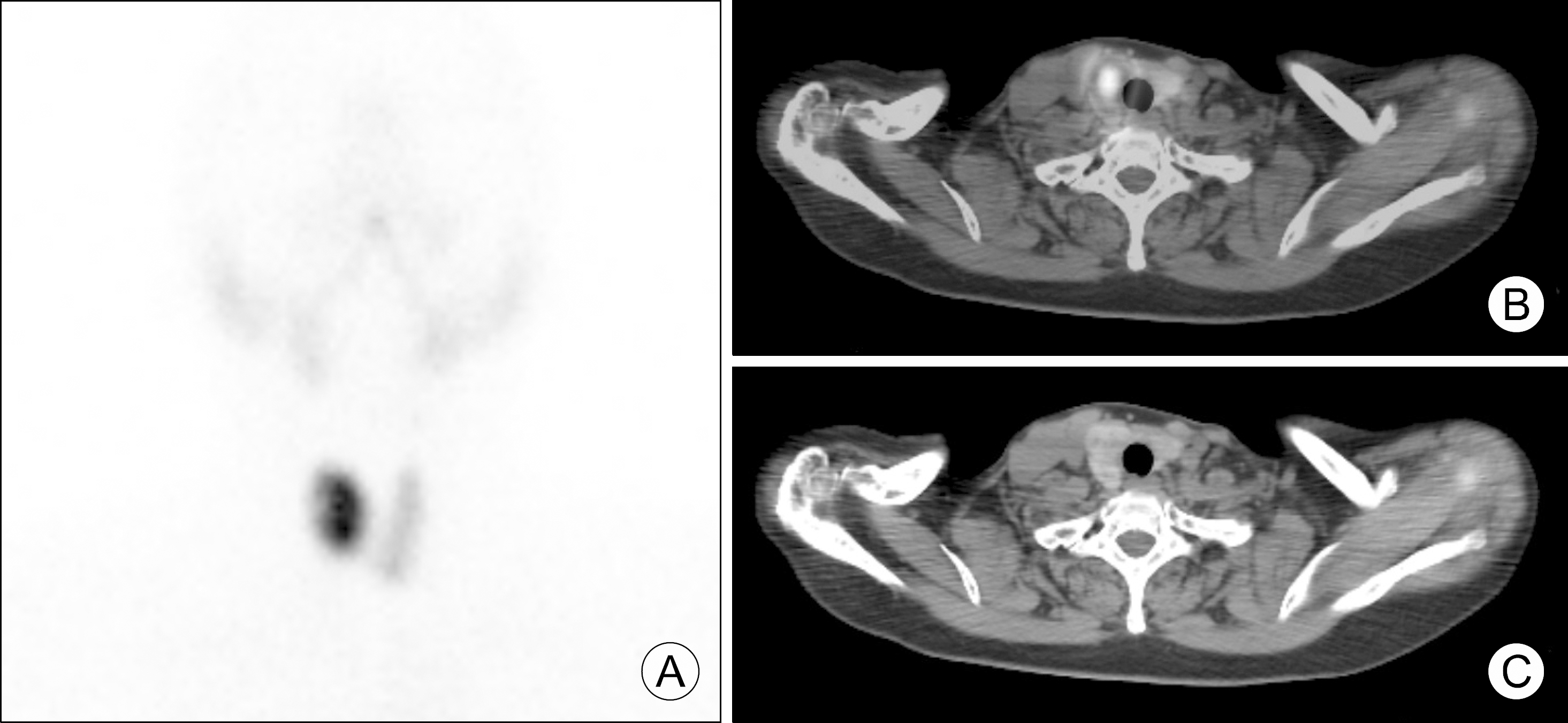
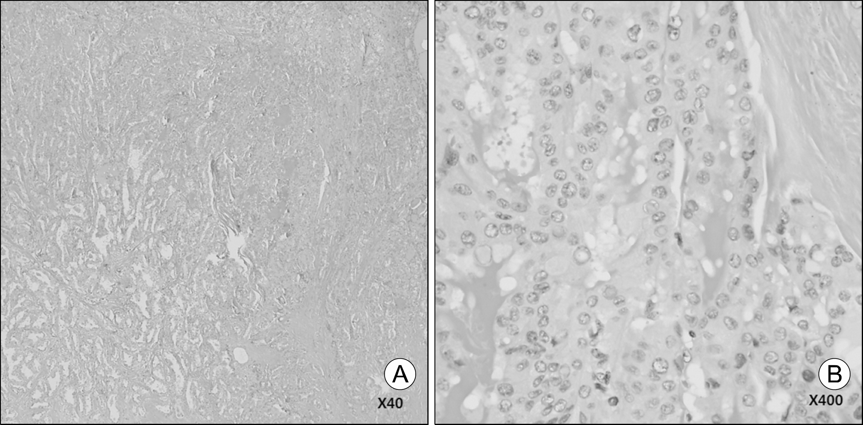
We report a case of a 74-year-old woman who was incidentally found to have a single thyroid nodule. Laboratory evaluation showed undetectable serum thyroid stimulating hormone and elevated free thyroxine levels.99m Tc thyroid scan showed a hyperfunctioning autonomous nodule in a right lobe of the thyroid. Thyroid ultrasonography showed a 2.2 cm sized nonhomogeneous spiculated nodule with microcalcification, and which99m Tc single photon emission was identical with the hyperfunctioning nodule confirmed in thyroid scan by computed tomography/computed tomography. Fine needle aspiration was done, and cytology reported as suspicious of malignancy. The patient underwent total thyroidectomy with central neck dissection, and pathology was consistent with papillary thyroid carcinoma. This case report demonstrates that diagnosis of a hyperfunctioning autonomous thyroid nodule does not preclude the possibility of thyroid cancer. Clinicians should consider further evaluation such as ultrasonography and fine needle aspiration in patients with hyperfunctioning autonomous nodules.
Go to : 
References
1. Hegedus L. Clinical practice. The thyroid nodule. N Engl J Med. 2004; 351(17):1764–71.
2. Haugen BR, Alexander EK, Bible KC, Doherty GM, Mandel SJ, Nikiforov YE, et al. 2015 American Thyroid Association Management Guidelines for adult patients with thyroid nodules and differentiated thyroid cancer: The American Thyroid Association Guidelines Task Force on thyroid nodules and differentiated thyroid cancer. Thyroid. 2016; 26(1):1–133.

3. Mirfakhraee S, Mathews D, Peng L, Woodruff S, Zigman JM. A solitary hyperfunctioning thyroid nodule harboring thyroid carcinoma: review of the literature. Thyroid Res. 2013; 6(1):7.

4. Lee ES, Kim JH, Na DG, Paeng JC, Min HS, Choi SH, et al. Hyperfunction thyroid nodules: their risk for becoming or being associated with thyroid cancers. Korean J Radiol. 2013; 14(4):643–52.

5. Mircescu H, Parma J, Huot C, Deal C, Oligny LL, Vassart G, et al. Hyperfunctioning malignant thyroid nodule in an 11-year-old girl: pathologic and molecular studies. J Pediatr. 2000; 137(4):585–7.

6. Tfayli HM, Teot LA, Indyk JA, Witchel SF. Papillary thyroid carcinoma in an autonomous hyperfunctioning thyroid nodule: case report and review of the literature. Thyroid. 2010; 20(9):1029–32.

7. Kim WB, Han SM, Kim TY, Nam-Goong IS, Gong G, Lee HK, et al. Ultrasonographic screening for detection of thyroid cancer in patients with Graves' disease. Clin Endocrinol (Oxf). 2004; 60(6):719–25.

8. Kim JH, Na GJ, Kim KW, Ko HJ, Jeon SW, Kim YJ, et al. Papillary thyroid carcinoma manifesting as an autonomously functioning thyroid nodule. Endocrinol Metab. 2012; 27(1):5962.

9. Hundahl SA, Fleming ID, Fremgen AM, Menck HR. A National Cancer Data Base report on 53,856 cases of thyroid carcinoma treated in the U.S., 1985–1995 [see commetns]. Cancer. 1998; 83(12):2638–48.
Go to : 
 | Fig. 1.Thyroid scan (A), 99m Tc SPECT/CT (B), and CT scan (C) showed about 2 cm sized hot nodule in upper pole of right lobe (99m TcO4 uptake: 3.3%). SPECT/CT: single photon emission com-puted tomography/computed tomography. |


 PDF
PDF ePub
ePub Citation
 Print
Print


 XML Download