

 PDF
PDF ePub
ePub Citation
 Print
Print


Abstract
A thyroglossal duct cyst (TGDC) is one of the most common causes of anterior midline neck mass. Successful management of a TGDC requires histopathology and an understanding of the embryogenesis of the thyroid. Traditional TGDC surgery uses a transcervical approach, which results in an external neck scar. In contrast to the surgical removal of a benign neck mass, TGDC surgery should include removal of the cyst, the hyoid bone, and the thyroid remnant track from the foramen cecum to the hyoid bone. Considering the embryological development of the TGDC, it was evident to us that an entirely transoral approach to the TGDC region was an option. Before its descent, the TGDC originates from the bottom of the tongue. The TGDC is located behind the strap muscles of the neck and the hyoid bone. Following this naturally predetermined access alongside the TGDC, we were able to develop a new surgical approach to the TGDC area and introduced the transoral TGDC excision.
References
1. ASGE; SAGES. ASGE/SAGES Working Group on Natural Orifice Translumenal Endoscopic Surgery White Paper October 2005. Gastrointest Endosc. 2006; 63(2):199–203.
2. Kim JP, Park JJ, Jeon SY, Ahn SK, Hur DG, Kim DW, et al. Endoscope-assisted intraoral resection of external dermoid cyst. Head Neck. 2012; 34(6):907–10.

3. Kim JP, Park JJ, Lee EJ, Woo SH. Intraoral removal of a thyroglossal duct cyst using a frenotomy incision. Thyroid. 2011; 21(12):1381–4.
4. Kalloo AN, Singh VK, Jagannath SB, Niiyama H, Hill SL, Vaughn CA, et al. Flexible transgastric peritoneoscopy: a novel approach to diagnostic and therapeutic interventions in the peritoneal cavity. Gastrointest Endosc. 2004; 60(1):114–7.
5. Marescaux J, Dallemagne B, Perretta S, Wattiez A, Mutter D, Coumaros D. Surgery without scars: report of transluminal cholecystectomy in a human being. Arch Surg. 2007; 142(9):823–6. ; discussion 6–7.
6. Woo SH, Jeong HS, Kim JP, Park JJ, Baek CH. Endoscopeassisted frenotomy approach to median upper neck masses: clinical outcomes and safety (from a phase II clinical trial). Head Neck. 2014; 36(7):985–91.
7. Woo SH, Park JJ, Hong JC, Wang SG, Park GC, Eun YG, et al. Endoscope-assisted transoral removal of a thyroglossal duct cyst using a frenotomy incision: a prospective clinical trial. Laryngoscope. 2015; 125(12):2730–5.
8. Woo SH, Jeong HS, Kim JP, Park JJ, Baek CH. Endoscopeassisted intraoral removal of ectopic thyroid tissue using a frenotomy incision. Thyroid. 2013; 23(5):605–8.
9. Woo SH. Endoscope-assisted intraoral removal of the thyroid isthmus mass using a frenotomy incision. J Laparoendosc Adv Surg Tech A. 2013; 23(9):787–90.
10. Woo SH. Endoscope-assisted transoral thyroidectomy using a frenotomy incision. J Laparoendosc Adv Surg Tech A. 2014; 24(5):345–9.
11. Kim JP, Park JJ, Park HW, Woo SH. Endoscopy-assisted resection of a submandibular gland mass via a thyroidectomy incision. Ear Nose Throat J. 2015; 94(9):E30–3.
12. Woo SH. Endoscopic-assisted total thyroidectomy via lateral keloid scar incision. Clin Exp Otorhinolaryngol. 2014; 7(4):338–41.
13. Woo SH. Endoscope-assisted transoral accessory parotid mass excision. Head Neck. 2016; 38(1):E7–12.
14. Woo SH, Kim JP, Baek CH. Endoscope-assisted extracapsular dissection of benign parotid tumors using hairline incision. Head Neck. 2016; 38(3):375–9.
Fig. 1.
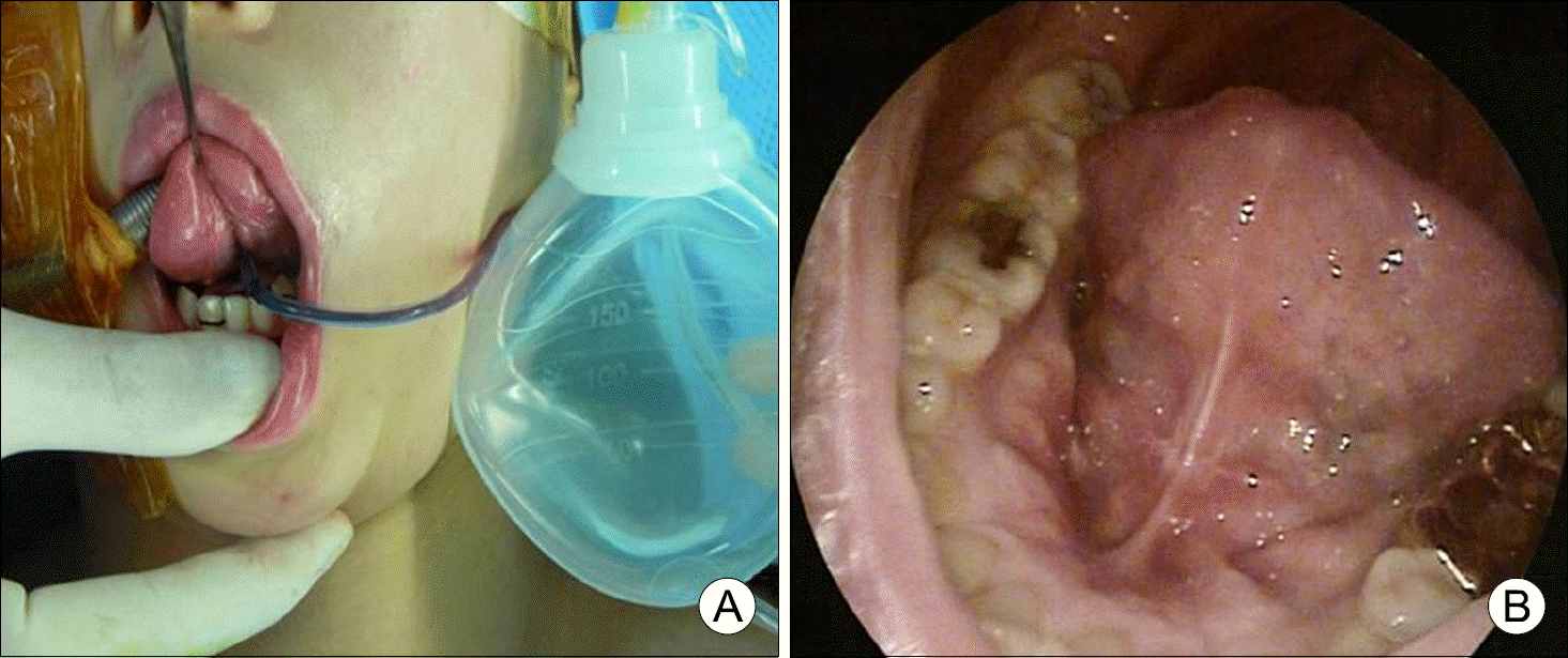
The presentation of transoral approach. After fre-notomy, we can find genio-glossus muscle and retrac-ted bilaterally. After that we can approach to the anterior neck area.
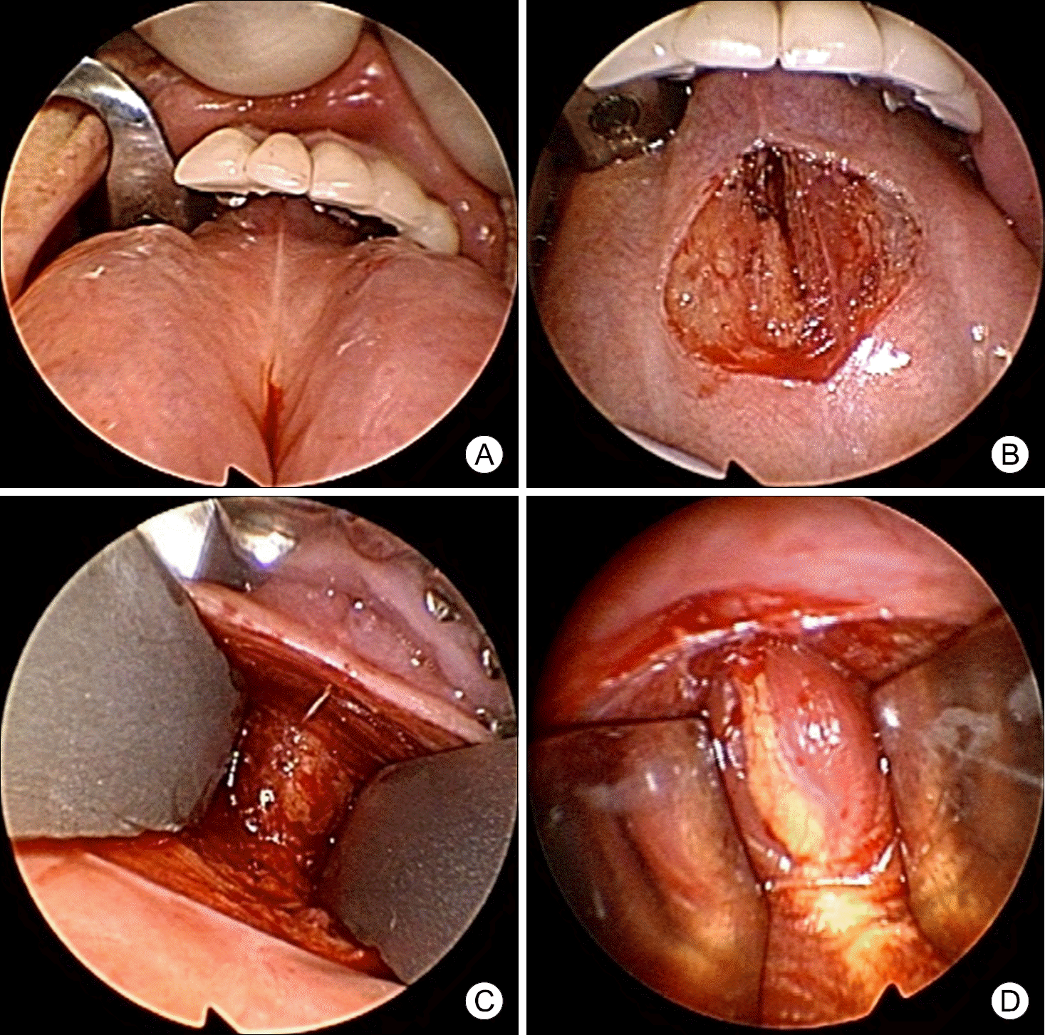
Fig. 2.
Transoral approach through a midline incision of the floor of the mouth. (A, B) CT scan confirmed the sub-mental mass between both digastric muscle and below mylohyoid muscle. (C) Verti-cal incision was made in the midline of the floor of the mouth through the frenulum. After dissection of the soft tissues in the floor of the mouth, the genioglossus muscles were separated in the midline, and retracted bilaterally. After re-traction of the genioglossus muscles, we exposed the my-lohyoid muscles. With the en-doscopic guidance, we identi-fied the mass like lesion after resection of the mylohyoid muscles. (D) After dissecting the soft tissue around the mass, we removed it.
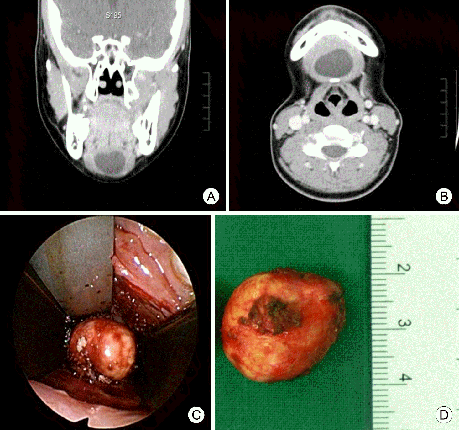
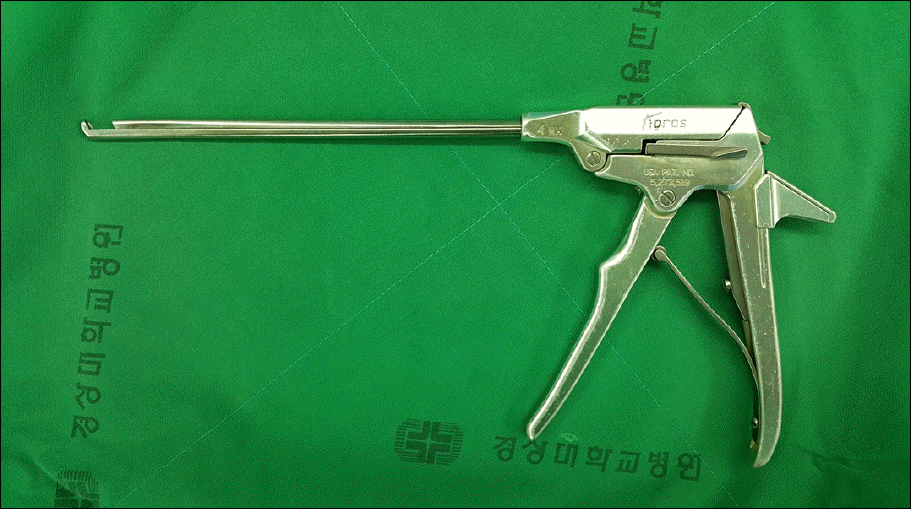
Fig. 4.
Computed tomo-graphy scan confirmed the 2-cm mass found just in-ferior to the level of the hyoid bone.
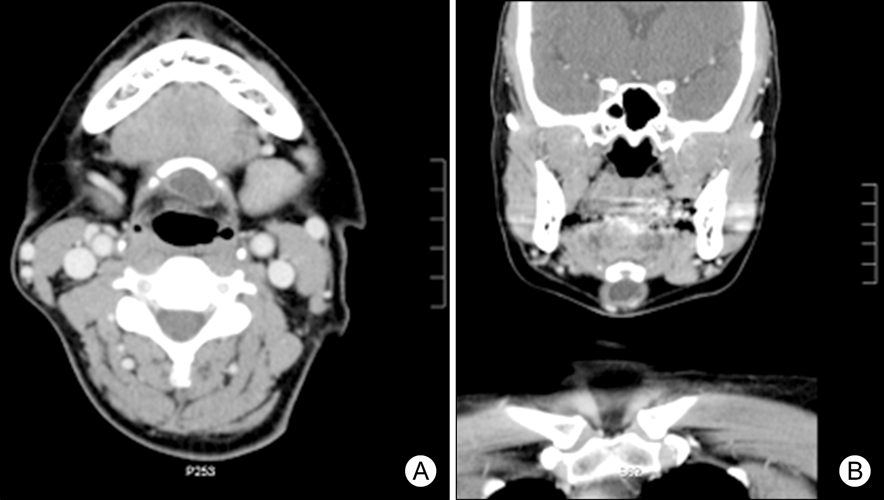
Fig. 5.
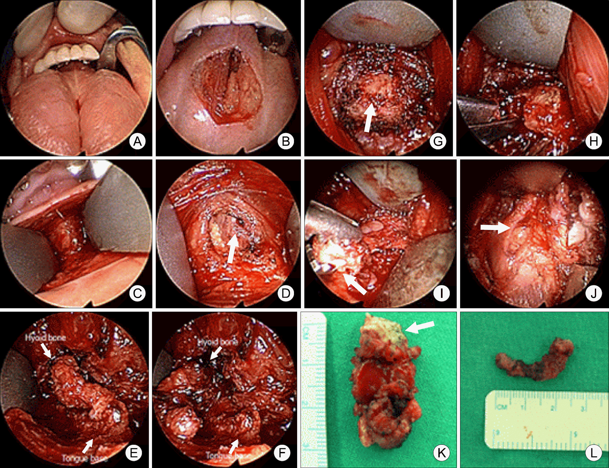
Transoral approach through a frenotomy incision of the mouth. (A, B) Vertical incision was made in the midline of the floor of the mouth through the frenulum. (C) After dissection of the soft tissues in the floor of the mouth, the genioglossus muscles were separated in the midline and retracted bilaterally. (D) After retraction of the genioglossus muscles, we found some midline longitudinal tissue (arrow). (E, F) This tissue extended from the midline of the hyoid bone toward the tongue base. After dissection, we cut the tissue between the tongue base and the hyoid bone. (G) With endoscopic guidance, we identified the hyoid bone (arrow). We dissected the soft tissue around the hyoid bone. (H) We cut the hyoid bone by 1 mm Osteo Punch Rongeur. (I) After cutting the hyoid bone (arrow), while pulling the hyoid bone upward, by careful cyst dissection we could remove the cystic mass attached to the hyoid bone en bloc by a stalk. (J) After removing the thyroglossal duct cyst, we found the thyroid cartilage (arrow). (K) Surgical specimen, arrow indicates the midportion of the hyoid bone. (L) Surgical specimen, the soft tissue between the tongue base and the hyoid bone.
 XML Download