Abstract
Thyroid rest is isolated deposit of normal thyroid tissue arising in the thyrothymic tract below the lower pole of thyroid gland. Malignant transformation of thyroid rest is very rare. We report an extremely rare case of papillary carcinoma arising from thyroid rest in a 56-year-old male. He presented with hoarseness due to vocal cord palsy. Paratracheal mass in the upper mediastinum was identified by the cause of vocal cord palsy on CT. During surgery, we identified that the mass invaded recurrent laryngeal nerve but had no connection to thyroid gland. Histopathologic examination revealed that the mass was primary papillary thyroid carcinoma and there was no evidence of malignancy in thyroid gland. The post-therapeutic I-131 whole body scan detected several focal hot uptake in lung and mediastinum, suggesting distant metastasis. We should have knowledge of developmental variations of thyroid gland such as thyroid rest and its malignant transformation.
References
1. Wang Z, Qiu S, Eltorky MA, Tang WW. Histopathologic and immunohistochemical characterization of a primary papillary thyroid carcinoma in the lateral cervical lymph node. Exp Mol Pathol. 2007; 82(1):91–4.

2. Sackett WR, Reeve TS, Barraclough B, Delbridge L. Thyro-thymic thyroid rests: incidence and relationship to the thyroid gland. J Am Coll Surg. 2002; 195(5):635–40.

3. De Jong SA, Demeter JG, Jarosz H, Lawrence AM, Paloyan E. Primary papillary thyroid carcinoma presenting as cervical lymphadenopathy: the operative approach to the "lateral aberrant thyroid". Am Surg. 1993; 59(3):172–6. ; discussion 176–7.
4. Fliegelman LJ, Genden EM, Brandwein M, Mechanick J, Urken ML. Significance and management of thyroid lesions in lymph nodes as an incidental finding during neck dissection. Head Neck. 2001; 23(10):885–91.

5. Cappellani A, Di Vita M, Zanghi A, Di Stefano B, La Porta D, De Luca A, et al. A case of branchial cyst with an ectopic thyroid papillary carcinoma. Ann Ital Chir. 2004; 75(3):349–51. ; discussion 352.
6. Kim KH, Kim HK, Kim JW, Lee SW. One case of a primary papillary thyroid carcinoma in the intrathoracic lymph node. Korean J Otorhinolaryngol-Head Neck Surg. 2011; 54(4):300–3.

7. Yoon JS, Won KC, Cho IH, Lee JT, Lee HW. Clinical characteristics of ectopic thyroid in Korea. Thyroid. 2007; 17(11):1117–21.

8. Noussios G, Anagnostis P, Goulis DG, Lappas D, Natsis K. Ectopic thyroid tissue: anatomical, clinical, and surgical implications of a rare entity. Eur J Endocrinol. 2011; 165(3):): 375–82.

9. Batsakis JG, El-Naggar AK, Luna MA. Thyroid gland ecto-pias. Ann Otol Rhinol Laryngol. 1996; 105(12):996–1000.
10. Tucci G, Rulli F. Follicular carcinoma in ectopic thyroid gland. A case report. G Chir. 1999; 20(3):97–9.
Fig. 1.
Preoperative imaging studies. (A) Axial CT scan showing an about 1.5-cm-sized well defined mass with peripheral enhancement in left upper paratracheal area (arrow). (B) Coronal CT scan showing well-encapsulated mass separated from main thyroid gland (arrow). (C) Ultrasonographic image showing about 1.2×1.5×1.4 cm sized hypoechoic mass in the left paratracheal area.

Fig. 2.
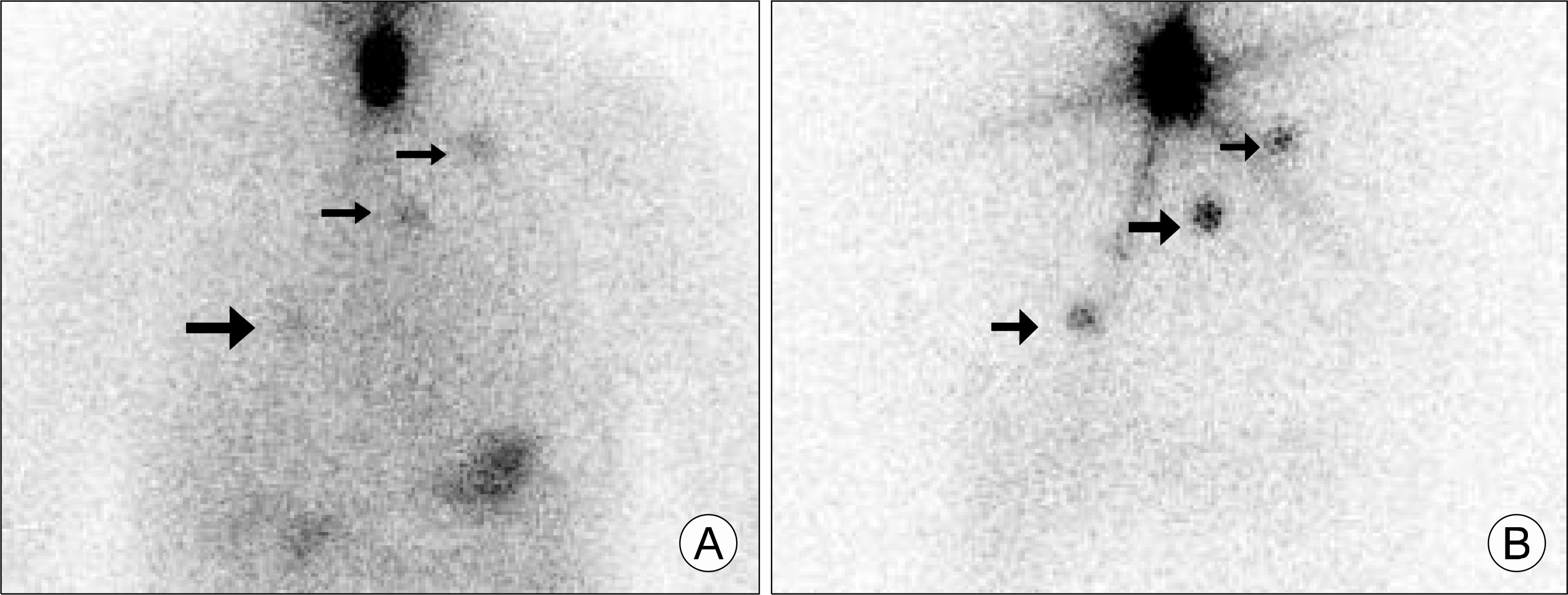
Post-therapeutic I-131 whole body scan. (A) Whole body scan with I-131 with 100 mCi at 2 days after administration showing multiple radioactive iodine uptakes at right hilar, left superior mediastinum and left supraclavicular region (arrows). (B) Whole body scan at 7 days after administration showing more intensive radioactive iodine uptake at the same region (arrows) compared with whole body scan at 2 days after administration.


 PDF
PDF ePub
ePub Citation
 Print
Print


 XML Download