

 PDF
PDF ePub
ePub Citation
 Print
Print


Abstract
Amyloidosis is an abnormal extracellular deposit of amyloid in various organs of the body. Amyloid goiter, defined by a clinically detectable thyroid enlargement due to amyloid deposition, is a rare cause of hyperthyroidism. We report the case of amyloid goiter mimicking Graves' disease in a 62-year-old woman. Graves' disease was diagnosed by diffuse goiter, hyperthyroidism, and positive TSH receptor antibody. Total thyroidectomy was planned due to progression of Graves' disease and respiratory distress. At surgery thyroid gland was very friable and fragmented like cobblestones when grasped with forceps. A diagnosis of amyloid goiter was established by the presence of diffuse amyloid deposits in the parafollicular areas. After systemic evaluation for amyloidosis, coexisting both multiple myeloma and systemic amyloidosis involving kidney and heart were detected. She underwent palliative chemotherapy but disease progressed. Amyloid goiter might be suspected in patient with thyroid enlargement and concomitant systemic disease such as renal or heart failure.
References
1. Villa F, Dionigi G, Tanda ML, Rovera F, Boni L. Amyloid goiter. Int J Surg. 2008; 6(Suppl 1):S16–8.

2. Kimura H, Yamashita S, Ashizawa K, Yokoyama N, Nagataki S. Thyroid dysfunction in patients with amyloid goitre. Clin Endocrinol (Oxf). 1997; 46(6):769–74.
3. Sethi Y, Gulati A, Singh I, Rao S, Singh N. Amyloid goiter: a case of primary thyroid amyloid disease. Laryngoscope. 2011; 121(5):961–4.
4. Chung R, Pilcher J. Primary amyloid goiter and review of imaging characteristics. Ultrasound. 2013; 21(2):98–101.
5. Tokyol C, Demir S, Yilmaz S, Topak N, Pasali T, Polat C. Amyloid goiter with hyperthyroidism. Endocr Pathol. 2004; 15(1):89–90.
6. Kyle RA, Gertz MA. Primary systemic amyloidosis: clinical and laboratory features in 474 cases. Semin Hematol. 1995; 32(1):45–59.
7. Pinto A, Nose V. Localized amyloid in thyroid: are we missing it? Adv Anat Pathol. 2013; 20(1):61–7.
8. Dinner S, Witteles W, Witteles R, Lam A, Arai S, Lafayette R, et al. The prognostic value of diagnosing concurrent multiple myeloma in immunoglobulin light chain amyloidosis. Br J Haematol. 2013; 161(3):367–72.
9. Gertz MA. Immunoglobulin light chain amyloidosis: 2014 update on diagnosis, prognosis, and treatment. Am J Hematol. 2014; 89(12):1132–40.
Fig. 1.
Preoperative imaging studies. (A) Ultrasonographic image showing enlarged thyroid gland (arrow) with heterogeneous echogenicity. (B) A thyroid scan (Tc-99m scintigraphy) showing diffusely enlarged thyroid gland with inhomogeneously decreased uptake in both thyroid lobes. (C) Non-enhanced axial CT scan showing symmetrically diffuse goiter without nodules.
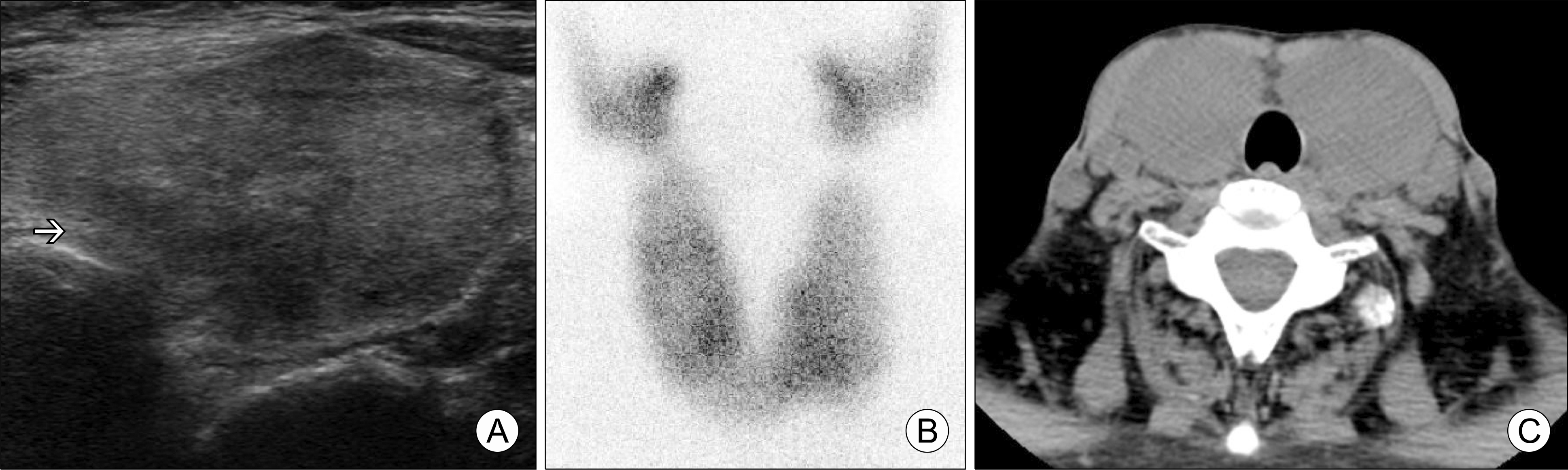
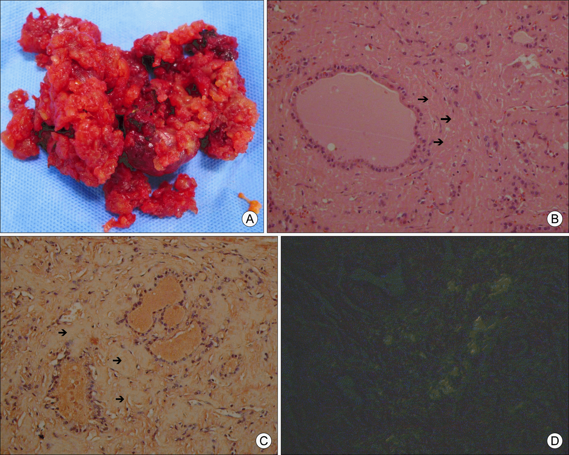
Fig. 2.
Thyroid pathology. (A) The thyroid gland was soft and fragile. Most of remnant thyroid gland tissue was removed by suction during operation. (B) Eosinophilic and amorphous substance accumulation (arrows) is seen in the interstitium (H&E, ×200).(C) Congo red staining marks the deposit (arrows) (Congo red, ×200). (D) Amyloid deposition showing apple-green birefringence under polarized light microscope (×200).
 XML Download