

 PDF
PDF ePub
ePub Citation
 Print
Print


Introduction
thyroid carcinoma (DTC), the most common type of endocrine malignancy, has an excellent prognosis and 10-year survival rate higher than 90%.12 This outcome has been ensured by increased diagnostic scrutiny. However, the DTC recurrence rate is 10–30%, and the potential for reappearance after several years34) indicates the need for prolonged follow-up.
According to the American Thyroid Association guideline,5) a disease-free status comprises all of the following: no clinical evidence of tumor, no imaging evidence of tumor (i.e., no uptake outside the thyroid bed on the initial post-treatment whole-body scan or, if uptake outside the thyroid bed is present, no imaging evidence of tumor on a recent diagnostic scan and neck ultrasound), and undetectable serum thyroglobulin (Tg) values during thyroid-stimulating hormone (TSH) suppression and stimulation in the absence of interfering antibodies.
The TSH-stimulated Tg level after successful thyroid ablation is known as a sensitive and reliable marker for surveilling tumor persistence and recurrence,67) particularly in patients with low or undetectable Tg values in the absence of TSH stimulation. Frequently, stimulated Tg cutoff values of >1–2.5 ng/mL are used to detect or predict persistent disease.89101112) Although some recent studies have reported that Tg stimulation with recombinant human TSH may be more beneficial in patients who have undetectable (unstimulated) Tg values, thyroid hormone withdrawal (THW) is more frequently used to measure stimulated Tg.
The adequate TSH value needed to achieve sufficient Tg stimulation after THW has not been determined, and the commonly used TSH cutoff has been derived from the value considered necessary for radioactive iodine (RAI) imaging. Edmonds et al.13) reported a study of seven patients in whom a TSH value >30 µU/mL was necessary for adequate uptake on an RAI whole-body scan. Other groups have reported that a TSH value of >25–30 µU/mL at 2–3 weeks after THW is sufficient.1314151617) Valle et al.18) reported that TSH values of >80–100 µU/mL would serve as a more appropriate cutoff, compared to >30 µU/mL. Although many institutions apply a TSH cutoff of >30 µU/mL, it remains unclear when TSH or Tg values might plateau and whether Tg values would continue to rise once the TSH cutoff has been achieved. In addition, the same patient might exhibit different TSH values after each THW, even when an identical THW protocol has been applied consistently. We thought that if the TSH values differed between two serial follow-up tests, a comparison of Tg values without considering TSH might not be clinically relevant, given the possibility of a change in Tg levels due to different levels of TSH stimulation.
The aim of this study was to evaluate the influence of TSH levels on Tg and to determine whether we could predict the exact trend in Tg change between two serial follow-up tests in each patient.
Materials and Methods
Patients
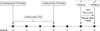
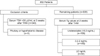
Fig. 1 illustrates the THW protocol used in this study, including a change of levothyroxine to liothyronine for 3 weeks followed by a period of no thyroid hormone treatment for the next 2 weeks. From October 2008 through September 2013, we consecutively enrolled 4967 patients with DTC who underwent THW, and selected 652 (13.1%) with limited TSH elevation (<30 µU/mL) at 2 weeks after THW and repeated TSH measurement at 3 weeks after THW. Only patients with a TSH value >30 µU/mL at 3 weeks after THW were then selected to effectively demonstrate the influence of elevated TSH levels on Tg levels. Subjects with pituitary or hypothalamic disease were also excluded (Fig. 2). Finally, 309 patients were evaluated in this retrospective study, which was approved by the Institutional Review Board at our institution (Kyungpook National University Medical Center and School of Medicine, Daegu, Korea). The requirement for written informed consent by participants for the use of their clinical records in this study was waived because patient information was de-identified prior to analysis.
Measurements of Tg and TSH
Radioimmunoassay commercial kits were used to measure serum Tg (Thyroglobulin IRMA; CIS Bio International, Gif sur Yvette, France). Seven calibrators, two controls, and patients' serum samples were mixed with buffer solution and incubated for 3 hours with agitation (400 rpm) at room temperature (18–25℃). The tubes were washed twice with washing solution, after which iodine-125–labeled anti-thyroglobulin was dispensed into each tube. The tubes were then incubated overnight (16–20 hours) at room temperature (18–25℃) without agitation. The tubes were again washed twice, and the remaining radioactivity bound to the tube was measured with a gamma scintillation counter for 1 minute. This Tg assay had a functional sensitivity of 0.7 ng/mL and analytical sensitivity of 0.2 ng/mL. The limit of detection for Tg in this study was ≥0.2 ng/mL.
Commercial immunoradiometric assay kits were used for TSH measurement (TSH IRMA, B·R·A·H·M·S GmbH, Hennigsdorf, Germany). Seven calibrators, two controls, and patients' serum samples were incubated with an excess of two anti-thyrotropin antibodies (mouse monoclonal) that recognized different binding sites on the antigen (TSH). One antibody was labeled with iodine-125, and the other was immobilized on the inner surface of the tube (coated tube system). The tubes were incubated in an orbital shaker (170–250 rpm) for 1 hour at room temperature (18–25℃), after which the remaining excess iodine-125–labeled anti-thyrotropin antibody was diluted and completely removed. The tubes were washed twice, and the radioactivity in each tube was measured for 1 minute in a gamma counter. The TSH assay had a functional sensitivity of 0.07 µU/mL.
Statistical Analysis
For analysis, patients were divided into three groups according to their Tg values at 2 weeks after THW (Fig. 2): <0.2 ng/mL (108 patients, 35%), ≥0.2 to <2 ng/mL (148 patients, 48%), and ≥2 ng/mL (53 patients, 17%). The paired-samples t-test was used to compare differences in TSH and Tg levels from 2 to 3 weeks after THW in each patient. In patients with Tg values ≥2 ng/mL, the absolute change in TSH values (ΔTSH=TSH at 3 weeks after THW-TSH at 2 weeks after THW) and percent change in Tg values [%ΔTg=(Tg at 3 weeks THW−Tg at 2 weeks THW)/Tg at 2 weeks THW×100] from 2 to 3 weeks after THW were calculated, and Pearson's correlation coefficients were calculated to determine the relationship between ΔTSH and %ΔTg. A Tg cutoff value of 2 ng/mL was used to represent clinical significance for diagnostic imaging or further evaluations. MedCalc Statistical Software version 15.6.1 (MedCalc Software bvba, Ostend, Belgium) was used for statistical analysis. Continuous variables are presented as means±standard deviations (SDs). A p value <0.05 was considered to indicate a statistically significant difference.
Results
Patient Characteristics
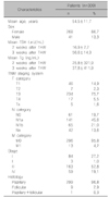
The characteristics of all 309 patients are presented in Table 1. The study cohort included 41 men and 268 women with a mean age of 54.5±11.7 years. All patients underwent total or near-total thyroidectomy, which was completed by central neck dissection or modified radical neck dissection in 86% of cases, resulting in the apparent complete resection of the neoplastic tissue. In addition, 93% of patients underwent THW for ablation of the thyroid remnant or treatment of persistent thyroid cancer, and 7% underwent THW for thyroid cancer surveillance.
Changes in TSH and Tg Values between 2 and 3 Weeks after THW
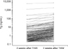
Fig. 3 and Supplementary Fig. 1 show the changes in serum Tg values in all 309 patients. In 227 patients (73.5%), the Tg values increased after an additional week of THW. The mean values of TSH and Tg at 2 weeks after THW were 16.9±7.7 µU/mL and 25.8±321.9 ng/mL, respectively, and the corresponding values at 3 weeks after THW were 56.6±14.3 µU/mL and 37.8±411.7 ng/mL, respectively. Tg values increased significantly after the additional week of THW (p=0.0233).
In a group of 108 patients with Tg values <0.2 ng/mL at 2 weeks after THW, the Tg values in approximately half (49.1%) of the patients remained below 0.2 ng/mL after the additional week of THW, whereas those in the other half (50.9%) increased but remained <2 ng/mL. Therefore, the Tg values at 3 weeks after THW had no clinical significance in patients with Tg values <0.2 ng/mL at 2 weeks after THW.
In a group of 148 patients with Tg values between 0.2 and 2 ng/mL at 2 weeks after THW, the Tg values in 121 (81.8%) patients increased after an additional week of THW, along with increases in TSH values (TSH: 19.1±8.0 to 55.1±13.8 µU/mL, p<0.0001; Tg: 0.5±0.3 to 2.9±3.2 ng/mL, p=0.0001). In these 121 patients, the Tg values of 57 (47.1%) patients increased to >2 ng/mL after an additional week of THW, accompanied by increases in TSH values (TSH: 15.0±7.6 to 56.8±14.5; Tg: 0.9±0.4 to 5.1±3.5, p<0.0001). We further dichotomized the 148 patients by their Tg values at 2 weeks after THW—122 patients with Tg values <1 ng/mL and 26 patients with Tg values ≥1 ng/mL—as a sub-analysis to determine the effect of Tg at 2 weeks after THW. Approximately 30% of 122 patients in the former group and approximately 77% of 26 patients in the latter eventually achieved a Tg value ≥2 ng/mL after the additional week. These results indicated that the patients with initially high Tg values achieved a relatively higher rate of increase in Tg after the additional week of THW (Table 2).
In a final group of 53 patients with Tg values ≥2 ng/mL, the Tg values of all but one patient remained in the same Tg category (≥2 ng/mL) after an additional week of THW (Fig. 4), and all but one of the 52 patients exhibited increased Tg values after the additional week (Table 3).
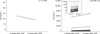
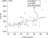
To evaluate the correlation between the changes in TSH and Tg values, we subjected 190 patients with detectable Tg values in both the first and second tests to a linear regression analysis and Pearson's correlation analysis. The %ΔTg was found to correlate positively with the ΔTSH (p=0.0001), with a correlation coefficient of 0.2881. We also performed the same analysis in the 53 patients with Tg values ≥2 ng/mL at 2 weeks after THW, a value that is suggestive of tum or recurrence or persistent tum or burden. The %ΔTg was also found to correlate positively with the ΔTSH (p=0.0001), with a correlation coefficient of 0.5037 (Fig. 5).
We performed a sub-analysis of 22 patients who underwent THW for thyroid cancer surveillance. The Tg values were undetectable in 12 (54.5%) of these 22 patients, and <2 ng/mL in the other 10 (45.5%) patients. Among the former 12 patients, three exhibited increased Tg values that remained <2 ng/mL after an additional week of THW. All but one of the latter 10 patients exhibited increased Tg values after the additional week of THW, and five (55.6%) of the nine patients' Tg values increased to >2 ng/mL after the additional week.
Discussion
Generally, serum Tg values are used to predict successful thyroid remnant ablation,19) assess responses to RAI therapy,20) detect recurrent tumors,621) and predict disease recurrence2223) and prognosis.2425) A few studies have demonstrated that Tg values could increase in accordance with increases in TSH values.18)
The current large-scale study aimed to determine the importance of considering the serum TSH value with regard to Tg stimulation by THW. Our results demonstrated increased Tg values in a majority (73.5%) of patients after an additional week of THW. Furthermore, 38.5% of the patients with Tg values between 0.2 and 2 ng/mL at 2 weeks after THW exhibited increases to >2 ng/mL, suggesting a need for diagnostic imaging or other evaluations. These results are consistent with those of a previous report.18) The current study demonstrated that stimulated Tg levels in response to THW must be interpreted in the context of the corresponding TSH level to avoid drawing false conclusions about the disease status.
Almost all patients with Tg values >2 ng/mL at 2 weeks after THW demonstrated a significant increase in Tg along with an increase in TSH values after an additional week of THW. As Tg synthesis and secretion is an active process requiring stimulation by TSH, TSH values should be carefully considered by clinicians when determining whether Tg levels suggest the need for further diagnostic evaluations. Furthermore, our results also revealed that Tg values did not increase to >2 ng/mL at 3 weeks after THW in patients with undetectable Tg values at 2 weeks after THW. This result signifies that further clinical evaluation may not be needed when Tg values are undetectable at 2 weeks after THW without reference to TSH values.
A previous study18) tried to predict final Tg values using serial evaluations of each patient's Tg values at different time points. However, accurate prediction was not possible because the rate of increase was quite diverse for each patient. The residual tumor volumes always differed among patients; therefore, the rate of Tg increase in accordance with the increase of TSH value would always differ in each patient. We defined ΔTSH as the absolute change in TSH values and %ΔTg as the percent change in Tg values. Accordingly, using %ΔTg, we could overcome the difference of the rate of increase in each individual patient. Finally, %Δ Tg was found to correlated significantly with ΔTSH. This result indicates that Tg values should be interpreted in light of the TSH value in each individual patient at each individual testing time point.
Even among the 22 patients who underwent THW for thyroid cancer surveillance, nine of 10 patients with Tg values <2 ng/mL exhibited increased Tg values after the additional week of THW, and more than half of these nine patients' Tg values increased to >2 ng/mL. This result indicates that TSH levels should carefully be considered even among patients in the surveillance group, who were thought to be disease-free.
Meanwhile, the current study was limited by a THW duration of only 3 weeks. It remains unknown how long the TSH and Tg would continue to increase. Although they used a different THW protocol, Valle et al.18) reported that 4 weeks of THW was not sufficient to reach a final Tg value. The mean TSH values of the current study were lower than the range of 80–100 µU/mL suggested by their study. Nevertheless, in actual clinical situations, 3–4 weeks is a common interval for levothyroxine withdrawal. If levothyroxine is withdrawn for ≥4 weeks, liothyronine may be substituted; in such circumstances, 2–3 weeks is a common time period for liothyronine withdrawal. The current study did not assess the clinical significance of an increase in Tg values to >2 ng/mL at 3 weeks after THW. In future studies, we will investigate the clinical significance of a Tg increase after an additional week of THW.
In conclusion, the Tg value increases significantly in accordance with the increase in TSH values in patients with initially detectable Tg values. Careful consideration should be given to the TSH value when interpreting the meaning of changes in Tg levels in patients with DTC, as TSH values can always vary, even within the same patient and when the values exceed the cutoff (30 µU/mL), and can affect every measurement of stimulated Tg.


 XML Download