

 PDF
PDF ePub
ePub Citation
 Print
Print


INTRODUCTION
Dental implants that are in direct contact with bone through osseointegration differ from natural teeth in terms of their biological structures and biomechanical characteristics [12]. Since they do not possess a periodontal ligament (PDL) or periodontal mechanoreceptors, implants display low shock absorption, a low tactile threshold, and a high stress concentration at peri-implant sites [34]. Therefore, the occlusal concept that applies to implants is different from the occlusal concept that applies to natural teeth in terms of the occlusal form and scheme design, including a rounded and diminished cusp tip, narrow occlusal table, smooth grooves and fossa, slight occlusal contact, and shallow or flat occlusal anatomy [56].
The modified occlusion applied to implants is designed to reduce the occlusal force during axial and nonaxial loading conditions, as well as the stress around the implant and supporting bone structures. In addition, this concept of occlusal morphology is considered to play a major role in increasing the likelihood of clinical survival and the success rates of implants without any mechanical, biological, or aesthetic complications [78]. However, due to the insufficiency of clinical studies and lack of evidence-based outcomes, the ideal occlusion for implants remains to be established [9].
Maintaining the occlusal stability of the existing dentition is a very important for avoiding traumatic occlusion [10]. In particular, the loss of the posterior teeth has the greatest influence on changes in dentition and occlusion [101112]. Including the first permanent molar teeth, which erupt first, the maxillary and mandibular molars are highly susceptible to dental caries and periodontal disease, and thus are associated with a high incidence of early loss [13]. Several treatment options exist in case of the early loss of molars. Among these options, dental implant therapy is often selected. However, this treatment strategy can sometimes result in traumatic occlusion or overloading in the adjacent premolars [1415].
Fremitus, thermal sensitivity, widening of the PDL space, tooth mobility, and fracture are the most common symptoms of traumatic occlusions that result from overloading of the premolars, which has been proposed to be associated with implant-protected occlusion [16]. However, few studies have been conducted of the physiological and biological effects of implants on the adjacent premolars, especially regarding traumatic occlusion. Therefore, the aim of this retrospective study was to characterize the association between implants in the posterior region and traumatic occlusion in the adjacent premolars over a mean follow-up of 5 years.
MATERIALS AND METHODS
This retrospective 14-year follow-up study assessed the associations between single or splinted implants in the molar region and traumatic occlusion in the adjacent premolars, using data collected from January 2002 to November 2015. The protocol was approved by the Institutional Review Board of the National Health Insurance Service (NHIS) Ilsan Hospital (Approval No. 2016-03-038) and was conducted in the Department of Periodontology and the Department of Prosthodontics, NHIS Ilsan Hospital.
Study design and population
Dental implant surgery and prosthesis placement were conducted by a registered and calibrated periodontist and prosthodontist, respectively, at NHIS Ilsan Hospital using internal-connection fixtures with a resorbable blasting material and a sandblasted, large-grit, acid-etched surface. Prosthetic occlusion was adjusted according to the concept of implant-protected occlusion, which was proposed for reducing the parafunctional force and protecting the implant structure [17]. All patients were encouraged to receive maintenance care at least every 6 months and have an intraoral radiograph taken every 12 months.
The following inclusion criteria were applied: 1) age of over 20 years; 2) placement of a single implant or multiple splinted implants in the molar region of the maxilla or mandible with the presence of opposing teeth (natural or implants); and 3) adjacent first and second premolars present with only natural teeth, excluding missing teeth but including restorative prostheses.
The following exclusion criteria were applied: 1) severe or uncontrolled systemic disease posing a constant threat to life; 2) uncontrolled or severe parafunctional activity such as clenching or bruxism (diagnosed by a self-administered questionnaire); 3) moderate to advanced or untreated periodontal infection or disease; 4) at the time of implant placement, adjacent premolars showing signs of inflammation or traumatic occlusion such as gingival bleeding, deep pocket depth, fremitus, widening of the PDL space, loss of the supporting bone, or tooth mobility; 5) adjacent first and second premolars including any missing teeth or restorative prostheses; 6) opposing occlusion with full or partial removable denture prosthetics; and 7) implants with mechanical complications (i.e., screw loosening and/or fracture, fixture fracture, or ceramic fracture), biological complications (peri-implantitis), or failed osseointegration.
Clinical and radiographic analysis
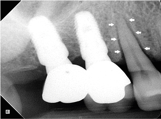
Each adjacent premolar was identified by examining the patient's electronic and paper dental records, as well as periapical and panoramic radiographs. Traumatic occlusion was assessed by examining clinical parameters (bleeding on probing, probing pocket depth, thermal sensitivity, tooth mobility, and fremitus) and radiographic parameters (loss of supporting bone and widening of the PDL space) at NHIS Ilsan Hospital by 2 periodontists who were specially trained and calibrated for the evaluation (Figure 1) [18]. Tooth mobility was evaluated and classified using Miller's classification, and the probing pocket depth was measured at 6 sites (mesiobuccal, midbuccal, distobuccal, mesiolingual, midlingual, and distolingual) around each tooth using a University of North Carolina (UNC) periodontal probe (Hu-Friedy, Chicago, IL, USA) [19]. All radiographic assessments were size-calibrated and performed using a picture archiving and communication system workstation (Centricity GE Healthcare, Waukesha, WI, USA).

Statistical analysis
The χ2 test, Fisher exact test, and t-test (2-tailed with independent samples) were performed to identify the relationship between implants in the posterior region and traumatic occlusion in the adjacent premolars using SPSS version 23 (IBM Corp., Armonk, NY, USA). Standard deviation values for the ordinary and continuous variables and the range of error at 95% confidence intervals (CIs) were calculated, and P<0.05 were considered to indicate statistical significance.
RESULTS
Study population
The baseline characteristics of the patients and implants are presented in Table 1. The 283 patients who met the inclusion criteria comprised 150 males and 133 females, with a mean age of 57.1 years (range, 23–91 years). The 347 implants investigated in these patients were distributed in the posterior region as follows: Mx. first molar, n=28 (8.1%); maxillary second molar, n=13 (3.7%); maxillary splinted implant prosthesis, n=84 (24.2%); mandibular first molar, n=47 (13.5%); mandibular second molar, n=85 (24.5%); and mandibular splinted implant prosthesis, n=90 (25.9%). There were 125 implants placed in the maxilla (36.0%), and 222 were placed in the mandible (64.0%). The mean duration of functional loading was 61.9 months (range, 8–165 months).
Table 1
Baseline characteristics of the patients and implants included in the study
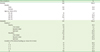
Relationship between adjacent premolar position and severity of mobility
The severity of tooth mobility according to position in the maxilla and mandible is shown in Figure 2. Tooth mobility and widening of the PDL space occurred in 12 maxillary first premolars (mobility [+], n=6; mobility [++], n=3; mobility [+++], n=3), 14 maxillary second premolars (mobility [+], n=8; mobility [++], n=5; mobility [+++], n=1), 1 mandibular first premolar (mobility [++], n=1), and 2 mandibular second premolars (mobility [+], n=2). No traumatic occlusion occurred at the same time in the same region in the first or second premolars.
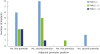
Relationship between adjacent premolar position and duration of functional loading
Symptoms of traumatic occlusion occurred in 21 (72.4%) of 29 premolars during the first 36 months after applying functional loading. Traumatic occlusion was only observed in the maxillary premolar after 36 months. Traumatic occlusion was observed in 1 premolar adjacent to a single implant and in 7 premolars adjacent to splinted implants in the molar region (Figure 3).
Relationship between mobility in the adjacent premolar and clinical factors
The χ2 test, Fisher exact test, and t-test were used to assess differences between gender, age, implant type, implant position, characteristics of the opposing teeth, and duration of functional loading with and without traumatic occlusion. As shown in Table 2, the incidence of traumatic occlusion in the adjacent premolars was significantly related to implant type (P=0.004), maxillary or mandibular position (P<0.001), and the characteristics of the opposing teeth (P<0.001). The other clinical factors of gender, age, and duration of functional loading were not significantly associated with traumatic occlusion.
Table 2
Incidence of traumatic occlusion in the adjacent premolars according to clinical factors
Data are presented as number of patients (%) or mean±standard deviation.
a)P values in this category were calculated using the χ2 test, Fisher exact test, and t-test (2-tailed with independent samples); b)Statistically significant (P<0.05); c)Included in the implant category if at least 1 of the opposing teeth was a dental implant.
DISCUSSION
Recent studies have found that mechanical overloading of implants did not result in either the loss of osseointegration or marginal bone loss, although this is still controversial [2021]. A limited number of studies have been carried out to investigate excessive loading affecting the adjacent teeth. In the present study, some premolars exhibited the symptoms of traumatic occlusion, which seemed to have been due to implant overloading. Signs of fremitus, thermal sensitivity, widening of the PDL space, tooth mobility, and root resorption are clearly associated with overloading of the natural teeth [22]. In particular, widening of the PDL space and increased tooth mobility are important considerations for avoiding or at least minimizing traumatic occlusal force [23].
Traumatic occlusion was more likely to occur in maxillary premolars (n=26, 20.8%) than in mandibular premolars (n=3, 1.4%; P<0.001). Alveolar bone in maxillary canines and premolars has the anatomical characteristics of having the thinnest cortical bone and little cancellous bone [24]. In addition, given that the maxillary premolars are located in the anterior area among teeth with lingual cusps, they are subjected to large occlusal forces and continuous stress during forward movement of the mandible [25]. Such anatomical and physiological characteristics reduce the tolerance of these premolars to traumatic occlusion, which in turn increases the frequency of dehiscence, fenestration, widening of the PDL space, and tooth mobility [26].
Traumatic occlusion did not occur in single implants in the second molar region. In contrast, traumatic occlusion in the adjacent premolars was present in 7 cases (4.0%) of single implants in the first molar region and in 22 cases (12.6%) of splinted implants in the molar region, and this difference was statistically significant (P=0.004). Based on these findings, it may be concluded that implants in the second molar region have a negligible influence on the incidence of traumatic occlusion in the adjacent premolars, while implants in the first molar region play a key role in inducing traumatic occlusion in the adjacent premolars.
The probability of traumatic occlusion in the adjacent premolars was higher for implants in occlusion with opposing teeth that include at least 1 dental implant (n=12, 4.6%) than when no implant was present (n=17, 20.2%; P<0.001). This is presumed to occur because the axial mobility is much smaller for opposing implants (3–5 μm) than for opposing natural teeth (25–100 μm). Greater axial mobility increases the ability of a tooth to offset overloading of the adjacent premolars [9].
A recent systematic review found that implants were associated with a 5-year cumulative survival rate of 97.2% (95% CI=96.3%–97.9%), a soft-tissue complication rate of 7.1% (95% CI=4.4%–11.3%), a mechanical complication rate of 8.8% (95% CI=5.1%–15.0%), and an aesthetic complication rate of 7.1% (95% CI=3.6%–13.6%) [27]. Various dental implant systems are currently being investigated with the aim of reducing mechanical, biological, and aesthetic complications, and this is consistent with a decreasing trend in the incidence of complications [2829]. In the present study, the 14-year cumulative incidence of traumatic occlusion was 8.4%, which is similar to the rates of other types of complications. Of all possible etiological factors of biological and mechanical complications, traumatic occlusion is one that may be directly and actively managed at the chairside after the final prosthesis is placed [30]. We reduced the widening of the PDL space and tooth mobility and increased the stability in 27 out of 29 premolars with traumatic occlusion by performing occlusal adjustment of the implant. Additional occlusal splint therapy can be applied if it is deemed necessary to more actively prevent overloading of both the implant and the adjacent premolars [31].
Other aspects to consider when checking and adjusting implant occlusion is the mechanically locking friction fit and the patient's perceptions [3233]. A report has indicated that the tactile threshold level, tactile function, and friction fit of the oral stimulation and occlusal force increased gradually after implant placement, although this is controversial [3435]. It has therefore been proposed that the type of occlusion that occurs for an implantation with immediate loading may increase the risk of overloading in the early stage, which would in turn increase the frequency of traumatic occlusion of the adjacent premolars. However, future studies are needed to further evaluate this possibility.
The limitations of this study include the retrospective data collection, selection bias, absence of a control group, nonstandardized observation period, and lack of definitive confirmation of traumatic occlusion. We conducted a retrospective clinical and radiographic investigation and found that the risk of traumatic occlusion in the adjacent premolars increased when splinted implants were placed in the maxillary molar region and when implants were present in the opposing teeth. These findings indicate the importance of conducting periodic occlusion checks and addressing possible etiological factors in order to prevent overloading of the adjacent teeth. If fremitus, thermal sensitivity, widening of the PDL space, tooth mobility, or root resorption occur in the adjacent premolars, implant occlusion should be carefully considered prior to the start of periodontal treatment.
Within the limitations of this study, we found that the risk of traumatic occlusion in the adjacent premolars increased when splinted implants were placed in the maxillary molar region and when implants were present in the opposing teeth. Insufficient evidence was obtained to establish ideal guidelines for implant occlusion, and future well-designed studies are therefore required to identify how best to minimize implant occlusion.
 XML Download