

 PDF
PDF ePub
ePub Citation
 Print
Print


INTRODUCTION
The presence of keratinized mucosa around dental implants is a critical component for the maintenance of their health. Interestingly, there is a lack of consensus in the literature regarding the relationship between the width of the keratinized mucosa and the health of peri-implant tissues. Wennström et al. [1] reported that there is no correlation between implant success rate and the presence of keratinized tissue in the peri-implant soft tissue. On the other hand, Warrer et al. [2] reported that experimentally ligated implants without keratinized mucosa accumulated more dental plaque and showed significantly more recession with attachment loss than implants where keratinized mucosa was present. Recently, a number of studies have reported that a lack of adequate keratinized mucosa around implants seems to be significantly associated with more plaque accumulation, tissue inflammation, mucosal recession, and attachment loss [34].
In contemporary implant treatment, the necessity of guided bone regeneration (GBR) has increased due to the concept of contour augmentation, and frequent GBR procedures usually lead to the reduction of keratinized mucosa around implants, since the flaps have to be advanced for tensionless primary closure [56]. To correct these mucogingival issues, a free gingival graft (FGG), an apically positioned flap, or a connective tissue graft is required if there is a minimal width of keratinized tissue, in order to increase the width [7]. If there is a sufficient amount of keratinized tissue near the implants, apically or laterally positioned flaps can be applied using the pre-existing keratinized mucosa [89], or the combination of an apically positioned flap (APF) and FGG can be used [1011]. However, these techniques have distinct limitations, such as a limited quantity of donor tissue, common infections, morbidity of patients, bleeding from the donor area, and unpredictable failure. Also, these techniques are highly technique sensitive and time consuming, and therefore general clinicians may not favor them [1213].
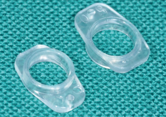
To overcome these disadvantages, a prefabricated implant-retained stent (Figure 1) clipped on provisional abutments was developed and evaluated in our previous study [14]. Using this prefabricated stent, pre-existing keratinized mucosa can be displaced in the bucco-apical direction and the width of the keratinized mucosa can be substantially preserved. Also, vertical pressure can be additionally applied over the displaced buccal flap, reducing the dead space under the flap, helping the displaced gingiva firmly attach to the underlying periosteum, and preserving the width of the pre-existing keratinized mucosa.
The aim of this study was to perform a prospective case control study to clinically assess the preservation of keratinized mucosa around implants after implant surgery using a prefabricated implant-retained stent clipped over healing abutments and to compare it with the application of horizontal external mattress sutures.
MATERIALS AND METHODS
Patient selection
This study was conducted according to guidelines from the Institutional Review Board (IRB) of Dankook University Dental Hospital (No. H-1503/003/004). All patients >18 years of age requiring first-stage implant surgery or second-stage implant surgery were enrolled after obtaining informed consent. The criteria for exclusion were as follows: 1) the presence of severe or aggressive untreated periodontal disease; 2) a history of systemic disease that would contraindicate surgical treatment; 3) reported long-term steroidal or antibiotic therapy; 4) current pregnancy; 5) unwillingness to return for the follow-up examination; and 6) smoking of >10 cigarettes per day.
A total of 50 patients (25 controls and 25 tests) were enrolled in this study (Table 1). Each patient was randomly allocated to a test group or control group.
Table 1
Demographic information of the enrolled patients
SD, standard deviation; Min, minimum; Max, maximum.
a)P values (statistically significant at the level of P<0.05) with a χ2 test for differences in means between groups in categorical variables; b)P values (statistically significant at the level of P<0.05) with an independent-samples t-test for differences in means between groups.
![]()
Surgical procedures and measurements
Following local anesthesia, a lingualized incision was made to mobilize the keratinized mucosa to the buccal aspect. Implants were placed and healing abutment was connected to the implant fixture. In the test group, a prefabricated implant-retained stent (Louis Button®, Dentis Co., Daegu, Korea) was clipped on the healing abutment and adjusted to apply adequate pressure to the displaced flap (Figure 2). Additional sutures were minimally performed if there was a risk of slippage of the flaps from the stent. In the control group, horizontal external mattress sutures were applied to adapt flaps around the healing abutment and to apply vertical pressure (Figure 3). After surgical procedures, clinical photographs were taken and the amount of buccal keratinized mucosa was measured in the mesial, middle, and distal sites of the healing abutment using a periodontal probe (Williams®, Hu-Friedy, Chicago, IL, USA). The average of these measurements was used to represent the width of buccal keratinized tissue. Medication prescribed to all subjects consisted of 500 mg of amoxicillin plus sulbactam (Sultamox®, Kunwha, Seoul, Korea) and 370 mg of talniflumate (Somalgen®, Kunwha, Seoul, Korea), twice a day for 5 days, and mouthwash (Gum Gargle®, Sunstar, Tokyo, Japan). Patients were directed to use mouthwash, twice a day, and not to brush the surgical sites for 10 days. Stents and sutures were removed 10 days after surgery. Patients were recalled after 1 and 3 months. The amount of buccal keratinized mucosa was measured at these time points.
 | Figure 2Clinical photograph of #37 implant first-stage surgery, which was adopted with the horizontal external mattress suture technique (control group). (A) The flap was secured using horizontal external mattress sutures and vertical pressure was applied over the flaps. (B) Ten days post-operation. (C) Three months post-operation. The healing was uneventful and the width of keratinized mucosa was well preserved.
|
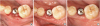 | Figure 3Clinical photograph of #35 implant first-stage surgery using a prefabricated implant-retained stent (test group). (A) The flap was placed bucco-apically using the stent. Dead space could be eliminated by vertical pressure from the stent. (B) Ten days post-operation. (C) Three months post-operation. Soft tissue healing is uneventful and the width of keratinized mucosa was well preserved.
|
Statistical analysis
Chi-square tests and t-tests were performed for demographic analysis. Mean values and standard deviations (SD) were calculated for each width assessed in the 2 treatment groups. Initially, changes of the width over the observation periods were determined with the use of the Friedman test. The Wilcoxon signed rank test was used to evaluate the differences between baseline and 1 month, and baseline and 3 months, while the Wilcoxon rank-sum test was used to evaluate the differences between the 2 groups at each time point. All evaluations were performed using SPSS software version 18.0 (IBM Corp., Armonk, NY, USA). The statistical significance was set at P<0.05.
RESULTS
The test and control groups were composed of 14 male, 11 female patients, and 18 male, 7 female, respectively (Table 1). The mean age of the study patients was 54.28±7.15 years in the test group and 54.40±7.09 years in the control group. The surgical sites were divided into maxilla (52% in the test group and 60% in the control) and mandible. A total of 6 patients were excluded during the study since they failed to show up for the recalls. In the end, 44 patients were successfully treated and healing was uneventful in all patients.
At the end of the study, the width of keratinized mucosa had decreased over time in both groups. At baseline, the mean width of the keratinized mucosa around the healing abutment was 5.05±0.31 mm in the test group and 4.22±0.28 mm in the control, and there was no statistical difference between groups (Table 2). After 1 month, the mean width of keratinized mucosa was 4.79±0.31 mm and 3.48±0.23 mm, respectively. The difference of width between baseline and 1 month was −0.26±0.85 mm in the test group without any statistical significance (P=0.137). Meanwhile, the corresponding difference in the control group was −0.74±0.73 mm and it showed statistical significance (P<0.001). These changes between the 2 groups were compared and the amount of reduction of keratinized tissue was greater in the control group, with statistical significance (P=0.034), suggesting that the sutures failed to preserve the width of keratinized tissue in the early healing phase at 1 month. The mean width of keratinized mucosa after 3 months was 4.79±0.31 mm in the test group and 3.48±0.23 mm in the control group. The difference of width between baseline and 3 months was −0.57±0.97 mm in the test group (P=0.011) and −0.86±0.71 mm in the control group (P<0.001). These amounts of reduction were statistically significant compared to the baseline. The reduction was substantially greater in the control group; nonetheless, there was no statistical difference. After these observations, all of the patients were treated with prostheses successfully, and they were routinely recalled for checkup thereafter.
Table 2
Mean values and SD of the width of keratinized mucosa around the healing abutment with group differences

SD, standard deviation.
a)Statistically significant difference compared to the baseline at 3 months in the test group (P=0.011); b)Statistically significant difference compared to the baseline at 1 month (P<0.001); c)Statistically significant difference between test and control groups at 1 month (P=0.034); d)Statistically significant difference compared to the baseline at 3 months in the control group (P<0.001).
![]()
DISCUSSION
The original idea of using stents for the mobilization and stabilization of displaced keratinized tissue around dental implants came from a number of previous studies, which used acrylic stents following periodontal plastic surgery [151617181920212223]. After the soft tissue has been grafted, the secure immobilization of the graft for successful vascularization and healing, and the protection of the surgical area to prevent discomfort for patients, are crucial factors to consider in a clinical setting. For these purposes, acrylic stents have been utilized in various cases with perimandibular sutures [15161718192021], fixation bone screws [2223], and adhesive [2124] or osseointegrated dental implants [2526]. Although these attempts have shown predictable and satisfying results, the time for the production of these stents is extensive, since these surgical stents are fabricated in the laboratory in advance before the surgery using altered casts, which also introduces additional cost. To overcome these shortcomings, the Loma Linda stent has been proposed, which can be fabricated chairside using photopolymerized resin [27]. However, the Loma Linda stent still takes additional processing and this may hinder clinicians from applying the tool in daily practice. The prefabricated implant-retained stent utilized in this study can be very useful, since it is of minimal cost and it significantly reduces chair time. Also, the size of the stent is as small as the healing abutments and the patient rarely feels any discomfort. The removal of the stent is also as easy as its connection, which can be very beneficial to clinicians in daily clinics.
In the present study, the impact of prefabricated implant-retained stent connection on the preservation of keratinized mucosa was compared to the horizontal external mattress suture technique following implant surgery. It was shown that the stent could effectively provide vertical pressure and stabilize the displaced flap during the initial healing period. Meanwhile, horizontal external sutures required meticulous effort and a significant amount of time to achieve the same purpose. Width reduction of the keratinized mucosa was similarly observed in both groups; however, it should be noted that the change in keratinized mucosa at 1-month follow-up was significantly greater in the control group than in the test group.
Because of less vascular supply and fewer fibroblasts, tissue around the implant is more susceptible to the development of inflammation and bone loss when exposed to plaque accumulation. For these reasons, the preservation of keratinized mucosa is one of the most important factors in implant maintenance. Several studies have indicated that the adequate width of keratinized mucosa for healthy tissues around dental implants is at least ≥2 mm [282930]. Although a lack of keratinized mucosa may not influence the long-term survival rate of implants [1], many studies have commonly shown that the presence of keratinized mucosa is more important around restorations and prostheses than natural tooth, irrespective of oral hygiene status [3132]. Also, implants surrounded by keratinized mucosa are less prone to plaque accumulation and gingival recession, without regard to whether patients engage in sufficient oral hygiene or receive adequate supporting periodontal therapy [4]. Therefore, clinicians should consider that it is essential to provide and maintain adequate keratinized mucosa for the successful long-term maintenance of implants.
Using a prefabricated implant-retained stent was shown to be equally effective for preserving the keratinized mucosa around a healing abutment compared to the control group after 3-month follow-up of the initial healing period. In the case of the conventional suturing method, it is time-consuming, and it is also difficult to apply enough vertical pressure over the displaced flap, leading to the creation of a dead space under the flaps. This dead space could be related to the delayed healing process or necrosis of the keratinized tissue in the worst cases. In contrast, the utilization of stents has the following benefits [14]: 1) the flap can be displaced in the bucco-apical direction with the existing keratinized mucosa, securing its position (Figure 4); 2) the flap can be compressed and stabilized over the underlying periosteum and can prevent the reduction of the keratinized mucosa; 3) the application of sutures on vertical incision lines is not required; and 4) the stent can be used off-the-shelf conveniently, since it is standardized according to the size of the healing abutments the clinician used.
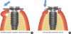 | Figure 4Schematic illustration showing the 2 different approaches. (A) Following the lingualized incision, the keratinized tissue was mobilized bucco-apically, and the prefabricated implant retained stent was applied to the healing abutment (test group). The flap is pressed down and displaced to preserve the keratinized tissue. (B) The external mattress suture is applied and the displaced flap is pushed down and apically (control group). However, there is limitation in the displacement in comparison to that of using the stent.
|
In sum, use of a prefabricated implant-retained stent was shown to be effective in the preservation of the keratinized mucosa around implants in comparison to the conventional external mattress suture technique, and it substantially reduced time and effort.
 XML Download