Journal List > J Periodontal Implant Sci > v.46(2) > 1082499

Cho, Kim, Koo, Seol, Lee, Rhyu, and Ku: Rescue of a periodontally compromised tooth by non-surgical treatment: a case report

Abstract

Purpose

This article describes a case of the successful non-surgical management of a periodontally compromised maxillary premolar.

Methods

A combination therapy, including root planing, occlusal adjustment, and tooth splinting, was applied. Clinical and radiographic examinations were performed during the 16-month follow-up period.

Results

All periodontal parameters were improved. There were dramatic decreases (3–6 mm) in the probing pocket depth, tooth mobility, and marginal bone loss. Interestingly, gradual resolution of the periapical radiolucency and alveolar bone regeneration were observed in the radiographs, and the periodontal condition was maintained during the follow-up period.

Conclusions

Within the limits of this study, these results demonstrate the importance of natural tooth preservation through proper periodontal treatment and occlusal adjustment of the periodontally compromised tooth, which is typically targeted for tooth extraction and dental implantation.

Graphical Abstract

jpis-46-128-abf001

INTRODUCTION

Chronic periodontitis is a destructive periodontal disease that involves gingival inflammation and bone destruction caused by specific microorganisms, and it is an indication for tooth extraction [123]. Chronic periodontitis is further classified as localized or generalized depending on whether ≤30% or >30% of the sites are involved [4]. The symptoms of chronic periodontitis usually progress without any episode of subjective pain, worsening the periodontal condition [5]. The specific aim of periodontal therapy is to arrest the inflammatory process. Various parameters, including the probing pocket depth (PPD), the clinical attachment level (CAL), tooth mobility, and bleeding on probing (BOP), are routinely used to evaluate the periodontal status. Periodontitis is typically treated by mechanical removal of the supra- and subgingival calculus and improvement of periodontal health through reductions of the PPD, BOP, and tooth mobility and an increase of the CAL [3]. Periodontists must regularly manage periodontitis with a diverse range of disease severity and extent. The available treatments are divided into two types, namely non-surgical and surgical therapies. Additionally, adjunctive treatments (antimicrobials or laser therapy) are also available [6789]. The American Academy of Periodontology (AAP) suggests that the most minimally invasive and cost effective periodontal treatment should be adopted, and this is usually performed through non-surgical treatments, such as scaling and root planing (SRP). Furthermore, the American Dental Association Council on Scientific Affairs presented an evidence-based clinical practice guideline on the non-surgical treatment of patients with chronic periodontitis by means of SRP with or without adjuncts [89]. They evaluated four adjunctive therapies such as systemic subantimicrobial-dose doxycycline, systemic antimicrobials, chlorhexidine chips, and photodynamic therapy with a diode laser, which are beneficial in the non-surgical treatment of chronic periodontitis with a moderate level of certainty [9]. However, when the periodontal condition does not recover with a non-surgical treatment, surgical treatment is considered to restore periodontal health.
In the periodontally compromised tooth, the normal occlusal force could affect the periodontal tissue and cause further attachment loss [10]. First, the signs of inflammation must be resolved. When the periodontal condition becomes stable, occlusal adjustment can be considered [11]. Trauma from occlusion (TFO) refers to an injury to the periodontal attachment components caused by abnormal occlusal force. TFO is divided into three categories: primary, secondary, and combined occlusal trauma [12]. Primary occlusal trauma results from excessive occlusal force with normal periodontal support. On the other hand, secondary occlusal trauma is caused by excessive or premature contact on teeth with a damaged periodontium. Many animal experiments and clinical studies have investigated the role of occlusion in the pathogenesis of periodontitis [1213141516]. The results suggest that premature contact or excessive occlusal force could be an etiologic factor in the progression of periodontitis through inflammation that spreads into the deep portion of the periodontium. Based on these findings, vertical alveolar bone resorption and infrabony defects should be considered a result of occlusal trauma [1718]. Here, we describe a successful non-surgical treatment with adjunct therapy in a periodontally compromised tooth with a secondary TFO showing alveolar bone regeneration. The use of the patient records was approved by the Institutional Review Board (ERI15007) of Seoul National University Dental Hospital (SNUDH).

CASE DESCRIPTION

Patient information and diagnosis

A 38-year-old man with a non-contributory medical history presented at the Department of Periodontology, SNUDH, Seoul, Korea, with a chief complaint of continuous buccal sinus tract formation of the maxillary left 1st premolar (#24). A clinical examination revealed a sinus tract with pus discharge at the buccal surface and hyper-tooth mobility, and Angle class III malocclusion and anterior cross-bite were observed. In the periapical radiograph, extensive radiolucency and active bone resorption around the root were noteworthy, and subgingival calculus deposition was observed (Figure 1A). The PPD was approximately 9–10 mm at the distal portion and 7–8 mm at the mesial portion of the #24 tooth. The diagnosis was localized chronic periodontitis with secondary TFO. Because of a periapical lesion, endodontic treatment was also considered. However, the tooth was responsive to the electric pulp tester (EPT), and periodontal treatment and occlusal adjustment were planned first.
Figure 1
Periapical radiography: (A) Initial radiography showing a periapical radiolucency and severe bone resorption of the maxillary left first premolar, which has subgingival calculus deposition. (B-F) Follow-up, 3 (B), 6 (C), 9 (D), 12 (E), and 16 months (F) after periodontal treatment and occlusal adjustment.
jpis-46-128-g001

Treatment

Subgingival calculus was removed by root planning, and inflamed soft tissue was managed by subgingival curettage. Minocycline ointment was locally applied to the deep gingival pocket, and antibiotics (100 mg cefdinir 3 times daily) and anti-inflammatory analgesics (650 mg acetaminophen 3 times daily) were prescribed for 5 days. At the next visit 2 weeks later, the patient reported that the gingival condition was more comfortable than before the treatment. The gingival inflammation was dramatically reduced and showed no sinus tract or pus discharge upon oral examination. However, the tooth mobility was still severe, and occlusal interference during canine guidance and premature occlusal contact were observed. Tooth splinting with a dead soft wire and composite resin on the palatal side and occlusal adjustment were performed. The premature contact was marked as the ‘high spot’ with a check bite (AccuFilm II, Parkell, Edgewood, NY, USA), and it was simply removed with selective grinding. After tooth splinting and occlusal equilibration, root planing was performed once more, and antibiotics (500 mg amoxicillin, 3 times daily) and anti-inflammatory analgesics (100 mg aceclofenac, 2 times daily) were prescribed for 5 days. At a re-check after 2 weeks, the PPD was slightly reduced and the inflammatory signs were nearly resolved.

Follow-up and results

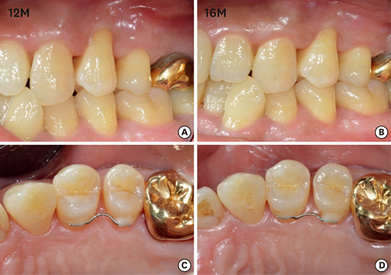
The patient was recalled periodically (3, 6, 9, 12, 16 months), and a follow-up radiographic examination showed noteworthy improvement of the periapical radiolucency (Figure 1B-F). Interestingly, alveolar bone regeneration was observed and maintained, showing new periodontal ligament attachment at the root surfaces including the surrounding teeth, as indicated with arrows in Fig. 1F. The clinical examinations included a pulp vitality test with an EPT, recordings of the PPD, tooth mobility, and a percussion test performed 16 months after treatment (Table 1). The tooth (#24) vitality was maintained, PPD was recovered within a normal range around 3–4 mm, and there was no symptom in the percussion test. As wire was attached at both sides of the #24 tooth, a proper tooth mobility check was not performed until the 12-month examination. The wire that was attached to the tooth with a composite resin was partly detached after 12 months of follow-up, and the mobility was examined. Tooth mobility was dramatically reduced, and it was assumed that this was the result of alveolar bone regeneration (arrow, Figure 1F). The gingival condition was well maintained without any signs of inflammation (Figure 2); however, buccal gingival recession was observed (Figure 2).
Table 1

Clinical examinations of left maxillary first premolar during the 16-month follow-up period

jpis-46-128-i001
Initial Follow-up (3M) Follow-up (6M) Follow-up (9M) Follow-up (12M) Follow-up (16M)
PPD (mm) jpis-46-128-i002 - jpis-46-128-i003 jpis-46-128-i004 jpis-46-128-i005 jpis-46-128-i006
Tooth mobility (Miller classification) III - - - - I
BOP + + - - - -
Pulp Vitality + + + + + +
PPD, probing pocket depth; M, mesial; B, buccal; D, distal; P, palatal; BOP, bleeding on probing.
Figure 2
Healing states of the gingiva around upper left first premolar on 12 months (A, C) and 16 months (B, D) after treatment.
jpis-46-128-g002

DISCUSSION

The relationship between periodontal condition and occlusal trauma has been discussed for a long time [19]. A healthy periodontal condition endures occlusal force and inhibits periodontal pocket formation; otherwise, periodontitis combined with occlusal trauma leads to periodontal destruction or vice versa [20]. In the presented case, subgingival calculus deposition was observed; however, this might not be a critical factor involved in the severe periodontium destruction surrounding tooth #24, considering the generalized subgingival calculus deposition. Instead, inappropriate canine guidance by the anterior crossbite and Angle class III malocclusion might be critical factors in the destruction of the periodontium. The lack of canine guidance induces contact with the molars in ways that generate sideways forces. These unfavorable forces can cause tooth mobility, possibly accompanied by alveolar bone resorption, progressing to tooth extraction. In the case of secondary TFO, including the degraded periodontium, occlusal adjustment/tooth splinting and periodontal treatment are necessary to eliminate the occlusal trauma caused by occlusal interference.
In the present case, the periodontal condition was diagnosed with a panoramic view in the first oral examination. Usually, in the panoramic view, the condition of tooth #24 is not remarkable, and distinguishing the apical radiolucency is difficult because of superimposition with other anatomical structures, as in this case (Figure 3). In the oral examination of a patient, radiographic examination is a critical aspect of diagnosis, and currently digital panoramic radiography is widely used due to its convenience and ability to achieve broad coverage of the alveolar bones and teeth [21]. Despite the many advantages of the panoramic view, anatomical overlap or image distortion might miss important features such as periapical pathology, bone resorption, and caries [22]. In this case, the exact diagnosis of the periodontal condition of the teeth was difficult using the panoramic view; on the other hand, the periapical view offered more detail with a focus on the specific teeth. Therefore, periapical radiography is essential for the proper diagnosis of the periodontal condition and evaluation of the patient’s records.
Figure 3
Panoramic radiography at the initial examination.
jpis-46-128-g003
In every practice, dentists are faced with the decision of whether or not to save a tooth. Although the maintenance of natural teeth has been accepted as a primary goal of dentistry, with the remarkable development of dental implant systems, tooth extraction and implant placement have become an easy treatment option in patients with periodontally compromised teeth. On the other hand, there is a risk of overuse of this approach without an effort to conserve the natural dentition. Therefore, a prudent approach to ensure accurate diagnosis and treatment planning is needed before finalizing the decision for extraction.

ACKNOWLEDGEMENTS

This research was supported by a grant from the Korea Health Technology R&D Project through the Korea Health Industry Development Institute (KHIDI), funded by the Ministry of Health & Welfare, Republic of Korea (HI14C2681).

Notes

Conflict of Interest No potential conflict of interest relevant to this article was reported.

References

1. Armitage GC. Development of a classification system for periodontal diseases and conditions. Northwest Dent. 2000; 79:31–35.
crossref
2. Heitz-Mayfield LJ, Schätzle M, Löe H, Bürgin W, Anerud A, Boysen H, et al. Clinical course of chronic periodontitis. II. Incidence, characteristics and time of occurrence of the initial periodontal lesion. J Clin Periodontol. 2003; 30:902–908.
3. Heitz-Mayfield LJ, Trombelli L, Heitz F, Needleman I, Moles D. A systematic review of the effect of surgical debridement vs non-surgical debridement for the treatment of chronic periodontitis. J Clin Periodontol. 2002; 29:Suppl 3. 92–102.
crossref
4. Wiebe CB, Putnins EE. The periodontal disease classification system of the American Academy of Periodontology--an update. J Can Dent Assoc. 2000; 66:594–597.
5. Loesche WJ, Giordano JR, Soehren S, Kaciroti N. The nonsurgical treatment of patients with periodontal disease: results after five years. J Am Dent Assoc. 2002; 133:311–320.
6. Sanz-Sánchez I, Ortiz-Vigón A, Matos R, Herrera D, Sanz M. Clinical efficacy of subgingival debridement with adjunctive erbium:yttrium-aluminum-garnet laser treatment in patients with chronic periodontitis: a randomized clinical trial. J Periodontol. 2015; 86:527–535.
crossref
7. Skurska A, Dolinska E, Pietruska M, Pietruski JK, Dymicka V, Kemona H, et al. Effect of nonsurgical periodontal treatment in conjunction with either systemic administration of amoxicillin and metronidazole or additional photodynamic therapy on the concentration of matrix metalloproteinases 8 and 9 in gingival crevicular fluid in patients with aggressive periodontitis. BMC Oral Health. 2015; 15:63.
crossref
8. Smiley CJ, Tracy SL, Abt E, Michalowicz BS, John MT, Gunsolley J, et al. Evidence-based clinical practice guideline on the nonsurgical treatment of chronic periodontitis by means of scaling and root planing with or without adjuncts. J Am Dent Assoc. 2015; 146:525–535.
crossref
9. Smiley CJ, Tracy SL, Abt E, Michalowicz BS, John MT, Gunsolley J, et al. Systematic review and meta-analysis on the nonsurgical treatment of chronic periodontitis by means of scaling and root planing with or without adjuncts. J Am Dent Assoc. 2015; 146:508–24.e5.
crossref
10. Branschofsky M, Beikler T, Schäfer R, Flemming TF, Lang H. Secondary trauma from occlusion and periodontitis. Quintessence Int. 2011; 42:515–522.
11. Davies SJ, Gray RJ, Linden GJ, James JA. Occlusal considerations in periodontics. Br Dent J. 2001; 191:597–604.
crossref
12. Jin LJ, Cao CF. Clinical diagnosis of trauma from occlusion and its relation with severity of periodontitis. J Clin Periodontol. 1992; 19:92–97.
crossref
13. Glickman I, Smulow JB. Further observations on the effects of trauma from occlusion in humans. J Periodontol. 1967; 38:280–293.
crossref
14. Ericsson I, Lindhe J. Effect of longstanding jiggling on experimental marginal periodontitis in the beagle dog. J Clin Periodontol. 1982; 9:497–503.
crossref
15. Hallmon WW. Occlusal trauma: effect and impact on the periodontium. Ann Periodontol. 1999; 4:102–108.
crossref
16. Harrel SK, Nunn ME. The effect of occlusal discrepancies on periodontitis. II. Relationship of occlusal treatment to the progression of periodontal disease. J Periodontol. 2001; 72:495–505.
crossref
17. Gher ME. Changing concepts. The effects of occlusion on periodontitis. Dent Clin North Am. 1998; 42:285–299.
18. Lindhe J, Nyman S. The role of occlusion in periodontal disease and the biological rationale for splinting in treatment of periodontitis. Oral Sci Rev. 1977; 10:11–43.
19. Harrel SK. Occlusal forces as a risk factor for periodontal disease. Periodontol 2000. 2003; 32:111–117.
crossref
20. Hallmon WW, Harrel SK. Occlusal analysis, diagnosis and management in the practice of periodontics. Periodontol 2000. 2004; 34:151–164.
crossref
21. Bourgeois M. Panoramic radiography for the general practitioner. Ont Dent. 1994; 71:29–30.
22. Rushton VE, Horner K, Worthington HV. Routine panoramic radiography of new adult patients in general dental practice: relevance of diagnostic yield to treatment and identification of radiographic selection criteria. Oral Surg Oral Med Oral Pathol Oral Radiol Endod. 2002; 93:488–495.
crossref
TOOLS
ORCID iDs

Young-Dan Cho
https://orcid.org/http://orcid.org/0000-0002-4694-5988

Sungtae Kim
https://orcid.org/http://orcid.org/0000-0001-6361-4104

Ki-Tae Koo
https://orcid.org/http://orcid.org/0000-0002-9809-2630

Yang-Jo Seol
https://orcid.org/http://orcid.org/0000-0002-2076-5452

Yong-Moo Lee
https://orcid.org/http://orcid.org/0000-0002-5619-3847

In-Chul Rhyu
https://orcid.org/http://orcid.org/0000-0003-4110-6381

Young Ku
https://orcid.org/http://orcid.org/0000-0001-5682-0604

Similar articles