

 PDF
PDF ePub
ePub Citation
 Print
Print


INTRODUCTION
Successful osseointegration of titanium (Ti) implants is partly determined by how the implanted materials influence bone responses at the cell-biomaterial interface [12]. Such events occurring between the bone and implant surface are influenced by a variety of specific surface properties, including topography, structure, chemistry, surface charge, and wettability [3456]. Of these, surface topography has been particularly well studied. Researchers have developed numerous additive and subtractive surface modification techniques to improve osseointegration by altering implant surface topography, thus enhancing bone-to-implant contact (BIC) and increasing biomechanical interlocking with bone [78]. The clinical introduction of a novel implant surface has also helped to advance the field [9]. This product aims to influence surface charge and wettability in animals via ultraviolet light irradiation [910].
Researchers have developed surfaces that are supposedly based not only on micrometre morphology, but also on other characteristics such as hydrophilicity, chemical bonding, and nanostructures [11]. Reports on the wetting behavior of rough surfaces have increased our understanding of the conditions surface topography has to satisfy to induce satisfactory hydrophilicity during contact with bone [12131415]. Extensive hydroxylation/hydration of the oxide layer, together with high wettability, improves interactions between the surface and the water shells around delicate biomolecules such as proteins [16]. Studies using modified sandblasted, large-grit, acid-etched (modSLA) surfaces that enhance hydrophilicity have indicated that bone apposition during the early stages of regeneration is higher after implantation compared with its predecessor (the SLA surface) [171819].
Anodic oxidation creates a thickened, porous, and moderately roughened titanium oxide layer [2021]. The anodized titanium surface shows superior osteogenic properties both in vitro and in vivo despite being hydrophobic [2021222324]. Although many studies have examined the longer-term impacts of surface roughness and topography on bone fixation over the long term, there has been relatively little work investigating the effects of these hydrophilic characteristics on the initial bone response [17192526]. To the best of our knowledge, there were not many studies that have evaluated the effects of hydrophobic oxidized and hydrophilic modSLA surfaces on early bone response in vivo [27]. Although the positive effects of the modSLA implants could be easily explained by their hydrophilicity, the clinical relevance needs to be further investigated [12].
We performed histomorphometric analyses to investigate the combined effects of physical and chemical surface factors on in vivo bone responses by comparing a modSLA surface and an anodized implant surface in a rabbit tibia model.
MATERIALS AND METHODS
Surface characteristics
Five modSLA (SLActive®, Institut Straumann AG, Basel, Switzerland) and five anodized (TiUnite®, Nobel Biocare AB, Göthenburg, Sweden) implants were used in this study. Both implants were 3.3 mm in diameter and 10.0 mm in length. We performed three surface analyses on each of three implants from both groups: field emission scanning electron microscopy (FE-SEM), energy dispersive spectroscopy (EDS), and confocal laser scanning microscopy (CLSM). The FE-SEM (model S-4700, Hitachi, Tokyo, Japan) was used to produce detailed images of the implant surfaces. The EDS (model EX220, Horiba Ltd., Kyoto, Japan) was used to analyze the element content and components of the modified surfaces; calibrations were performed four times each at four different points. The CLSM (model LSM 5-Pascal, Carl Zeiss AG, Oberkochen, Germany) enabled us to measure the surface roughness of four screw sides (measurement area: 300 µm × 300 µm on a 200× optically and 1.5× digitally magnified image), which were randomly selected from each implant. We measured two roughness parameters: average surface deviation (Sa) and developed surface area ratio (Sdr) [21].
In vivo surgery
This study was approved by the Animal Research Committee of Seoul National University Bundang Hospital (IACUC protocol approval number: BA1101-076/001-01). All procedures, including animal selection, management, preparation, and subsequent surgical protocols, were performed in accordance with the Institute of Laboratory Animal Resources guidelines of Seoul National University Bundang Hospital.
Five male New Zealand white rabbits (each about 6 months of age and weighing 2.5-3 kg) were implanted with a modSLA and an anodized implant; the location of each implant (left or right tibia) was chosen at random. The rabbits showed no sign of illness or disease prior to the study. Prior to surgery, all study subjects were anesthetized with an intramuscular injection of tiletamine/zolazepam (15 mg/kg; Zoletil 50, Virbac Korea Co. Ltd., Seoul, Korea) and xylazine (33 mg/kg; Rompun, Bayer Korea Ltd., Seoul, Korea). The skin of each proximal tibia area was shaved and washed with povidone iodine solution, and each rabbit received an intramuscular injection with 33 mg/kg of Cefazolin (Yuhan Co., Seoul, Korea), a preoperative prophylactic antibiotic. The local anesthetic lidocaine (1:100,000 epinephrine; Yuhan Co.) was injected into each surgical site. The skin was incised with a surgical blade, and each tibia was exposed via full-thickness periosteal flap reflection. The implant sites were prepared on the flat tibial surface using a dental implant drill and profuse sterile saline irrigation.
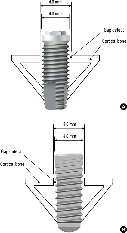
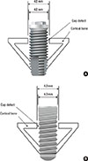
We performed bicortical drilling as described in a previous study [20]. For the 3.3-mm implants, we used a drill that was 2.8 mm in diameter; gap defects were created with a cortical drill (Astra Tech, Mölndal, Sweden) with a 4.0 mm diameter (Fig. 1). The cortical drill was used monocortically and created a 4.0-mm hole in the upper cortex only. After implant insertion, cover screws were securely fastened and the surgical sites were closed in layers. Muscle and fascia were sutured with absorbable Vicryl sutures (Vicryl 4-0, Polyglactin 910, Ethicon, Johnson & Johnson, Somerville, NJ, USA) and the outer dermis was closed with a silk suture (Mersilk 4-0, Ethicon, Johnson & Johnson). Rabbits were housed in separate cages for 1 week post-surgery, after which they were anesthetized and sacrificed by intravenous administration of potassium chloride.
Histomorphometric analysis
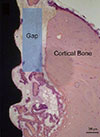
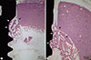
The tibiae of the sacrificed rabbits were exposed so that implants could be surgically removed en bloc with an adjacent collar of bone, which was immediately fixed in 10% neutral formaldehyde. For histomorphometry, the specimens were embedded in light-curing resin (Technovit 7200 VLC, Kultzer, Wehrheim, Germany) prepared as previously described [28]. Undecalcified, cut, and ground sections were prepared using the Exakt® system (Exakt Apparatebau, Norderstedt, Germany) according to the method described by Donath and Breuner [29]. The specimens were ground to a thickness of approximately 50 µm and stained with hematoxylin and eosin (H&E). Histological examinations of specimens were performed under a light microscope (Olympus BX, Olympus, Tokyo, Japan). BIC and BA percentages were defined and measured in the range of 2 mm below the upper bone crest, as shown in Fig. 2. Histomorphometric analyses were performed on both the right and left sides of each specimen using image analysis software (Kappa PS30C Imagebase, Kappa Opto-electronics GmbH, Gleichen, Germany).
Statistical analyses
The Mann-Whitney U test was used to assess the statistical significance of the difference in surface roughness parameters (Sa and Sdr) between the test and control implants. The Wilcoxon signed-rank test was used to determine statistically significant differences in BIC and BA between the groups. Significance was defined as P<0.05.
RESULTS
Analysis of surface characteristics
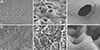
The modSLA surface comprised 70.3% titanium, 29.3% oxide, and no phosphorous, while the anodized surface was 26.1% titanium, 69.8% oxide, and 3.5% phosphorus (Table 1). The FE-SEM images of each surface are shown in Fig. 3. Magnification at 5,000× revealed the anodized surface to be scattered with many volcano-like porous structures. The modSLA surface had a sharp, irregular pattern produced by the sandblasting and acid-etching processes. At 50,000× magnification, the anodized implant was characterized by a relatively smooth surface composed of large micro-pores and small nanopores. This contrasted with the relatively rough surface observed on the hydrophilic modSLA implant, which exhibited beadings of approximately 1-2 µm diameter, along with 0.1-0.2 µm millet-like prominences.
Surface roughness data collected during the CLSM analysis are shown in Table 2. Anodically oxidized surfaces had significantly lower values for both Sa (1.22±0.17 µm) and Sdr (26.27±4.14%) than observed for chemically modified surfaces (2.53±0.07 µm and 139.82±7.59%, respectively) (both P<0.001). Although the modSLA implant screw design is flatter and thus has less total surface area, the modSLA screws are smaller and result in greater osseointegration because of the higher percentage of BIC.
Histomorphometric analysis
All experimental animals healed without complications and at the time of sacrifice all implants were submerged and covered by a healthy ridge of skin. For both types of implant, a favorable bone response was observed on the implant surface; a small amount of new bone formation was found both within a thread and in the old bone after only 1 week of healing. Osseointegration within the gap defect was notable in some specimens; however, the distinguishing feature of most samples was a visible growth pattern along the border of the gap defect toward the implant surface (Fig. 4). This was observed in both treatments with a statistically significant greater growth pattern seen in the modSLA group. Although the 1-week post-surgical period was insufficient for new bone formation, all specimens showed at least the beginnings of new bone formation or active bone formation on the inner cutting side of the cortical bone and the inner portion of bone marrow. Both implant surfaces were surrounded by small, newly formed trabeculae of woven bone. Although histomorphometric light microscopy revealed that the BIC ratio was significantly higher around the modSLA implants than around the anodized implants (P=0.02; Table 3), there was no significant difference in BA between the two types of implant because of a large standard deviation (P=0.09; Table 3).
DISCUSSION
We set out to determine whether modSLA implants would possess clinical superiority over anodically oxidized surface implants. Given the results of our in vivo experiment, hydrophilicity seems to be very important in bone response.
Thread density but not thread geometry is known to have an effect on BIC [3031]. As the anodized implant had a shorter thread pitch than the modSLA implant in this study (Fig. 4), it was considered to have an advantage in BIC. We also found the modSLA surface to be significantly rougher than the anodized surface. Although the modSLA surface is classified as rough, the anodized surface is considered moderately rough, which is known to be more advantageous for bone responses than a "rough" surface [32]. However, we found a significantly higher BIC in response to the modSLA implant. In fact, in terms of the surface treatment based on Sa and Sdr, several studies concluded that modSLA surface showed better performance than conventional SLA surface as well as different surface treatment [171921]. Although the mechanism linking surface properties and osteoblast production is not yet sufficiently understood, the hydrophilic property of the modSLA surface may have a stronger influence on bone response than either the surface configuration of the implant or the surface features resulting from anodic oxidation [19]. These results correlated with the previous study concluding that surface hydrophilicity rather than microtopography affected soft and hard tissue integration [33].
Since the dental implant was first introduced, there have been a huge number of studies of which the purposes were to find the factors for the improvement of osseointegration. Eventually, we now have diverse dental implants at out hand with various designs, configurations, surface treatment, and modifications. Hugh leap of development was already made in the field of dental implants. Therefore, the scientific investigation on only one factor for better dental implant by well-controlled experiments can be so stereotyped but the intuitive comparison among the several dental implants considering each one as an independent entity of the experiment, although somewhat obscure and less scientific, can be more practical and clinically helpful. The well-controlled study on one factor could be even more redundant [621]. In such a way, a previous study showed that hydrophilic SLA group showed higher BIC after 10 days than Nobel Biocare Replace Select implant group with oxidized TiUnite surface [34].
We observed many different stages of new bone formation, ranging from no osseointegration at all to a very thin layer of new bone around the original cortical bone, to marked growth toward the implant surface, to complete osseointegration with new bone, which is why such a large standard deviation was observed on histomorphometric analyses. One week of post-surgery recovery time is too short to truly evaluate osseointegration at the bone-implant interface, although the findings of this study support previous results indicating that this process is initiated within the first week of wound healing [3536]. Further studies are needed to more clearly determine significant histomorphometric differences by controlling the healing period after implant insertion.
Our results suggest that the hydrophilic modSLA surface may have a stronger affinity for bone than the anodized surface during the initial healing period. Somewhat similar results have been indicated by previous studies reporting that a hydrophilic surface implant is associated with better initial bone response [1737]. Although both surface configuration and hydrophobic properties of the implant surface were found to affect early bone formation, the latter appears to have a more significant effect on BIC for reasons that are yet to be fully elucidated. Various experiments including animal models and immunohistochemistry have reported that blood clots may be formed within 24 hours of implant insertion and that formation of capillaries preceded and accelerated new bone formation [3738]. We found blood clots close to the hydrophilic modSLA surface, while a previous study demonstrated that the coagulum was partially collapsed at the conventional SLA surface [37]. Within the limitations of this study, the hydrophilicity of the modSLA surface may have a stronger effect on in vivo bone healing than optimal surface roughness and surface chemistry of the anodized surface. Further investigations are required to elucidate the interactions between the implant surface factors such as hydrophilicity and the physiology of blood and bone with large sample size at the various different stages during the early healing period.


 XML Download