

 PDF
PDF ePub
ePub Citation
 Print
Print


INTRODUCTION
Methimazole is the drug of choice for treating hyperthyroidism. This drug blocks thyroid hormone synthesis by inhibiting thyroid peroxidase, an enzyme involved in the production of thyroxine (T4) and triiodothyronine (T3). When anti-thyroid drugs (ATD) are administered, it is important to monitor the patient for any symptoms of neutropenia, such as fever and a sore throat, even though these side effects are uncommon. Methimazole-induced agranulocytosis occurs in only 0.2%-0.3% of patients [1,2]. Although this frequency is quite low, agranulocytosis may cause life-threatening complications. There are several case reports of patients with methimazole-induced agranulocytosis in the medical literature [1,2,3,4,5]. However, few reports have presented the oral manifestations of methimazole-induced agranulocytosis, and only one previous case report has described oral complications caused by methimazole-induced neutropenia [6]. In our case, the patient had been taking methimazole for three months due to hyperthyroidism and had signs and symptoms of methimazole-induced neutropenia, the oral manifestations of which were severe gingival necrosis and sequestration of the alveolar bone. As a result, our patient required a longer healing period than the patient who was described in the previous case report [6].
CASE DESCRIPTION
A 31-year-old female was referred to the Department of Periodontics from the Department of Endocrinology of Kyung Hee University Hospital at Gangdong, Seoul, Korea, complaining of generalized gingival pain and whitish gingival lesions. She had been hospitalized five days previously due to a sore throat, fever, and gingival pain. Intravenous antibiotic injections (1.2 g of amoxicillin/clavulanic acid and 200 mg of isepamicin sulfate, twice a day) were initiated to treat the infection.
Her past medical history revealed that she had been diagnosed with hyperthyroidism at a local clinic nine months previously and started on propylthiouracil. She had a complete blood cell count test taken every month, and the dose of propylthiouracil was decreased as her symptoms improved. However, she developed a rash and itching sensation after six months of taking propylthiouracil. Therefore, methimazole was substituted for propylthiouracil, and the patient continued taking methimazole for three months prior to admission to the hospital.
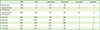
While hospitalized, she was diagnosed with methimazole-induced neutropenia based on blood test results (Table 1), clinical signs and symptoms, and her medical history. Methimazole was immediately discontinued, and intravenous antibiotic injections were continued to prevent further infections until she was discharged. Granulocyte colony stimulating factor (G-CSF; 75 µg of filgrastim, once a day) was injected subcutaneously on the second and third days of hospitalization, in order to increase the neutrophil count and reduce the recovery time [4,7]. Dexamethasone disodium phosphate (5 mg, once a day) was also injected intravenously on the first and third days, and then hydrocortisone sodium succinate (33 mg, three times a day) was administered on the sixth and seventh days to control severe inflammatory reactions. Lugol's solution was administered orally to treat the patient's hyperthyroidism. The white blood cell (WBC) count of the patient began to increase and her general condition began to improve. Two weeks after hospitalization, her WBC count had stabilized in the normal range and the patient was discharged.
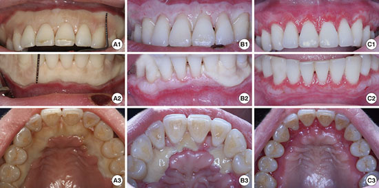
Her first dental visit was on the second day after the G-CSF injections were administered, in order to treat the whitish gingival lesions and pain. The maxillary and mandibular gingiva showed whitish necrotic lesions from the free gingival margin to the nearby mucogingival junction. Whitish necrotic lesions were also observed in the palatal and lingual tissue, encompassing almost the entire dentition (Fig. 1 A1, A2, and A3). As the WBC count of the patient was severely lowered as a result of methimazole-induced neutropenia, biopsy of the gingival tissue was avoided due to concerns that the healing process would be compromised. Periodontal treatment was delayed until the WBC count increased. Instead, the lesions were dressed with a diluted 50% policresulen solution (Albothyl® concentrate, Pacific Pharma Co., Seoul, Korea) to remove the necrotic tissue and a 0.1% chlorhexidine gluconate mouthwash was prescribed for its bactericidal effect.
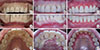
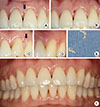
Two days after the first dental visit, the necrotized gingival tissue began to shed, exposing the root surfaces under the cementoenamel junction and showing prominent interdental spaces. The WBC count at this stage was 13.2×10³ cells/mm3. Periodontal treatment was delayed until the WBC count stabilized. The lesions were dressed with hydrogen peroxide and chlorhexidine gluconate to a limited extent in order to irrigate the necrotic tissue and debris. Five days after the first dental visit, the size and whiteness grade of the necrotized gingiva had decreased remarkably, and re-epithelialization was observed on the labial, palatal, and lingual sides (Fig. 1 B1, B2, and B3). However, a large whitish necrotized area was still present in the palatal and left mandibular buccal regions. Two weeks after the first dental visit, the gingiva had regenerated compared to the previous visit, and had a pale pink color with reddish gingival margins (Fig. 1 C1, C2, and C3). However, the interdental spaces and exposed root surfaces were still visible. The patient reported that she was in less pain. Since the increased WBC count had stabilized, initial periodontal treatment was started and plaque control was performed at every follow-up visit. Eight weeks after the first dental visit, the gingiva had a shiny, reddish appearance, and had become more epithelialized (Fig. 2A). At that time, the patient reported that she could consume hot and spicy food. At 18 weeks of follow-up, stippling near the gingival margin was observed along with a reddish band were observed, because the gingiva was not fully keratinized (Fig. 2B). The root surfaces were almost covered, but reduced interdental spaces were still present. At nine months of follow-up, the embrasure spaces were filled with gingiva, even though calculus and plaque retention were evident (Fig. 2C). The gingiva appeared almost normal, except for a reduced red band. At the one-year follow-up visit, her maxillary and mandibular gingiva had a normal appearance (Fig. 2D), and the keratinized gingiva was repaired, extending almost to the cementoenamel junction. At a 21-month follow-up visit, sequestra of the necrotic alveolar bone were observed at the interdental gingiva of the maxillary left central incisor (Fig. 3A) and canine (Fig. 3C). The sequestra were removed through the gingiva (Fig. 3B, D, and E) and uneventful healing of the tissue occurred. At a three-year (34-month) follow-up visit, the patient showed healthy gingiva with a normal appearance, although some interdental space was still present between the maxillary central incisors (Fig. 3F).
DISCUSSION
A patient who had been taking methimazole to treat hyperthyroidism was hospitalized due to a sore throat, fever, and gingival pain. Based on the results of blood tests, clinical symptoms, and her medical history, she was diagnosed with methimazole-induced neutropenia. Neutrophils are the first line of defense against infections, acting as primary phagocytic cells, especially during the acute phase of an infection, and comprise 50%-70% of the circulating WBCs [8,9]. Infections that are easily localized or overcome in healthy individuals can spread rapidly and systemically and/or cause more aggravated responses within a very short time in patients with neutropenia or agranulocytosis. The absolute neutrophil count (ANC) is the real number of neutrophils and is calculated from the percentage of neutrophils in the differential WBC count multiplied by the WBC count. The percentage of neutrophils is calculating by aggregating the segmented mature neutrophils and bands that indicate immature neutrophils. A normal ANC is > 1,500 cells/mm3. Generally, an ANC<1,000 cells/mm3 indicates a moderate risk of infection, and an ANC<500 cells/mm3 is defined as neutropenia with a severe risk of infection. Although the terms agranulocytosis and neutropenia are often used interchangeably, agranulocytosis is a term used for cases with an ANC<100 cells/mm3. It takes about 14 days for neutrophils to differentiate and proliferate in the bone marrow. They then circulate through the peripheral blood system and migrate into tissues, where they survive for one to two days. The average half-life of neutrophils in the circulation is therefore about 12 hours.
The patient showed symptoms of neutropenia three months after taking methimazole. The onset of methimazole-induced neutropenia is abrupt and idiosyncratic [1]; agranulocytosis usually develops within the first 2-12 weeks of ATD therapy, but can occur as late as 20 weeks after ATD therapy is initiated [2]. The timing of the onset of neutropenia in the present case is consistent with previous reports. The cause of neutropenia was suppression of the bone marrow by methimazole. Drug toxicity results in the decreased production of the rapidly growing progenitor cells of the marrow. The extent of marrow suppression is generally dose-related. Many other drugs can cause non-chemotherapy drug-induced agranulocytosis, such as dipyrone (an analgesic), ticlopidine and captopril (cardiovascular drugs), and phenytoin and carbamazepine (anticonvulsants) [7].
Detecting the oral manifestations of drug-induced agranulocytosis is critical for further diagnosis and treatment. Neutropenia can be a severely dangerous condition at the time of diagnosis. Therefore, routine WBC monitoring is recommended for patients taking ATD, especially during the first three months [10,11]. However, the importance of routine WBC monitoring for the early detection of ATD-induced agranulocytosis remains controversial [2]. ATD-induced agranulocytosis sometimes has a very sudden onset, and routine WBC counts, even conducted at intervals of every one or two weeks, cannot predict the appearance of all cases of ATD-induced agranulocytosis. Therefore, it is important to inform patients taking these drugs that they should be alert to the onset of symptoms such as fever, sore throat, or other infections, even when WBC counts are normal, and that they should obtain WBC counts immediately if symptoms occur [2,7].
Gingival necrosis, oral ulceration, infectious pharyngitis, and tonsillitis are the most common oropharyngeal features of drug-induced agranulocytosis [6]. These symptoms may be the first signs of neutropenia [8]. Compared to the previous report by Hou and Tsai [6], our present case showed more extensive gingival necrosis that almost covered the entire dentition, as well as a much lower WBC count, before systemic treatment was started. This report includes a description of the long-term observation of the patient over the course of three years, and suggests that alveolar bone sequestration can be associated with methimazole-induced neutropenia, because alveolar bone sequestration was found at the 21-month follow-up visit. In general, the severity of clinical signs is proportionate to the severity of agranulocytosis [6,9]. The gingival crevice is the main source of leukocytes, and the crevicular granulocytes respond to local bacterial stimuli. In neutropenic conditions, bacterial infections are not contained and progress rapidly. Thus, cells and connective tissue elements disintegrate, and rampant necrotizing gingival lesions appear in the oral cavity [8].
For oral lesions, a policresulen dressing was applied in a sextant-wise manner to the necrotic gingiva at every visit in order to exfoliate the injured cells, induce wound cleansing and reactive hyperemia in the treated area, and facilitate rapid re-epithelialization. Policresulen is a topical hemostatic and antiseptic with a highly acidic pH, which results in a marked bactericidal action on the most common pathogens as well as efficacy against Candida albicans [12]. The favorable effects of policresulen are attributed to its highly acidic characteristics, which cause the selective coagulation of necrotic or pathologically altered tissues while leaving healthy tissues unaffected. Therefore, policresulen is used to treat infections of the mucous membranes and eruptions on the tongue. However, policresulen is a polycondensation product of metacresol sulfonic acid and formaldehyde. Although no reports have yet indicated that policresulen is carcinogenic, and the formaldehyde content is 0.36 µg per 1 mL in the Albothyl® concentrate solution, which is far below the threshold of 0.5 mg/mL used by the registration authorities in Germany according to the manufacturer, policresulen may have potential carcinogenic effects. In addition, a 0.1% chlorhexidine gluconate mouthwash was administered twice a day during hospitalization and at home after discharge. Chlorhexidine gluconate is a bactericidal agent that is effective against both Gram-positive and Gram-negative bacteria. Hence, it is an effective antiplaque and antigingivitis agent that reduces gingival inflammation [13]. After the ANC reached a normal level, the patient received scaling and root planing.
In addition to fever and/or a sore throat, which are the best known and typically the earliest presentations of drug-induced neutropenia, oral manifestations, such as gingival necrosis and ulcerations, may be the first signs of neutropenia. Even if oral manifestations are not the first signs of neutropenia, their occurrence is clinically significant. Therefore, dentists need to be aware of the oral complications of non chemotherapy drug-induced neutropenia in order to make an accurate diagnosis and ensure that prompt medical intervention is provided.

 XML Download