

 PDF
PDF ePub
ePub Citation
 Print
Print


INTRODUCTION
Staphylococcus aureus is a major causative agent of major human disease; although it is associated with mild symptoms in healthy skin or soft tissue, in some environments, such as hospitals, it is the main source of infections accounting for more than 80% of pyogenic diseases. In the oral tract, S. aureus has been associated with dentoalveolar infections, and oral mucosal lesions. Moreover, staphylococcal colonization has been demonstrated from the tongue, saliva, mucosal surfaces, supragingival tooth surfaces and the periodontal pocket [12]. The wide range of virulence factors of S. aureus associated with infection and diseases can be classified into surface-associated factors, degradative enzymes and superantigenic toxins; the diversity and great variability in these genes may affect the course of an infection [3]. The enterotoxins of S. aureus are heat-resistant toxins causing diarrhea and vomiting in humans, and serological analysis has identified five different types based on their antigenicity. Although new types of enterotoxin have recently been reported, their association with food poisoning is not clear [4]. S. aureus has four hemolysins, hla dissolves cells and causes hemolytic necrosis, hlb affects the lung and the cornea, and hld which has been reported to be produced in 97% of S. aureus samples, dissolves the inner cell structures of red blood cells and various mammalian cells [5]. Due to the increase of the indiscriminate use of penicillin, which was discovered in 1928 by Alexander Fleming, more penicillin-resistant strains had emerged by approximately 1950, when the resistance rate reached 80%, leading to a gradual loss of the effectiveness of penicillin. The vicious cycle of misuse and abuse of antimicrobials and the emergence of antimicrobial-resistant bacteria has become a problem that has not shown meaningful improvement from the early days of antimicrobial use through the present days [6].
Periodontitis involves the progressive loss of alveolar bone around the teeth, and if left untreated, can lead to the loosening and subsequent loss of teeth. Periodontitis is caused by microorganisms that adhere to and grow on the surfaces of teeth, along with an over-aggressive immune response to those microorganisms. Studies of the causative pathogens of periodontal diseases isolated from Korean periodontitis patients have demonstrated that S. aureus plays a role in exacerbating dental diseases by forming a biofilm with the causative pathogens of periodontal diseases [78910]. The frequency of antimicrobial use in oral surgical procedures and minor procedures such as implant placement has recently increased, which is a trend that can be directly associated with the problem of increased antimicrobial resistance. This study characterized the distribution of antimicrobial resistance, antibiotic resistance genes, and virulence genes in S. aureus cultures isolated from Korean periodontitis patients. The findings of this study are relevant for the ongoing treatment and prevention of periodontitis.
MATERIALS AND METHODS
Sample collection
From July 2015 to August 2015, oral saliva was collected from a total of 112 patients diagnosed with periodontitis, including 80 outpatients in dental hospitals and 32 patients of dental clinics located in Seoul and Cheonan. The subjects of this study were patients who visited a dental hospital or dental clinic and were diagnosed with periodontitis, and were mostly chronic periodontitis patients (K05.3 in the International Classification of Diseases-10th Revision). They had periodontal pockets deeper than 4 mm and alveolar bone loss visible on X-ray films. Scaling only is unlikely to be an adequate treatment, so all patients who required periodontal treatment were screened. Samples were collected in the form of oral saliva prior to scaling and medication and in the form of plaque from the tooth surface obtained with sterile swabs when patients were in the hospital, and the samples were stored in sterile containers. This study was approved by the Institutional Review Board of Dankook University (IRB No: DKU 2015-06-003-001).
Isolation and identification of S. aureus
The isolation of S. aureus was performed in accordance with the methods described by the Korea National Research Institute of Health [11] and Murray et al. [12]. Polymerase chain reaction(PCR) was used to identify S. aureus [1314]. A Bio-Rad (Hercules, CA, USA) C1000 thermal cycler was utilized and the final product was subjected to electrophoresis followed by confirmation of the electrophoresis pattern using a transilluminator(Model: Gel Doc XR+ Bio-Rad, Hercules, CA, USA). A 100-base pair ladder (Bioneer, Daejeon, Korea) was used as a standard size maker.
RESULTS
Isolation rates of S. aureus from the oral cavity of patients with periodontitis
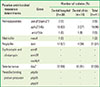
The results regarding the isolation of S. aureus from outpatients in dental hospitals and clinics located in Seoul and Cheonan are presented in Table 1. 41 strains of S. aureus (36.6%) were isolated from the test samples of oral saliva collected from a total of 112 patients diagnosed with periodontitis which include 80 outpatients in dental hospitals and 32 outpatients in dental clinics located in Seoul and Cheonan from July to August of 2015, and 30 strains (37.5%) were isolated from the outpatients in dental hospitals and 11 strains (34.4%) from the patients of dental clinics.
Antimicrobial susceptibility of S. aureus isolated from patients with periodontal diseases
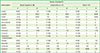
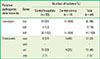
Data regarding the antimicrobial susceptibility of S. aureus isolated from the oral cavity of outpatients in dental hospitals and clinics are presented in Table 2. Among the total number of 41 S. aureus isolates, the susceptibilities to vancomycin, chloramphenicol, rifampin, clindamycin, sulfamethoxazole, and imipenem were all(100%). High susceptibility was found to oxacillin, cefepime, and cefotetan 40 isolates(98%), ciprofloxacin 39 isolates(95%), and erythromycin and tetracycline 37 isolates(90%). In contrast, 11 isolates (27%) were susceptible to gentamicin and 5 isolates (12%) were susceptible to ampicillin and penicillin. The antimicrobial resistance rate was 88% (36/41) each for ampicillin and penicillin, 10% (4/41) for erythromycin, 7% (3/41) for tetracycline, 5% (2/41) for gentamycin, and 2% (1/41) each for cefepime and cefotetan.
Table 2
Antimicrobial resistance rates in samples of Staphylococcus aureus isolated from the oral cavity of patients with periodontitis.
Patterns of multiple antimicrobial resistance
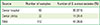
The results regarding multiple antimicrobial resistance for the 41 strains isolated from the oral cavity of patients with periodontal disease are presented in Table 3. Of the 41 strains, four (10%) showed susceptibility to all antimicrobials tested, while 37 (90%) were resistant to at least two antimicrobials. Resistance to two antimicrobials was found in 29 strains (71%), seven strains (14%) showed resistance to three antimicrobials, one strain (2%) showed resistance to four antimicrobials, and one strain (2%) was resistant to six antimicrobials.
Table 3
Patterns of multiple antimicrobial resistance in Staphylococcus aureus isolated from the oral cavity of patients with periodontitis.
Analysis of antimicrobial resistance genes
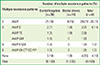
In order to determine the presence or absence of antimicrobial resistance genes in the 41 S. aureus strains isolated from patients with periodontal disease, PCR was performed, and the results are presented in Table 4. The aac(6′)/aph(2″) gene was identified in two strains (5%), the aph(3′)-IIIa gene in 19 strains (46%), and the ant(4′)-Ia gene was isolated in one strain (2%). The methicillin resistance gene mecA was isolated from one strain (2%) and tem, an ampicillin resistance gene, was isolated from 21 strains (51%), but ampicillin resistance was found in 36 strains (58%). Resistance to erythromycin is derived from the ermB, ermTR and mefA/E genes, none of which was detected. The β-lactamase resistance gene blaZ, was detected in 37 strains, which was consistent with the results of the disk expansion method. Moreover, the gene encoding penicillin-binding protein was not detected.
Table 4
Antimicrobial resistance genes in Staphylococcus aureus samples isolated from periodontitis patients in dental hospitals and dental clinics.
Analysis of pathogenic genes
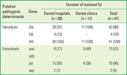
In order to determine whether the virulence genes encoding for hemolysins and enterotoxins were present in the 41 S. aureus strains isolated from patients with periodontal disease, PCR was performed and the results are presented in Table 5. The hemolysin gene hla was present in 40 strains (97.6%), hlb was present in 4 strains (10%), and hld was present in 41 strains (100%). The enterotoxin gene sea was present in 13 strains (32%), seb was present in no strains (0%), sei was present in 19 strains (46%), and seq was present in two strains (5%).
Table 5
Distribution of hemolysin and enterotoxin gene in Staphylococcus aureus samples isolated from periodontitis patients in dental hospitals and dental clinics.
DISCUSSION
Periodontal disease represents a possible reservoirs for opportunistic bacteria in the oral cavity. The use of antibiotics to treat periodontal disease or other infections may lead to the increase of Staphylococcus spp. in the oral cavity. Strains of S. aureus may easily become resistant to antibiotics, potentially resulting in periodontitis that persists despite antibiotic treatment. The presence of a higher proportions of S. aureus in the oral cavity may lead to a significant increase in the incidence of periodontal infections and the difficulty of treating those that do occur.
The isolation rate of S. aureus from patients with periodontitis in this study was lower than the figure of 41.9% reported by Kim [20], but higher than the rate of 4.8% reported by Min et al. [21], who investigated the nasal cavities of hospital clinical staff; the figure of 20% reported by Han et al. [22]; and the rate of 23% reported by Kang [23], who studied dental students. The rate that we found was higher than the rate of 30% reported by Kim [24] among patients with oral infections, but similar to the rate of 36% that they observed in the general population. Notably, the rate of 60.4% reported by Lee et al. [25] was much higher than the results of the present study. The previously published literature indicates that S. aureus is thought to play an important role in causing periodontal diseases by forming a biofilm on dental plaque. Previous studies have suggested that S. aureus exhibits a diverse range of pathogenicity. In dental patients, S. aureus is thought to be transmitted through the nasal cavity and the oral cavity, where it is most highly distributed in the human body. Kim et al. [24] raised the possibility of cross-infection with multiple strains of S. aureus in the dental field, mediated either by contact of effusions, skins, and saliva between dental care staff and patients or by contact with S. aureus floating in the form of aerosols and contaminating the medical instruments and unit chairs. The general use of antimicrobials is therefore necessary to treat nosocomial S. aureus infections. Dental patients take antibiotics primarily to prevent postoperative infections and to prevent secondary infections, for which purpose antimicrobials are administered prior to surgery. Antimicrobials are primarily used therapeutically to treat abscess formation, osteomyelitis of the jaw, inflammation of the maxillary sinus, and acute orofacial infections that are caused by dental infections; in such cases, antimicrobials have been reported to be prescribed for an average of approximately 6.9 days [26]. Kim [20] reported that S. aureus strains isolated from dental patients with periodontal disease showed antimicrobial resistance rates of 92.6% to ampicillin, 90.7% to penicillin, 11.1% each to oxacillin and cefotetan, and 5.6% to erythromycin, which is consistent with the results of our study. This profile of antimicrobial resistance may have resulted from the fact that the samples were isolated from the oral cavity of patients with periodontal disease. In contrast, Jung and Lee. [27] reported that strains of S. aureus isolated from the nasal cavity of students and hospital clinical staff showed resistance rates of 90% to penicillin, 43% to tetracycline, 37% to erythromycin, 10% to cephalothin, and 13% to clindamycin and vancomycin. These findings were different from those obtained in the present study, and may have been related to the fact that nasal cavity is more favorable for the survival of S. aureus than the oral cavity. In contrast, Kim et al. [28] reported that the antimicrobial resistance rates of S. aureus collected from pyogenic lesions of outpatients were 97.6% to penicillin, 9.4% to oxacillin, 8.1% to erythromycin, and 2.4% to clindamycin, and Kim [24] reported that resistance rates of S. aureus in patients who were hospitalized due to acute oral infections were 100% to penicillin, 68.1% to oxacillin, and 88.1% to erythromycin, which are drastically different from the resistance rates observed in this study. It is possible that those higher resistance rates were due to the higher likelihood that lesions in acute oral infections and pyogenic lesions may come into contact with antimicrobials, thereby acquiring resistance.
In the study performed by Kim[20] on S. aureus isolated from patients in dental hospitals and clinics, 3.7% of strains were susceptible to all antimicrobials, 5.6% showed resistance to a single antimicrobial, 74.1% showed resistance to two antimicrobials, 3.7% were resistant to three antimicrobials, 3.7% were resistant to four antimicrobials, 3.7% were resistant to five antimicrobials, 3.7% were resistant to six antimicrobials, and 1.9% were resistant to seven antimicrobials. In these results, strains with resistance to two antimicrobials comprised the majority, accounting for 70% of the samples, which was consistent with the findings of the present study. However, the rate of strains with resistance to three antimicrobials in the present study was much higher than that reported by Kim [29], indicating that S. aureus isolated from patients with the same periodontal disease may show strikingly different patterns of multidrug resistance, which is a topic that requires further research. Kim [20] showed that the detection rates of enterotoxin genes in S. aureus isolated from patients with periodontal disease were 11.6% for sea, 88.9% for seb, and 44.4% for sei, in sharp contrast to the 2006 findings of Jung et al. [30], indicating that methicillin-resistant S. aureus strains isolated from patients in university hospitals had both sea and seb. This discrepancy may have resulted from the different characteristics of strains originating from dental patients in comparison to patients from general hospitals. Peck et al. [19] reported seg and sei to be the main enterotoxins in their study; Hwang et al. [31] reported that the main enterotoxins were seg, seh and sei; Cho et al. [32] found that seg, sei and she were the main enterotoxins; Kim et al. [33] identified sea as the main enterotoxin; Baik et al. [34] reported seg, sei, and sec to be the main enterotoxins; Nashev et al. [18] identified seg, sei and seb as the main enterotoxins; and Bania et al. [35] reported that sec and seg were dominant, suggesting that enterotoxins are very diverse, and vary according to the location where the strains and specimen were isolated. S. aureus produces five toxins that cause damage to various membranes, including four hemolysins and leukocidin. Alpha hemolysin is secreted by S. aureus and dissolves bacterial cells and red blood cells by forming complexes on the membranes of the target cells. In addition, it reacts with platelets and white blood cells to induce inflammation responses and the secretion of cytokines. The role of beta hemolysin in disease is not yet clear, but its high prevalence in strains derived from animals suggests that producers of beta hemolysin accumulate selective advantages in toxin secretion. 97% of S. aureus strains have been found to produce delta hemolysin. In this study, the hla and hld genes were detected in 97.6% and 100% of samples, respectively, confirming that they are important pathogenic factors.
In summary, S. aureus isolated from patients with periodontal diseases showed a significant prevalence of antimicrobial resistance and virulence factors. The expression of various pathogenic factors by the virulence genes of S. aureus aggravates periodontal disease and increases a patient’s tolerance to antibiotic treatment by generating a biofilm with the bacteria that cause periodontitis. This challenge to periodontal treatment can meaningfully exacerbate the condition of periodontitis patients. S. aureus can currently exacerbate periodontal diseases by secreting a variety of pathogenic factors. In particular, antimicrobial-resistant bacteria make treatment difficult, can result in a poorer prognosis, can cause unexpected bacterial infections, and can cause the growth of opportunistic pathogens. Therefore, it is very important to characterize patterns of antimicrobial susceptibility, multidrug resistance, and pathogenic factors in S. aureus.
 XML Download