

 PDF
PDF ePub
ePub Citation
 Print
Print


INTRODUCTION
Transplantation and replantation of teeth are methods that were developed to resolve tooth loss, especially in situations where dental implants and other prosthetic treatments are contraindicated [123]. Nevertheless, due to the frequent development of ankylosis and root resorption, the outcomes of these methods become less favorable over time, and hence, they are not considered the first-line treatment for tooth loss. However, the proportion of patients who want to preserve their own teeth is increasing, and the use of tooth transplantation and replantation is increasing due to the efforts of many clinicians to reduce ankylosis and root resorption.
Andreasen [4] and Löe et al. [5] reported that substantial ankylosis and inflammatory root resorption develop if the periodontal ligament (PDL) is exposed for some time (≥18 minutes) prior to tooth transplantation or replantation. This type of root resorption is referred to as replacement resorption (ankylosis), inflammatory resorption, or superficial root resorption, and the pattern of root resorption is determined by factors including the extent of injury to the PDLs attached to the root and in the presence of infection in the pulp [6]. Replacement resorption occurs when PDLs are lost in a wide area and results in the fusion of bone tissue and roots. In contrast, inflammatory resorption develops in teeth with an infected pulp cavity. Furthermore, when the PDL is partially lost, even dentin is resorbed.
If the injury to the PDL is minor, it may frequently occur that the area of ankylosis disappears and the affected PDL is restored by adjacent PDLs and cells adjacent to the alveolar bone [7]. Nevertheless, ankylosis and root resorption are unavoidable when the area of PDL loss exceeds approximately 2 mm2 [8] and when some time has passed prior to replantation, which results in the PDL drying out over a wide area. Furthermore, ankylosis can develop when replantation is performed in cases where a wide area of damage to the PDL and cementum has occurred due to periodontal disease.
A method of transplanting soft tissue to injured PDL regions has been introduced in order to treat wide areas of PDL damage [9], and good clinical outcomes have been reported for the transplantation of PDL tissue directly to areas where the PDL and cementum have been injured [10]. Current research programs have been directed towards the development of cell-based techniques for periodontal regeneration. Furthermore, the application of tissue engineering to periodontal regeneration provides excellent models for study [11]. The choice of the stem cell population is the most critical component of tissue engineering [12]; of the various types of stem cells, mesenchymal stem cells hold special promise for tissue regeneration due to their accessibility, capacity for growth, and multipotentiality. In addition, endogenous mesenchymal stem cells and/or progenitor cells are believed to exist within PDL tissue, suggesting that PDL cells might be useful for the treatment of periodontal disease [1314].
Recently, some researchers have tried to develop new techniques to accelerate the regeneration of periodontal tissue by applying human recombinant cytokines locally to stimulate the proliferation and differentiation of undifferentiated mesenchymal cells among PDL cells. To date, the direct local applications of various growth factors, such as platelet-derived growth factor, insulin-like growth factor-1, bone morphogenetic protein-2, transforming growth factor-β, and fibroblast growth factors (FGFs), have been reported to stimulate and promote the regeneration of regional periodontal tissue.
Fibroblast growth factor (FGF)-2 is a heparin-binding protein with several physiological functions. It is produced primarily by fibroblasts and endothelial cells, enhances PDL cell proliferation, and has a dose-dependent migratory effect on PDL cells and gingival fibroblasts [1516]. FGF-2 also potently stimulates the angiogenic and mitogenic activities of mesenchymal cells. Previous studies have shown that the application of FGF-2 enhances the healing of periodontal tissue without ankylosis, root resorption, or epithelial downgrowth in experimental alveolar bone defects in beagle [17] and primate models [18]. In addition, studies employing animal models with artificial periodontal tissue defects or surgically induced periodontitis have found that FGF-2 effectively promotes the regeneration of periodontal tissues [1819]. However, the degree to which FGF-2 promotes PDL regeneration after tooth replantation remains unclear.
The purpose of this study was perform a histological and histomorphometric investigation of the effect of FGF-2 on injured PDL and cementum after tooth replantation in dogs.
MATERIALS AND METHODS
Animals
This experimental study was performed according to the guidelines approved by the Institutional Animal Care and Use Committee, Yonsei Medical Center, Seoul, Korea (2012-0251-1). Six mongrel dogs (12 to 16 kg, six to eight months old) were used for this study.
Study design
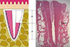
Twenty-four mandibular third and fourth premolars were selected and extracted. Any tooth found to be fused, fractured, or damaged on the root surface during hemisection was excluded from the experiment. A total of 36 roots were used in this study. The roots were randomly divided into three groups: (1) a positive control group (n=12), in which the PDL was retained; (2) a negative control group (n=12), in which the PDL and the cementum between the notches were removed; and (3) an experimental group (n=12), in which the PDL and the cementum between the notches were removed and the roots were soaked in FGF-2 (Fig. 1). Each group was subdivided into a four-week group and an eight-week group as the experiment was conducted, with six roots in each subgroup.
Surgical protocol
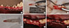
Before the experimental operation, scaling and oral hygiene were performed. During the surgical procedures, animals were placed under general anesthesia by Gerolan (Choongwae Pharmaceutical, Seoul, Korea) inhalation along with an intravenous injection of Zoletil (5 mg/kg; Virbac, Carros, France) and xylazine (0.2-0.5 mg/kg; Rompun, Bayer Korea, Seoul, Korea). In addition, lidocaine hydrochloride containing 1:80,000 epinephrine was administered for local anesthesia. The third and fourth mandibular premolars were extracted from the mandibles with forceps using rotary movements, after separating the roots (Fig. 2A). Four notches were formed on the coronal parts of the root surfaces at intervals of approximately 5 mm with a #1/2 round bur (Fig. 2B). In the negative control group and the experimental group, the PDL and the cementum between the notches were removed with a sharp-edged Gracey curette (Fig. 2C). The extraction sockets were also curetted. Next, the roots in the experimental group were soaked in 0.1 mL saline containing 30 µg of recombinant human FGF-2 (Genoss Institute, Suwon, Korea) for two minutes (Fig. 2D). The roots in the control group were covered with saline solution for two minutes to prevent dryness. The extraction sockets were curetted. After treating the root surfaces, the extracted roots were replanted into their respective sockets and the tooth crowns were severed using a carbide bur (Fig. 2E). The roots were completely covered with coronally repositioned flaps (Fig. 2F). For three days after surgery, 20 mg/kg of cefazoline sodium (Yuhan, Seoul, Korea) was injected intramuscularly, and soft foods were given. One week after surgery, the sutures were removed. Oral hygiene was maintained through the weekly application of 0.2% chlorohexidine solution (Hexamedin, Bukwang Pharmaceutical, Seoul, Korea) for infection control.
Histological processing
The dogs were euthanized with an overdose of pentobarbital sodium (90-120 mg/kg, intravenously) at four or eight weeks. The jaws were removed and the specimens were placed in 10% neutral buffered formaldehyde, decalcified in 5% formic acid, trimmed, and embedded in paraffin. Sections were cut in the mesiodistal plane, and serial 5-µm sections were stained with hematoxylin and eosin or Masson's trichrome.
Histological and histomorphometric evaluations
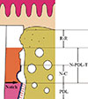
All measurements were obtained using a light microscope (LEICA DM750, Wetzlar, Hesse, Germany) equipped with a digital camera (LEICA ICC50, Wetzlar, Hesse, Germany). Measurements were made using the i-SOLUTION Lite® processing and analysis program (IMT i-Solution Corporation, Burnaby, British Columbia, Canada) on a personal computer. Six randomly chosen sections from each experimentally treated tooth were used for morphometric evaluation. The following measurements were obtained (Fig. 3): (1) newly formed PDL-like tissue (N-PDL-T), reflecting the length of the connective tissue attached to the root-planed surface; (2) replacement resorption (ankylosis) (R-R), comprising the combined longitudinal height of areas with ankylosis; and (3) newly formed cementum (N-C), reflecting the longitudinal length of the regenerated cementum or cementum-like deposits on roots. The values of N-PDL-T and R-R were expressed as percentages of planed root lengths, and values of N-C were expressed in percentages of the N-PDL-T lengths. The results are presented in the format of mean±standard deviation.
Statistical analysis
The mean and standard deviation for each measurement were calculated for each group. The Kruskall-Wallis test, the Mann-Whitney U test, and the Bonferroni correction were used to assess whether the differences between the control groups and the experimental groups were statistically significant. P-values < 0.05 were considered to indicate statistical significance, and the analysis was performed using SPSS version 17.0 (SPSS Inc., Chicago, IL, USA).
RESULTS
Clinical observations
Eight weeks after surgery, the extraction wounds had healed and were covered by healthy mucosa, although gingival recession was observed in one root of the positive control group at eight weeks and one root of the experimental group at four weeks. Extensive inflammatory root resorption also occurred in these roots.
Histologic observations
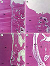
The positive control group
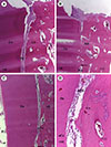
At four and eight weeks, normal PDL covered the roots (Fig. 4A, 5A, 6A). Superficial root resorption was observed in some areas. Root resorption was the predominant feature in the cervical portions of the roots (Fig. 4A, 5A). However, the PDL close to the dentin surface in the regions of root resorption was partially occupied by newly formed cementum (Fig. 5A).
The experimental group
At four weeks, PDL-like tissue partially separated the root surfaces and the newly formed bone (Fig. 4C). The connective tissue fibers of the PDL-like tissue appeared to consist of thin and randomly oriented fibers. Some of these fibers attached to dentin surfaces and newly formed cementum-like tissue (Fig. 4D).
At eight weeks, PDL-like tissue partially separated the root surfaces and the newly formed bone, and newly formed bone covered the entire area of the roots of the replanted teeth (Fig. 5C, 6C). Newly synthesized cementum-like tissue formed from the edges of the notches. In some areas, newly formed cementum-like tissue had formed in regions of superficial root resorption. In addition, cementoblast-like cells were visible in newly formed cementum-like tissue (Fig. 5D).
Histomorphometric analysis
The histomorphometric results are summarized in Tables 1 and 2. One root of the positive control group at eight weeks and one root of the experimental group at four weeks were excluded from the analysis because they displayed extensive inflammatory root resorption. Ankylosis was observed in part of the positive control group. However, four weeks after surgery, the mean length of the PDL tissue on the roots of the positive control group was significantly greater than that of the newly formed PDL-like tissue on the roots treated with FGF-2. At four and eight weeks after surgery, the mean length of newly formed cementum-like tissue on the roots treated with FGF-2 was significantly greater than on the positive control roots (four weeks, P=0.010; eight weeks, P=0.027) and the negative control roots (four weeks, P=0.008; eight weeks, P=0.042) (Tables 1, 2). The mean length of newly formed PDL-like tissue on the roots treated with FGF-2 was greater than on the negative control roots, and the mean length of replacement resorption on the roots treated with FGF-2 was lower than on the negative control roots. However, no significant differences were observed between the roots treated with FGF-2 and the negative control roots with respect to newly formed PDL-like tissue and replacement resorption (Tables 1, 2).
DISCUSSION
Root resorption is the major complication after tooth transplantation or replantation. Of the three categories of root resorption, replacement resorption is the most irreversible because it is a component of the physiological process of normal bone remodeling. Furthermore, ankylosis is largely responsible for the low five-year survival of teeth after tooth injury [20], and for this reason, it is important to prevent ankylosis in replanted teeth. In the present study, the PDL and cementum in the coronal portion of replanted roots were removed to model root surface damage. After healing, roots treated with FGF-2 were found to have shown favorable healing with respect to newly formed cementum-like tissue. Furthermore, FGF-2 induced the formation of newly formed PDL-like tissue in previously denuded coronal areas. However, the roots in the negative control group frequently showed ankylosis. This pattern of favorable healing and reduction in replacement resorption (ankylosis) in teeth treated with FGF-2 suggests that FGF-2 effectively promotes the regeneration of the injured PDL and cementum.
Many attempts have been made to prevent ankylosis and to encourage the production of new PDL tissue and newly formed cementum tissue after the transplantation or replantation of teeth. Seshima et al. [21] reported that the incidence of ankylosis in their FGF-2 treatment group was significantly lower than in the control group (3.6%±4.0% of total root length versus 25.4%±1.0% of total root length; P<0.01) at eight weeks after surgery. Shiratani et al. [22] reported that roots treated with FGF-2 exhibited the formation of new PDL-like tissue and new cementum with inserting collagen fibers after delayed transplantation, and Sato et al. [23] reported that teeth treated with 1 µg of basic FGF in collagen gel exhibited the formation of dense fibers bound to alveolar bone and new cementum at eight weeks. Similarly, the histomorphometric analysis conducted in the present study showed that the amount of replacement resorption in the FGF-2-treated group was less than in the negative control group at four and eight weeks (four weeks, 34.7%±11.0% of the denuded surface of the roots versus 65.8%±27.8% of the denuded surface of the roots; eight weeks, 36.9%±30.0% of the denuded surface of the roots versus 72.1%±22.7% of the denuded surface of roots). No significant difference was observed between the FGF-2-treated group and the negative control group. However, the amount of newly formed cementum-like tissue in the FGF-2-treated group was significantly greater than in the negative control group (four weeks, 81.1%±22.5% of the denuded surface of the roots versus 11.5%±16.8% of the denuded surface of the roots; eight weeks, 55.0%±37.2% of the denuded surface of the roots versus 14.1%±27.0% of the denuded surface of the roots, P<0.05). Although the amounts of newly formed cementum-like tissue in the FGF-2-treated group at four and eight weeks were not significantly different, these findings enable us to confirm that FGF-2 cytokine therapy prevents ankylosis and promotes cementogenesis and the regeneration of PDL-like tissues on injured root surfaces.
However, the proportion of roots showing ankylosis in the negative control group and the experimental group of this study was considerably higher than in other studies. Moreover, the width of the newly formed PDL-like tissue on roots treated with FGF-2 was less than that observed in other studies. Three reasons for this difference may be suggested. First, a lower amount of remaining PDL was present, because curettage of the remaining PDL tissue was performed on extraction sockets and on experimental roots. Second, the distances between the root surfaces and the extraction sockets were small because replantation was performed without further reduction of the extraction sockets. Third, the roots were submerged to prevent exposure to occlusal forces, and thus, the activities of PDL tissue might have been blocked.
The best method for increasing the amount of remaining PDL is to preserve vital PDL cells on the root surfaces and the extraction socket walls. In order to maintain the viability of the PDL cells, the extra-alveolar time of an avulsed tooth should be less than five minutes prior to replantation [24]. Soder et al. [25] reported that the number of visible PDL cells decreases in proportion to extra-alveolar time, and Andreasen [4] reported a greater incidence of replacement resorption in teeth with an extra-alveolar time of 18 minutes versus 0 minutes. However, Iqbal and Bamaas [26] reported no significant observable differences in the incidence of root resorption for extra-alveolar times of 15, 30, and 60 minutes, but a decreased incidence of periodontal healing was found corresponding to increased extraoral dry time. In the present study, the roots were replanted within five minutes to maintain PDL cell vitality. This explains why intact PDL tissue was found on a large proportion of the root surfaces in the positive control group. However, we also found that short-term replantation had no effect on the regeneration of injured PDL and cementum in the negative control group. Although it is important to maintain the viability of PDL cells during short-term replantation, the histologic results of FGF-2-treated roots showed that a signaling factor, such as FGF-2, was required for the regeneration of injured PDL and cementum.
The ability of storage media to maintain tooth viability during transplantation and replantation is considered to be more important than extra-alveolar time [2728]. Different types of wet storage media, such as Hank's balanced salt solution, minimum essential medium, saline, water, saliva, bovine milk, propolis, and green tea, have been investigated [29]. In the present study, saline was used to avoid tooth dehydration. However, although saline is a physiological match in terms of osmolality and pH, it does not contain essential ions and glucose, which are fundamental requirements of cells [3031]. Moreira-Neto et al. [32] evaluated the viability of cultured cells in saline, finding that 55% of cells remained alive after storage in saline for four hours. Consequently, saline is not a suitable medium for long-term storage, but is probably suitable for short periods of time, as demonstrated by the fact that intact PDL tissue was observed in many areas in the positive control group of this study.
The distance between recipient bone tissue and the root surface of a transplanted tooth is another important consideration. Optimal contact with the recipient site can improve the supply of blood and nutrients to PDL cells, thereby improving success rates after tooth transplantation [33]. In fact, good blood supply has been shown to be important for wound healing [34]. Furthermore, optimal contact between donor teeth and recipient bone ensures immobilization. However, to our knowledge, no study has been performed to determine the optimal distance between transplanted root surfaces and alveolar bone during tooth transplantation. In an effort to provide better blood supply and make the seating of donor teeth easier, Nethander [35] advocated a two-stage transplantation procedure. Promising results were obtained after a follow-up period of up to five years, and it was found that this two-stage surgical technique made it possible to transplant autogenous teeth with little risk of root resorption or other complications. For transplantation, it has been suggested that the donor site should be made 1-2 mm wider than the transplanted tooth in order to preserve the PDL [36]. Hence, in the present study, a small gap between the root surface and the socket wall promoted higher rates of replacement resorption, because the osteogenic action of granulation tissue on the surface of the extraction socket wall is activated soon after the tooth is extracted.
Furthermore, in the present study, tooth crowns were removed and the roots were submerged for protection. However, several reports have suggested that masticatory stimulation during the healing period may activate the function of the periodontal membrane area, promote the proliferation of PDL cells, and thus improve the prognosis of transplantation [3738]. Yang et al. [39] concluded that the application of orthodontic forces promotes PDL healing and possibly prevents dentoalveolar ankylosis. However, they did recommend a rest period of at least two weeks before loading the transplants, because the outcomes of regenerative procedures are critically dependent on initial wound stability [24]. This fact shows that complete coverage by a flap may have a beneficial influence on the wound healing of the PDL and cementum during the initial two weeks, while subsequently disrupting the stimulation of PDL cells. This may explain our finding that N-PDL-T did not increase and R-R did not decrease from four to eight weeks.
Moreover, in the present study, the application of FGF-2 did not completely prevent replacement resorption, and more instances of ankylosis were observed in roots treated with FGF-2 than has been reported in other experiments [2122]. Nonetheless, the results of this study clearly show that FGF-2 stimulates adjacent PDL tissue on the root surface and exerts a positive influence on the regeneration of the PDL and cementum.
FGF-2 enhances PDL cell proliferation, but inhibits cell differentiation by downregulating intracellular alkaline phosphatase activity [17]. However, the effects of FGF-2 on hard tissue formation and mineralization are probably cell-type dependent [40]. In addition, FGF-2 plays a significant role in the migration and proliferation of PDL fibroblasts, and their migration into areas of healing is key during the initial phase of periodontal regeneration. Murakami et al. [19] reported that FGF-2 expression could be detected in immature granulation tissue one week after flap surgery, and suggested that the aggregates formed by FGF-2 during the early phase of wound healing constituted a favorable environment for periodontal cell proliferation.
In present study, the standard deviation was high because the sample size was small, and healing dynamics vary considerably among individuals. Further studies are required with larger samples.
In summary, the histological and histomorphometric results obtained in the present study demonstrate that the use of FGF-2 reduces ankylosis and promotes the formation of newly formed periodontal tissue on injured PDLs and cementum after tooth replantation in dogs.
 XML Download