Journal List > J Periodontal Implant Sci > v.44(1) > 1082444

McCrea: Advanced peri-implantitis cases with radical surgical treatment

Abstract

Purpose

Peri-implantitis, a clinical term describing the inflammatory process that affects the soft and hard tissues around an osseointegrated implant, may lead to peri-implant pocket formation and loss of supporting bone. However, this imprecise definition has resulted in a wide variation of the reported prevalence; ≥10% of implants and 20% of patients over a 5- to 10-year period after implantation has been reported. The individual reporting of bone loss, bleeding on probing, pocket probing depth and inconsistent recording of results has led to this variation in the prevalence. Thus, a specific definition of peri-implantitis is needed. This paper describes the vast variation existing in the definition of peri-implantitis and suggests a logical way to record the degree and prevalence of the condition. The evaluation of bone loss must be made within the concept of natural physiological bony remodelling according to the initial peri-implant hard and soft tissue damage and actual definitive load of the implant. Therefore, the reason for bone loss must be determined as either a result of the individual osseous remodelling process or a response to infection.

Methods

The most current Papers and Consensus of Opinion describing peri-implantitis are presented to illustrate the dilemma that periodontologists and implant surgeons are faced with when diagnosing the degree of the disease process and the necessary treatment regime that will be required.

Results

The treatment of peri-implantitis should be determined by its severity. A case of advanced peri-implantitis is at risk of extreme implant exposure that results in a loss of soft tissue morphology and keratinized gingival tissue.

Conclusions

Loss of bone at the implant surface may lead to loss of bone at any adjacent natural teeth or implants. Thus, if early detection of peri-implantitis has not occurred and the disease process progresses to advanced peri-implantitis, the compromised hard and soft tissues will require extensive, skill-sensitive regenerative procedures, including implantotomy, established periodontal regenerative techniques and alternative osteotomy sites.

Graphical Abstract

jpis-44-39-ab001

INTRODUCTION

The consensus report from the first European Workshop on Periodontology [1] originally defined the term peri-implantitis as a clinical diagnosis that requires the assessment of inflammation in the peri-implant tissue as well as the loss of supporting bone. As a clinical term, peri-implantitis is used to describe an 'inflammatory process' affecting the physiological function of soft and hard tissues around an osseointegrated implant in function, which may lead to peri-implant pocket formation and loss of supporting bone. However, the lack of a precise definition of the degree of peri-implantitis has resulted in a wide variation of its reported prevalence. For example, a prevalence of up to 10% of implants and 20% of patients during a 5- to 10-year period after implantation has been reported. In addition, variations in the reporting and recording of bone loss, bleeding on probing (BOP), and pocket probing depth (PPD) have led to a large variation in the reported prevalence [2]. Other researchers have also stated that the prevalence and incidence of peri-implantitis depends on the specific definition of the disease [3].
The following clinical and retrospective studies have reported a considerably high prevalence of peri-implantitis among their subjects:
  • In a cohort study by Roos-Jansaker et al. [4] 14%-16% of the patients had at least one implant with radiographic bone loss of at least 3 threads and a pocket depth of ≥6 mm after being followed for 9-14 years.

  • With the threshold of bone loss defined as the presence of at least three threads in at least one implant, Fransson et al. [5] reported that 28% of their patients showed radiographic bone loss over 5-20 years. Further, out of the 3,413 implants included in the study, 12.4% demonstrated progressive bone loss.

  • Ferreira et al. [6] defined peri-implantitis as the presence of a PPD >4 mm in association with peri-implant bleeding and/or suppuration with radiographic confirmation of bone loss; 9% of their patients were diagnosed with peri-implantitis.

  • Koldsland et al. [7] found a wide variation in peri-implant prevalence, 11%-47%, dependant on the radiographic interpretation of peri-implant bone loss and PPD threshold limit. With a PPD ≥4 mm, BOP, and radiographic bone loss ≥2 mm, 20.4% of their patients had peri-implantitis. When the threshold was increased to a PPD ≥6 mm with BOP and radiographic bone loss ≥3 mm, the prevalence of peri-implantitis decreased to 11.3%.

  • In a systematic review by Jung et al. [8] on the 5-year survival and complications of peri-implantitis, the incidence of the disease at single crowns was 9.7% . Further, bone loss >2 mm occurred in 6.3% of all implants over the 5-year observation period.

  • A recent systematic review [9] considering the biological and technical complications in implant dentistry revealed that the incidence of peri-implantitis increased with time; the 5- to 10-year incidence was greater than the 0- to 5-year incidence.

Because of the wide variation in threshold levels, the reported incidence and prevalence of peri-implantitis are not precise measurements. In an attempt to clarify the definition of peri-implantitis, Froum and Rosen [10] recently proposed a simple and logical classification system as follows:
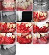
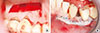
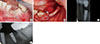
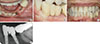
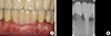
  • Early peri-implantitis, a PPD ≥4 mm with bleeding and/or suppuration on probing. Bone loss <25% of the implant length.

  • Moderate peri-implantitis, a PPD ≥6 mm with bleeding and/or suppuration on probing. Bone loss 25%-50% of the implant length.

  • Advanced peri-implantitis, a PPD ≥8 mm with bleeding and/or suppuration on probing. Bone loss >50% of the implant length.

The bone loss was to be measured using radiographs taken from the time of definitive prosthesis loading throughout follow-up. However, from the moment of definitive implant loading, physiological remodelling of the peri-implant bone will occur; therefore, bone loss may result from the physiological remodelling process. Therefore, crestal bone levels should be recorded from standardized periapical radiographs (using an individualized fim holder) taken at baseline and 6 months following definitive loading. That film-holder should be stored with the patient records and used whenever radiographs are taken to allow for a systematic and accurate evaluation of the crestal bone margins.
The treatment of peri-implantitis should then be determined according to its severity. However, improving the treatment of peri-implantitis within the field of periodontology is challenging. Exposed root surfaces in periodontitis may or may not be of visible concern. The form and colour of the exposed implant thread in a patient suffering from peri-implantitis may change the original aim of surgery, which is typically the establishment of not only function, but also a crown-form of high aesthetic value. For example, the previously described case of 'advanced peri-implantitis' will likely suffer extreme implant exposure resulting in a loss of soft tissue morphology and keratinized gingival tissue [10]. That same loss of bone at the implant surface may also lead to bone loss in adjacent natural teeth and/or implants. Thus, if the early detection of peri-implantitis fails and the disease progresses to that of the advanced condition, the patient may demand the removal of the implant and a remedial solution to achieve the original goal of implant therapy. Unfortunately, a patient with advanced peri-implantitis will likely have extremely compromised hard and soft tissues that require extensive, skill-sensitive regenerative procedures to be performed. This report presents two cases of advanced peri-implantitis, as defined by Froum and Rosen [10], where extreme peri-implant bone loss required both hard and soft tissue regeneration to re-establish dental morphology and aesthetics. The techniques used emphasize the importance of preoperative diagnosis and evaluation so that the desired results may be attained.

CASE REPORTS

Case 1

A 60-year-old Caucasian woman was referred to a private practice specializing in periodontics, dental implants and advanced restorative techniques to replace a 'failed' implant. The author informed the patient that the soft and hard tissues around her implant (tooth position 34 according to the World Dental Federation notation) showed evidence of peri-implantitis. The implant had been placed 20 months prior to her visit (to the current practice), and the crown fitted 4 months after the implant surgery. She reported a problem-free implant surgery and subsequent healing period; however, she reported that from the day the crown was fitted, the crown and adjacent gum had never felt 'comfortable.' The patient completed normal hygiene visits during her dental check-ups, but since the crown was fitted onto the implant in position 34, she had visited a hygienist every 3 months for help with this implant site. During visits to the hygienist, topical antibiotics were inserted on several occasions into the pocket to treat infections and pocket/implant surface debridement was carried out. The referring dentist had already warned the patient that the implant should be removed. Upon visiting our office, the patient described feeling very unsatisfied with the loss to her buccal tissue and bone at the implant site and asked if the implant could be replaced. Additionally, the tooth anterior to the implant (position 33) had also suffered extreme recession, which she felt was attributed to the development of peri-implantitis at position 34. The patient had no contraindicating medical condition. Clinical examination showed a very pronounced loss of attachment at positions 33 and 34 (Fig. 1A and B). The patient was diagnosed as having a mixed 'thin and thick' gum phenotype. The width of keratinized gingivial tissue (KGT) in the lower arch varied between 0 and 6 mm. We performed a radiographic examination that produced periapical, panoramic and linear tomograms (Fig. 1C-E). The initial diagnosis was controlled chronic adult periodontitis with horizontal bone loss affecting both the upper and lower dental arches. Peri-implantitis was also present at implant position 34 with no buccal KGT present. The aetiology of the problem was described to the patient, and a suggested treatment plan that involved the elective removal of the implant affected by peri-implantitis was suggested. In addition, we explained that bone loss was present at not only the implant site but also the anterior tooth and that both teeth should be removed to allow for the re-establishment of normal crestal bone at the site. With the elective removal of both the affected implant and anterior tooth, we had to create a stable peri-implant gingival apparatus. As a result, the width of the KGTs required reestablishing. The advantages of using a free split gingival graft was explained to the patient. Advantages included the presence of a stable, wide band of KGT conducive to ease of surgical handling and the carrying out of oral hygiene techniques. Because of the extreme bone loss suffered at site 34, new implants had to be placed at positions 33 and 35. The surgery was divided into three separate interventions as follows:
  1. The removal of tooth 33 and the implant at 34 followed by Guided Bone Regeneration GBR to re-establish the width and height of the hard tissue

  2. A free split gingival graft to reconstruct the width of KGT to extend from position 33 to 35

  3. Placement of implants into positions 33 and 35

One hour prior to surgery, the patient received systemic coverage of 2-g amoxicillin, 400-mg ibuprofen and 2-mg dexamethasone. Additionally, a mouth rinse of 2% chlorhexidine gluconate was administered 20 minutes before surgery. The surgery was carried out under intravenous sedation (midazolam) and local anaesthesia (4% articaine, Septodont, Saint-Maur-Des-Fosses, France). The mucoperiosteal flap was raised and tooth 33 and implant 34 were removed with periotomes to preserve the socket walls (Fig. 1F and G). The sockets were debrided of all inflammatory and granulation tissue, but the remaining sulcular epithelium from the soft tissue margin was left intact (Fig. 1H). The lingual soft tissue was then reflected to allow the submucosal placement of a resorbable membrane (Bio-Gide, Geistlich Pharma, Wolhusen, Switzerland) (Fig. 1I). The vacated sockets were then packed with xenograft Bio-Oss (Geistlich Pharma, Wolhusen, Switzerland) with a particle size of 0.25-1.0 mm and covered with Bio-Gide (Fig. 1J and K), thus attempting to re-establish a normal height in the crestal hard tissue. Last, 4.0 Prolene sutures were used to close the wound (Fig. 1L).
Four months postoperatively, a periapical radiograph was taken to display the newly augmented crestal bone (Fig. 1M). Afterward, the second stage of surgery was carried out under the same surgical protocol. The free split gingival graft was carried out using donor tissue from the left palatal vault (Fig. 2A), and the wound was covered with an eugenol-free pack for 7 days. Preparation of the recipient site involved the removal and apical reflection of the supraperiosteal mucosa, and this was sutured into the sulcus with 6.0 Prolene sutures (Ethicon, Johnson and Johnson, Wokingham, Berks, UK). Next, the 'free-graft' from the donor was sutured into the recipient site with 6.0 Prolene sutures (Fig. 2B).
Two months later, the third stage of surgery was performed under the same surgical protocol. The implants were placed by centralizing them to the 'bony ridge' (MIS, SEVEN, Tel Aviv, Israel). The primary stability of all of the inserted implants was high; both implants had an initial seating torque of 50 Ncm as calibrated on the seating apparatus (Implantmed, W&H Dentalwerk, Buermoos, Germany). The final placement of the implants (torque value>50 Ncm) was carried out using a hand wrench. The crestal bone was further augmented with the xenograft (Fig. 3A and B), and periapical radiographs were taken immediately postoperation (Fig. 3C and D).
Five months later, the implants were exposed via a crestal incision displaced to the lingual side, and healing abutments were fitted. The final fixed bridge was fitted eight weeks later (Fig. 4A-C). Periapical radiographs were taken systematically at the bridge fitting and one, six and 12 months postloading (Fig. 4D). All treatments proceeded uneventfully, and the results showed a marked improvement in marginal gingival tissue levels and bone levels (Fig. 5A and B). Every six months, the patient has been examined and radiographs taken, which will continue for two years. After two years, observational radiographs will be taken every 12 months. The patient remains in supportive periodontal therapy.

Case 2

A 44-year-old Caucasian man was referred to our office to replace implants that were expected to be imminently lost. Over the past five years, the patient reported receiving multiple implant therapies where many of the implants were removed and replaced. There were sites where the implants had been replaced on three occasions during that period. The patient did not understand the aetiology of the loss, had never received oral hygiene instruction or other forms of long-term supportive periodontal therapy and had no concept of peri-implantitis. The initial diagnosis was uncontrolled chronic adult periodontitis with horizontal bone loss affecting both the upper and lower dental arches. Additionally, peri-implantitis was present in varying degrees of aggression at all implant positions. Multiple photographs and radiographs were included in the initial diagnosis. The aetiology of the problem was described to the patient. Supportive periodontal therapy was initiated, and various surgical designs for implantotomywere applied with great success. Because of the degree of bone loss associated with the implant in position 42 (Fig. 6A), the corrective surgical therapy was complicated and divided into two stages as follows:
  1. The removal of implant 42 followed by GBR to re-establish the width and height of the hard tissue

  2. Placement of a 'remedial' implant into position 42 accompanied by a subepithelial connective tissue graft to replace the missing width of KGT at the site and further GBR (if necessary)

The same surgical protocol as in the preceding case was followed.
A mucoperiosteal flap was raised to allow full visualization of the affected site (Fig. 6B and C). The site was partially debrided and implant 42 was removed by luxation, which minimizes trauma to the socket walls (Fig. 6D). The socket was debrided of all inflammatory and granulation tissue, but any remaining sulcular epithelium was kept intact in the soft tissue margin (Fig. 6E). The lingual soft tissue was then reflected to allow the submucosal placement of the Bio-Gide membrane (Geistlich Pharma, Wolhusen, Switzerland). The vacant void was then packed with xenograft Bio-Oss (Geistlich Pharma) with a particle size of 0.25-1.0 mm and covered with Bio-Gide. These efforts attempted to re-establish a crestal hard tissue of a normal height. Last, 5.0 Prolene sutures were used to close the wound (Fig. 6F).
Five months postoperatively, a periapical radiograph was taken to display the newly augmented crestal bone (Fig. 7A and B). After that, the implants (MIS, SEVEN) were placed by centralizing them to the 'bony ridge' and following the same surgical protocol (Fig. 7C). The primary stability of both inserted implants was high; both implants had an initial seating torque of 50 Ncm as calibrated on the seating apparatus (Implantmed). Final seating of the implants (torque value>50 Ncm) was carried out using a hand wrench (Fig. 7C). The crestal bone was further augmented with the xenograft and covered with Bio-Gide. Connective tissue was harvested from the right palatal vault and sutured to the underside of the extended, reflected labial mucosal flap (Fig. 7D-F).
Five months later, the implants were exposed via a crestal incision displaced to the lingual side, and healing abutments were fitted. The final fixed bridge was fitted four weeks later (Fig. 8A-D). All of the treatments proceeded uneventfully, and the results showed a marked improvement in marginal gingival tissue levels and bone levels (Fig. 9A and B). To date, the patient has received periodic examinations and radiographs over two years. After two years, observational radiographs will be taken every 12 months. The patient remains in supportive periodontal therapy.

DISCUSSION

Controversy exists as to whether a history of periodontitis predisposes the future implant to peri-implantitis. In a review by Karoussis et al. [11], patients with a history of chronic periodontitis had no statically different outcomes for short or long-term implant survival when compared to periodontally healthy patients. Both cases in this article had a history of periodontitis. Case 1 presented as a controlled supportive periodontal therapy (SPT) case. The evident peri-implantitis may have been due to the ill-fitting abutment and equally ill-fitting crown; her oral hygiene was at a high standard even from the initial presentation for consultation. However, over-loading of the bridge is also a possible cause. Regardless of both topical antibiotic and systemic antibiotic use, bone loss increased to such an extent that she had lost nearly 75% of the bone support for the implant as well as a substantial amount of the bone support for the adjacent tooth within 18 months. Nonetheless, this patient successfully received new implants that have been in use for 48 months with no recurrence of peri-implantitis. She is also enthusiastically attends appointments for SPT.
Serino et al. [12] reported that local factors such as accessibility for oral hygiene at the implant sites were related to the presence or absence of peri-implantitis. Further, they suggested that peri-implantitis is frequently seen among subjects with no signs of periodontal disease in their natural dentition. These findings agree with Lindquist et al. [13] who reported that marginal bone loss was found on implants that had been functional for 10 years. They also concluded that a patient's knowledge of oral hygiene reduced bone loss surrounding implants. However, case 2 had a prosthesis that made it nearly impossible to carry out adequate oral hygiene procedures at the site of the implant (position 42), even if the patient had known how to perform oral hygiene. Thus, accessibility to the implant must be incorporated into the prosthetic design; otherwise, the risk of peri-implantitis is increased.
The role of the width and volume of keratinized gingiva in maintaining periodontal health remains controversial. Some studies support the view that periodontal health can be maintained in sites if optimal plaque control is practised regardless of the amount of attached gingival tissue [14-16]. However, Lang and Loe [17] concluded that even tooth surfaces free of plaque demonstrated clinical inflammation if the surface had <2.0 mm of KGT. Additionally Serino et al. [12] found gingival recession, thin periodontium and root prominence in combination with reduced or missing attached gingival tissue to be factors that influence periodontal health.
The free or split gingival graft is an established technique used for gingival augmentation. Dorfman et al. [18] showed significant differences in the amount of KGT, attached gingiva, and recession between treated and untreated sites after 4 years of follow-up. When comparing two techniques for gingival augmentation at sites without attached gingiva that produced a significant increase in KGT, Agudio et al. [19] found the stability of the gingival margin to be maintained over a long period (10-25 years). In a study by the same authors [20] using a split-mouth design, gingival augmentation surgery improved the gingival conditions of sites with gingival recessions and minimal or absent attached gingiva. Additionally, their long-term data demonstrated that the improvements in the amount of KGT and recession reduction can last for ≥10 years. These results supported the Lang and Loe [17] hypothesis of a beneficial role of grafted KGT in reducing, halting or preventing the recession of the gingival margin over time. Thus, the multistaged surgery in both cases presented here produced an environment whereby supportive periodontal therapy and home care could be successfully maintained; the medium-term photographs and radiographs demonstrate this effect (Figs. 5A and 9A). The American Academy of Periodontology [20] agrees with these recommendations and stated that "gingival augmentation procedures should be carried out to prevent soft tissue damage in the presence of alveolar bone dehiscence.... to halt progressive recession of the gingival margin, to improve plaque control and patient comfort around teeth and (dental) implants, and to increase the insufficient dimension of gingival tissue in conjunction with fixed or removable prosthetic dentistry." Although this statement was intended for the maintenance of periodontal conditions around natural teeth, oral hygiene procedures for natural teeth have been applied to dental implants. Therefore, this statement could also be applicable to patients with dental implants.
In conclusion, the current aim of dental implant replacement therapy not only includes the re-establishment of occlusal function but also the satisfaction of patients with a level of aesthetic results that increase their sense of well-being. Therefore, a high level of aesthetics at a functional implant should remain a common goal among all periodontists. Systematic recall, supportive periodontal therapy, periodontal monitoring and radiographic follow-up minimize the risk of peri-implantitis when used together effectively. These presented cases uncover potential benefits that may be obtained from exercising the philosophy of supportive periodontal therapy for dental implant replacement therapy to avoid the development of advanced peri-implantitis.

Figures and Tables

Figure 1
(A, B) Initial clinical presentation showing the extent of the buccal marginal gingival recession and the exposure of both root and implant surfaces. (C) Diagnostic panoramic radiograph. (D) Initial preoperative/diagnostic periapical radiograph showing the extent of bone loss at the implant and the adjacent canine tooth. The crown on the implant does not fit. (E) Linear tomogram showing the lack of hard tissue on the buccal aspect of the implant. (F) Diagonal mesial relieving incision. No relieving incision was made distal to the wound site. This flap design was chosen in anticipation of the large wound and increase in volume that would result from the graft materials and the need for increased sulcus vascularity for wound repair. A mucoperiosteal flap has been elevated to expose the extent of the defect. (G) The canine tooth was luxated first to allow for a complete assessment of the extent of bone loss associated with the implant. (H) The implant after luxation. Debridement of the defect was carried out, yet all marginal tissue was left intact to assist in wound coverage. The extent of the bone loss/defect was apparent at the lingual surface. (I) Bio-Gide membrane was placed at the lingual side between the lingual bony wall, but under the periosteum, to contain the subsequent Bio-Oss granules. (J) Bio-Oss granules (size, 0.25-1.00 mm) were placed into the voids to reform the bony morphology. (K) The Bio-Gide membrane was then folded over buccally to cover the Bio-Oss granules. The marginal granulations were placed over the membrane again. (L) The wound closure was facilitated with 5.0 Prolene sutures. (M). Periapical radiograph taken at 4 months post-operatively displaying the re-attainment of crestal bony margins and the filled in former void.
jpis-44-39-g001
Figure 2
(A) At 4-month postimplantotomy, a split gingival graft was harvested from the left palatal vault. (B) Into the prepared buccal recipient site, the free split gingival graft was sutured into place using 6.0 Prolene sutures.
jpis-44-39-g002
Figure 3
(A) The clinical situation before implant surgery and six months postimplantotomy. (B) A simple crestal incision was made, and reflection of the mucosa allows the exposure of the hard tissue formation for evaluation. Osteotomies were prepared in a centralized position on the crestal bone, and the implants were inserted with an initial seating torque value of 50 Ncm; final seating was carried out with a hand wrench at >50 Ncm. Additional Bio-Oss and the covering membrane were used to enhance the hard tissue volume at the site. Wound closure was carried out using 4.0 Vicryl sutures. (C, D) Immediate post-operative periapicals of the implants in place.
jpis-44-39-g003
Figure 4
(A) Cervical margins of the implants just before the abutment fitting. (B) Cement-on fixed bridge in place. (C) Comparison of the surgically corrected left gingival margins with the natural tooth level on the right side. (D) Periapical radiograph taken 1-month postcementation.
jpis-44-39-g004
Figure 5
(A) Clinical presentation at 36 months after fitting the bridge. (B) Periapical radiograph 36 months after procedure completion.
jpis-44-39-g005
Figure 6
(A) Preoperative periapical radiograph. (B) Clinical situation immediately before implantotomy. (C) Mucoperiosteal flap reflection exposing the implant threads and fibrousfibrose tissue. (D) Fibrous tissue was removed to display the extent of the clinical bone loss. (E) The implant was removed and the site completely debrided prior to the placement of xenograft granules which were then covered with its dedicated membrane. (F) The wound was closed using 6.0 Prolene sutures.
jpis-44-39-g006
Figure 7
(A) Periapical radiograph taken 5 months postoperation. (B) Before the implant placement surgery and 5 months post implantotomy. (C) Osteotomies were prepared and the implants were placed centrally on the crestal bone in positions 41 and 31 (FDI-Notation), both with an initial seating torque of 50 Ncm; the final seating was carried out with a hand wrench at >50 Ncm. (D) Position 41 has been further 'grafted' with Bio-Oss granules and covered with its membrane to enhance the hard tissue volume at the site. Connective tissue was harvested from the palate and sutured to the underside of the mucoperiosteal flap using 6.0 Vicryl Rapide to prevent its exfoliation. (E) Wound closure via the coronal advancement of the labial flap using 5.0 Prolene sutures. (F) Periapical radiograph taken immediately postoperatively.
jpis-44-39-g007
Figure 8
(A, B) Clinical presentation of the soft tissues six months postimplantation. (C) The fixed bridge was cemented onto the abutments. (D) Periapical radiograph taken 1-month postcementation.
jpis-44-39-g008
Figure 9
(A) Clinical presentation at 24-months after cementation of the bridge. (B) Periapical radiograph at 24-month postcementation.
jpis-44-39-g009

Notes

No potential conflict of interest relevant to this article was reported.

References

1. Albrektsson T, Isidor F. Consensus report session IV. In : Lang NP, Kasrring T, editors. Proceedings of the First European Workshop on Periodontology. London: Quintessence;1994. p. 165–169.
2. Klinge B, Meyle J. EAO consensus report: peri-implant tissue destruction. In : The Third EAO Consensus Conference 2012; 2012 Feb 15-18; Pfäffikon, Switzerland.
3. Mombelli A, Muller N, Cionca N. The epidemiology of peri-implantitis. Clin Oral Implants Res. 2012; 23:Suppl 6. 67–76.
crossref
4. Roos-Jansaker AM, Lindahl C, Renvert H, Renvert S. Nine- to fourteen-year follow-up of implant treatment. Part II: presence of peri-implant lesions. J Clin Periodontol. 2006; 33:290–295.
crossref
5. Fransson C, Lekholm U, Jemt T, Berglundh T. Prevalence of subjects with progressive bone loss at implants. Clin Oral Implants Res. 2005; 16:440–446.
crossref
6. Ferreira SD, Silva GL, Cortelli JR, Costa JE, Costa FO. Prevalence and risk variables for peri-implant disease in Brazilian subjects. J Clin Periodontol. 2006; 33:929–935.
crossref
7. Koldsland OC, Scheie AA, Aass AM. Prevalence of peri-implantitis related to severity of the disease with different degrees of bone loss. J Periodontol. 2010; 81:231–238.
crossref
8. Jung RE, Pjetursson BE, Glauser R, Zembic A, Zwahlen M, Lang NP. A systematic review of the 5-year survival and complication rates of implant-supported single crowns. Clin Oral Implants Res. 2008; 19:119–130.
crossref
9. Berglundh T, Persson L, Klinge B. A systematic review of the incidence of biological and technical complications in implant dentistry reported in prospective longitudinal studies of at least 5 years. J Clin Periodontol. 2002; 29:Suppl 3. 197–212.
crossref
10. Froum SJ, Rosen PS. A proposed classification for peri-implantitis. Int J Periodontics Restorative Dent. 2012; 32:533–540.
11. Karoussis IK, Kotsovilis S, Fourmousis I. A comprehensive and critical review of dental implant prognosis in periodontally compromised partially edentulous patients. Clin Oral Implants Res. 2007; 18:669–679.
crossref
12. Serino G, Wennstrom JL, Lindhe J, Eneroth L. The prevalence and distribution of gingival recession in subjects with a high standard of oral hygiene. J Clin Periodontol. 1994; 21:57–63.
crossref
13. Lindquist LW, Carlsson GE, Jemt T. Association between marginal bone loss around osseointegrated mandibular implants and smoking habits: a 10-year follow-up study. J Dent Res. 1997; 76:1667–1674.
crossref
14. Wennström J, Lindhe J. Role of attached gingiva for maintenance of periodontal health. Healing following excisional and grafting procedures in dogs. J Clin Periodontol. 1983; 10:206–221.
crossref
15. Wennstrom J, Lindhe J. Plaque-induced gingival inflammation in the absence of attached gingiva in dogs. J Clin Periodontol. 1983; 10:266–276.
crossref
16. Miyasato M, Crigger M, Egelberg J. Gingival condition in areas of minimal and appreciable width of keratinized gingiva. J Clin Periodontol. 1977; 4:200–209.
crossref
17. Lang NP, Loe H. The relationship between the width of keratinized gingiva and gingival health. J Periodontol. 1972; 43:623–627.
crossref
18. Dorfman HS, Kennedy JE, Bird WC. Longitudinal evaluation of free autogenous gingival grafts: a four year report. J Periodontol. 1982; 53:349–352.
crossref
19. Agudio G, Nieri M, Rotundo R, Cortellini P, Pini Prato G. Free gingival grafts to increase keratinized tissue: a retrospective long-term evaluation (10 to 25 years) of outcomes. J Periodontol. 2008; 79:587–594.
crossref
20. Agudio G, Nieri M, Rotundo R, Franceschi D, Cortellini P, Pini Prato GP. Periodontal conditions of sites treated with gingival-augmentation surgery compared to untreated contralateral homologous sites: a 10- to 27-year long-term study. J Periodontol. 2009; 80:1399–1405.
crossref
TOOLS
Similar articles