

 PDF
PDF ePub
ePub Citation
 Print
Print


INTRODUCTION
Many types of graft materials have been used for sinus grafting [1,2]. Of these, thus far, an autogenous graft material with osteoinduction potential has been considered the gold standard. However, the limitation of harvest sites, the amount of graft material, and the additional surgical sites required for the grafts lead to patients' hesitation to agree to the use of autogenous bone grafts [3]. Another limitation of a sinus graft is the large amount of resorption of the autograft [4,5]. Xenografts are grafts derived from other species. Currently, anorganic bovine-derived bone, equine-derived bone, and coral skeleton have been marketed for periodontal regenerative and implant therapy. In fact, demineralized bovine bone material (DBBM) is the most widely used material on the market. The inorganic component of DBBM is crystalline hydroxyapatite, and the organic components in DBBM are removed by efficient chemical and thermal processes.
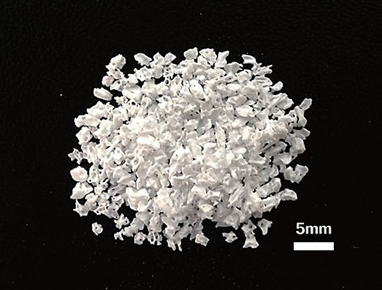
This new DBBM was produced for the present cases according to previously published methods [6]. Bone from the bovine femur was cut into 5-cm3 pieces. After the removal of blood, heterologous protein was removed using sodium hypochlorite instead of an amine group. Sodium hypochlorite is effective for the removal of animal-origin viruses including prion proteins. The bone was crushed to less than 0.7 mm. To remove the residual solvent, bone particles were irrigated with distilled water and sintered at over 400℃ for 3 hours. The size of the particles from the cortical bone was 0.2-1.0 mm/1.0-2.0 mm, and the nonuniform shape resembles that of white granules with a porous structure, which is similar to the human cancellous bone (Fig. 1).
This new DBBM showed a more homogenous surface texture in scanning electron microscope (SEM) [7]. The amount of residual protein calculated using Kjeldahl protein analysis was 0.053%, which was lower than that of two other DBBMs. Further, the residual heterologous protein could induce inflammation. In an in vivo study, DBBM particles were grafted into rat muscle to evaluate biocompatibility. During the 2 weeks of the graft, DBBM particles did not show any inflammatory cell infiltration and were encapsulated by a fibrous tissue [6]. In a rabbit calvarial defect model, new bone was formed and fused using DBBM particles after 2 weeks. The aim of this case report is to present the longitudinal results of sinus grafting using the new DBBM in human cases.
CASE DESCRIPTION
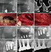
Case 1 (Fig. 2)
A 54-year-old Korean male missing his #26 and #27 teeth was referred to the Department of Periodontology at the Seoul National University Dental Hospital. The 2nd premolar on the upper left (#25) was diagnosed as hopeless and extracted. The patient wanted to have the missing space restored using dental implants. The residual bone height was 3-8 mm. A computerized tomography study was performed and evaluated. The lateral wall of the sinus was exposed by a mucoperiosteal buccal flap in the posterior maxilla. A window into the sinus was created with a round carbide bur under copious saline irrigation. The Schneiderian membrane was carefully detached from the sinus floor. The space was filled with bovine bone graft materials (OCS-B, NIBEC, Jincheon, Korea). A resorbable barrier membrane (BioGide, Geistlich Biomaterials, Wolhusen, Switzerland) was placed over the sinus window. The buccal flap was repositioned and sutured with 4-0 nylon.
In the postoperative phase, the patient was prescribed with systemic antibiotics and anti-inflammatory analgesics for 7 days. The patient was also instructed to rinse the surgical area twice a day with a 0.12% chlorhexidine digluconate gargling solution for 2 weeks. After a healing period of 6.5 months, three implants were placed (φ: 4.3 mm×11.5 mm; Oneplant, Warrantec Co., Seoul, Korea). The final restoration was completed at 9 months after the implant placement. The patient was periodically recalled and followed up after the prosthetic restoration for 5 years.
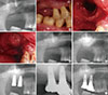
Case 2 (Fig. 3)
A 50-year-old Korean male missing his #16 and #17 teeth was scheduled for implant restoration after sinus grafting. The residual bone height was 2-6 mm. After a crestal incision and a periosteal flap reflection, the lateral wall of the sinus was exposed. An oval-shaped window was created with a round carbide bur under copious saline irrigation. The Schneiderian membrane was elevated, and a bovine bone graft material (OCS-B, NIBEC) was packed under the membrane. A resorbable membrane (BioGide, Geistlich Biomaterials) was also placed over the window opening. After suturing with 4-0 nylon, postoperative instructions were given including information on medication and mouth rinsing. After a healing period of 6 months, two implants were placed (φ: 5.3 mm×11.5 mm; Hexplant, Warrantec Co.). Seven months later, the final restorations were completed. They were screw-type porcelain fused gold splinted crowns. The patient was followed up for over 4 years after the sinus graft surgery.
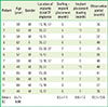
Both cases were handled by the same surgeon (Y.J.H.). Table 1 shows a summary of patients who underwent the sinus graft surgery using the new DBBM. Twelve partially edentulous patients, ranging in age from 40 to 65 years (average age, 55.7 years), were considered in this retrospective study. All patients had insufficient residual height in their maxillary posterior area and underwent maxillary sinus graft surgery to increase the height of their maxilla. The techniques described by Tatum Jr [8], and Boyne and James [9] were applied. In all, 27 fixtures were placed in an augmented bone area. On average, 8.6 months later, the implants were loaded using provisional or final restorations. The observation period ranged from 27 to 75 months (average, 43.3 months), and the patients did not show any severe resorption of the graft material or any infection during this time.
This study was approved by the Seoul National University Dental Hospital Clinical Dental Research Institute (IRB068/06-14).
DISCUSSION
A sinus graft is a predictable procedure for maxillary height augmentation in an edentulous area with maxillary insufficiency. Although a new crestal approach with osteotomes or newly developed devices has been developed [10], the lateral approach developed by Tatum Jr [8] and Boyne and James [9] is still widely used. Various types of graft materials have been used for maintaining the space below the elevated Schneiderian membrane [4,11,12,13,14]. Further, a sinus augmentation procedure without bone grafts has been proposed [15]. However, most of these procedures use an implant fixture to support the Schneiderian membrane.
DBBM has been extensively studied as a graft material histologically as well as radiographically [11,12,13,16,17,18]. DBBM is biocompatible and osteoconductive and is an appropriate graft material for the preservation of extraction sockets [16,17]. Lee et al. [11] have reported that a newly formed bone following the sinus graft with DBBM increases in volume and matures over time up to 12 months. Histologically, the newly formed bone increases from 18% to 26% from 6 months to 12 months after the sinus graft procedures. In a study by Valentini and Abensur [18], the newly formed bone also increased from 21% to 28% and the bovine bone graft material decreased from 39% to 27%, which implies that the newly formed bone had been increasing during 6 to 12 months after the sinus graft surgery. Son et al. [12] have also reported that newly formed bone increases by over 23% in 10 months after maxillary sinus grafting using bovine bone. These results imply that new bone formation increases as time passes, and the success rate of the implant also increases.
DBBM is also used as a combination of other materials. Hallman et al. [13] used a combination of bovine bone and autogenous bone to supplement the lack of osteoinduction. An autogenous bone graft can be substituted with bovine hydroxyapatite to 80% or 100% when used for maxillary sinus floor augmentation.
The new DBBM used in this study was used in mandibular defects of beagle dogs [19]. The defects sized 8 mm×6 mm×5 mm were filled with grafts. The average bone area fraction was increased over time from 26.08% to 32.33% at 4 to 6 weeks, respectively, after grafting. The use of a membrane over the graft materials did not show any additional effect. The new DBBM used in this study was used as scaffolding for synthetic peptides in animal studies [20,21]. Peptide-coated DBBM accelerated the cell attachment and proliferation of preosteoblastic cells in an in vitro model and the new bone formation in the rabbit calvarial model [20]. Synthetic oligopeptides corresponding to the bone morphogenetic protein (BMP) receptor domains were used for enhancing guided bone regeneration in beagles [21]. BMP receptor I (BMPRI) and BMP receptor II (BMPRII) binding domains were coated on the bovine bone mineral. In a study with beagle dogs, new bone formation was observed throughout the buccal dehiscence defect and a considerable ratio of the regenerated bone was found in the histologic section. Hieu et al. [22] compared the effects of two types of DBBMs in maxillary sinus graft surgeries. All sites were treated with two types of DBBMs with platelet-rich plasma. In both groups, the augmented bone height showed a decreasing tendency over time. The distance from the implant platform to the base of the maxilla decreased from 24.50 mm to 18.53 mm from the baseline to 25-48 months, respectively, in the case of the new DBBM (P<0.05). However, both graft materials showed no significant difference in their height change in 25-48 months after the graft.
Of the 12 patients, no patient showed any complications such as severe resorption, infection, mucosal thickening, or implant failure during the mean observation period of 43.3 months. Of the 27 implants, no implant showed a biologic failure. The interval between the sinus graft surgery and prosthetic loading was on average 8.6 months, which suggests that a sufficient amount of the newly formed bone was formed and could contact the implant surface during loading.
There were some limitations of this study. This study is neither a prospective study nor a consecutive case series. The prosthetic procedure was not standardized. Therefore, long-term follow-up studies including a histologic evaluation are necessary. Additional randomized controlled trials are also necessary to evaluate the longitudinal effect of this new DBBM. Our results show that the new DBBM is useful for a maxillary sinus graft procedure. Clinically good healing responses and reliable results were shown for a mean follow-up period of 43.3 months.
 XML Download