

 PDF
PDF ePub
ePub Citation
 Print
Print


INTRODUCTION
Currently, aggressive periodontitis is considered to be a well-defined inflammatory periodontal disease. It usually affects people who, in most cases, otherwise appear healthy. It tends to have a familial aggregation and there is a rapid rate of disease progression. Aggressive periodontitis occurs in localized and generalized forms. Localized aggressive periodontitis is frequently associated with Aggregatibacter actinomycetemcomitans while generalized aggressive periodontitis is frequently associated with A. actinomycetemcomitans and Porphyromonas gingivalis [1].
The specific attributes of the disease process are due to the virulence of the pathogens and the host susceptibility of patients. Since it is difficult to modify underlying immune defects, the goals of periodontal therapy are to alter or eliminate the microbial aetiology and contributing risk factors for periodontitis.
The prevalence rates of aggressive periodontitis in epidemiologic studies are variable. Prevalence ranges from approximately 1% to a maximum of 15% [2]. In Morocco, this form of periodontal disease has a reported prevalence of 7.6% [3].
Aggressive periodontitis, especially in its severe form, was traditionally considered to have an unfavourable prognosis, which often led to radical treatments. Eickholz et al. [4] reported that initial diagnosis was identified as a statistically significant influence on tooth loss. For aggressive and generalized severe chronic periodontitis, the risk for tooth loss was doubled in comparison with moderate periodontitis.
On the other hand, many studies [5,6,7,8,9,10] have demonstrated a favourable healing potential in lesions associated with localized aggressive periodontitis, but not in the severe generalized form. Other studies [11,12] have reported a stabilization of aggressive periodontitis cases achieved with both nonsurgical and surgical debridement. As well, many other studies have addressed only short-term therapeutic outcomes [13,14,15].
The aim of our study was to show clinical and radiographic outcomes following nonsurgical periodontal treatment alone in severe generalized aggressive periodontitis (SGAP) in the short and the long term.
MATERIALS AND METHODS
The present article reports on a retrospective cohort study of cases of SGAP treated with nonsurgical periodontal therapy. The study protocol was approved by the local Ethics Committee for Biomedical Research of the Faculty of Medicine and Pharmacy, Mohammed V Souissi University.
Patient population
Patients with advanced generalized aggressive periodontitis were included in this study. They were referred for treatment at the Department of Periodontology, Ibn Sina University Hospital-Rabat.
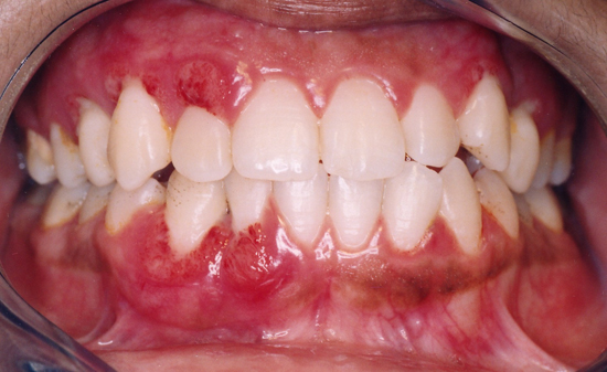
The initial diagnosis was based on severe periodontal tissue destruction and angular bone destruction in healthy subjects under 30 years of age (Figs. 1 and 2). There was generalized interproximal attachment loss affecting at least three permanent teeth other than the first molars and incisors.
The inclusion criteria were: (1) clinically healthy patients, except for the presence of periodontitis, with SGAP; (2) a periodontally compromised tooth with clinical attachment loss of ≥5 mm and/or probing depth (PD) of ≥7 mm; (3) presence of intrabony defect with at least 50% bone loss.
The noninclusion criteria were: (1) patients with systemic disease or drug therapy that could lead to impaired wound healing; (2) patients with periodontal risk factors (such as smoking and diabetes mellitus).
Patients were treated for deep periodontal pockets and angular bone loss. In order to demonstrate the favourable outcomes of nonsurgical periodontal therapy, sites with advanced lesions that were treated by periodontal surgery (open flap debridement) were not included in the study.
All the patients received nonsurgical periodontal treatment, including oral hygiene instructions and complete periodontal debridement followed by an antimicrobial therapy based on chlorhexidine (0.12%) mouth rinse and antibiotic combination of 1,000 mg of amoxicillin and 750 mg of metronidazole per day for seven days.
Patients were enrolled in a maintenance care program and were provided with supportive periodontal care every six months.
Clinical and radiographic assessment
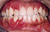
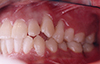
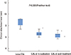
Estimations of samples in the study sample were based on a subject-level analysis. The following parameters were measured at severely affected sites with a millimeter calibrated periodontal probe, by the same examiner (A.B.) not blinded to the procedure: PD of pockets ≥7 mm, and clinical attachment level (CAL) of sites with attachment loss ≥5 mm. For each patient, the mean of multiple measurements was taken at baseline (prior to the treatment), at re-evaluation after the initial treatment (Fig. 3), and posttreatment (Fig. 4). Responding sites were defined as those showing at least 2-mm reduction in probing pocket depth or 1 mm of attachment gain after treatment. Resolution of inflammation and bone fill were recorded dichotomously as present or absent for each patient. The resolution of inflammation was determined by a decrease of gingival bleeding and the modification of gingival aspect at least to score one according to the Loe and Silness index. Pre- and postoperative radiographs were taken with the long cone parallel technique and bone fill was defined by bone densification and/or augmentation of bone level (Fig. 5).
Statistical analysis
The data were analyzed using the patient as a unit (subject based analysis). For each patient, mean values of PD and CAL were calculated before the analysis. The statistical analysis was performed with a statistical program (SPSS Inc., Chicago, IL, USA). The analysis compared baseline values with re-evaluation and posttreatment values.
The quantitative variables (PD, CAL) were expressed as medians and quartiles and were analysed between the three paired groups using the Friedman test followed by post hoc analysis. The Wilcoxon test with the Bonferroni correction was used for each paired group. A level of P<0.05 was accepted for statistical significance.
RESULTS
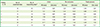
Seven patients with 266 sites were examined (mean age, 26.14±2.61 years; male/female sex ratio, 1/6). Two hundred and twenty-six of these sites had a favourable response to nonsurgical periodontal therapy. Clinical and radiographic data are shown in Table 1.
Re-evaluation after initial treatment was carried out at a mean of three months (3.71±1.4), with a maximum of 6 months after initial treatment. The posttreatment recall was performed at a median of five years (5 [1.5-7]). The results of the treated teeth in the seven patients are summarized in (Table 2).
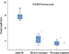
A significant difference was found between the values of PD at baseline (7.94 [7.33-8.19] mm) and both re-evaluation (4.33 [3.63-5.08] mm) and posttreatment (3.54 [3.33-4.11] mm) values (P=0.002). A significant difference was also found between the values of CAL at baseline (9.02 [7.5-9.2] mm) and both re-evaluation (6.55 [6.30-6.87] mm) and posttreatment (6.45 [5.70-6.61] mm) values (P=0.002) (Figs. 6 and 7).
The inflammation was resolved and angular bone defects were repaired in all cases. A radiographic analysis carried out on angular bone defects showed significant repair of prior angular bone defects.
DISCUSSION
Our study has demonstrated that SGAP can be successfully treated by nonsurgical therapy alone. This therapy resulted in the termination of disease progression, resolution of inflammation, decreased pocket depths, gains in clinical attachment and significant repair of alveolar defects associated with increased height of the supporting alveolar bone of the affected teeth. Aggressive periodontitis, especially in its severe generalized form, was previously considered to have an unfavourable prognosis. In some studies [11,12] the stabilization of periodontal health in aggressive periodontitis was achieved in sites with advanced disease after surgical therapy. Aimetti et al. [13], Aimetti et al. [14], Darby et al. [16], Hughes et al. [17], and Yek et al. [18], have showed that nonsurgical treatment can be a highly effective treatment for patients and sites affected by aggressive periodontitis. Their results dealt with short-term outcomes. Further studies [15,19,20,21,22] have demonstrated the short-term clinical outcomes of nonsurgical periodontal debridement in subjects with generalized aggressive periodontitis.
In the present study, the posttreatment exam demonstrated a statistically significant decrease in PDs of deep periodontal pockets and significant gains in CAL. Changes in PD and CAL were observed from baseline to posttreatment. There was no relapse of the disease at the posttreatment stage. This observation suggests that nonsurgical treatment may have a long-term positive effect on the periodontal status of patients with SGAP. This is in accordance with the results of a five year longitudinal study demonstrating favourable healing following surgical and nonsurgical treatment of juvenile periodontitis [23].
Our patients were very pleased to retain their own natural teeth despite the advanced, seemingly hopeless state of their teeth prior to treatment. Pocket depth and attachment loss continued to decrease in most of the patients after the completion of nonsurgical periodontal therapy while alveolar bone repair continued to increase. The postoperative healing was continuous in six of the seven patients and was not dependent on recall frequency. The posttreatment radiographs demonstrate a significant formation of the mineralized component of the supporting mineralized tissues. The reparative potential associated with this form of periodontitis can be explained by the good healing potential of the periodontium in SGAP and the effectiveness of antimicrobials in modifying the microbiological profile of this form of periodontitis. The architecture of periodontal lesions in the severe aggressive form of periodontitis can also explain the positive therapeutic outcomes. The rapid rate of disease progression often leads to angular lyses and deep periodontal pockets. Increases in pocket depth were associated with a significant reduction after periodontal therapy. Machtei et al. [24] demonstrated that the greater the initial pocket depth was, the greater the potential of regeneration. This could explain the striking improvement of severe periodontal lesions.
Our study has demonstrated favourable radiographic and clinical outcomes of nonsurgical periodontal debridement in subjects with SGAP. The results of this study suggest that nonsurgical periodontal therapy, based on oral instructions, periodontal debridement, and the use of antimicrobials, may be an effective procedure to improve the healing potential of severe aggressive periodontitis cases. The reparative potential associated with severe aggressive periodontitis may exceed that observed in moderate aggressive and chronic periodontitis and suggests a conservative therapeutic approach.
This study provides evidence of favourable outcomes following nonsurgical therapy alone of SGAP. However, the results of the study are limited by the small study population, the retrospective design of the study and the heterogeneity of the time points of posttreatment recalls.
Overall, although this study provided some indications of the beneficial effect of nonsurgical therapy on SGAP cases in the short and the long term, a broader comparative study is necessary to more fully assess the effects of nonsurgical periodontal therapy compared to surgical therapy.
The therapeutic result of SGAP cases suggests that this form of periodontitis can have positive outcomes. The reparative potential associated with SGAP should encourage clinicians to save seemingly hopeless teeth in this type of periodontitis cases.


 XML Download