

 PDF
PDF ePub
ePub Citation
 Print
Print


INTRODUCTION
Periodontal regeneration requires the attachment of a new connective tissue to the root surface, a process that involves the regeneration of periodontal fibers and the insertion of these fibers into a newly formed cementum. Several procedures are performed for periodontal regeneration, including guided tissue regeneration, topical application of an enamel matrix derivative, and the application of various growth factors [1,2]. However, previous studies have reported that these treatments are insufficient due to their limited regeneration of the periodontal tissue [3]. Since the regenerative events of periodontal wound healing require the recruitment of progenitor cells attempting to repair the periodontium infected by periodontitis, tissue engineering provides a new paradigm based on molecular and cell biology for periodontal regeneration [4]. Tissue engineering requires three essential components: (1) a population of stem/progenitor cells, (2) the presence of signaling molecules, and (3) a scaffold or extracellular matrix (ECM) [5]. Selection of the optimum types of cells, scaffold properties, and growth-factor combinations is a fundamental prerequisite for ensuring that the characteristics of the reconstructed tissue are close to those of the lost native tissue [6].
Suitable scaffolds for use in tissue engineering are classified as either natural or synthetic. Synthetic biomaterials are based on ceramics including hydroxyapatite (HA) and tricalcium phosphate, or on polymers such as polylactic acid, polyglycolic acid, and a copolymer thereof. However, many of these synthetic materials lack sites for cell adhesion and, therefore, have to be chemically modified to ensure that they will support stem-cell adhesion and culturing [7]. Furthermore, some inflammation responses to synthetic polymers have been observed when the polymers are degraded in vivo [8]. Natural scaffolds have recently received attention because natural polymers such as collagen, fibrin, and silk possess good biocompatibility and biodegradability [9,10]. Silk is a protein-based biomaterial that would be suitable as a natural scaffold for tissue-engineering applications involving cell differentiation and transplantation because one of the main functions of proteins is to provide structure to tissues [7]. In the present study, we conjugated nano-hydroxyapatite (nHA) to the silk scaffold in order to improve the osteogenic outcomes and the structural rigidity of the scaffold [11]. HA has been investigated for bone replacement since it mimics many of the features of natural bone minerals [12]. A silk scaffold incorporating HA was shown to enhance both the calcium deposition and the transcript levels of bone-specific markers such as bone morphogenic protein-2 (BMP-2), bone sialoprotein, and collagen I, and cell functionality such as alkaline phosphatase activity was ameliorated on mineralized nanofibers [13]. However, there has been no in vivo test regarding periodontal regeneration ability of HA-coated silk scaffolds.
A tissue-engineering strategy for periodontal regeneration exploits the regenerative capacity of cells residing within the periodontium, which are grown in a three-dimensional (3D) construct and subsequently implanted into the defect to overcome many of the limitations of the current regeneration modalities [4]. The present study used two kinds of dental cells-periodontal ligament (PDL) cells (PDLCs) and dental pulp cells (DPCs), which were isolated from extracted human third molars. It has been demonstrated that PDLCs and DPCs constitute a heterogeneous cell population that can differentiate into various cell types: PDLs can differentiate into either cementum-forming cells or bone-forming cells [14,15], while dental pulp can differentiate into odontoblast-like cells, osteoblast-like cells, chondrocytes, adipocytes, or neural-like cells [16,17]. The ability of PDLC and DPC populations to differentiate into diverse cell types within the periodontium implies that these cell populations include progenitor cells, and also possibly stem cells, which means that they may be effective in periodontal regeneration [18].
The present study cultured cells in a 3D scaffold. Conventional approaches use two-dimensional (2D) cell culture systems due to their convenience, ease, and high cell viability. However, a 2D substrate cannot accurately replicate the structure, function, and physiology of living tissues [19]; 3D cell culture matrices, also known as scaffolds, were introduced to overcome these limitations of 2D cultures. Moreover, they are more realistic since nearly all tissue cells reside in an ECM comprising a complex 3D network in the body, and these 3D matrices, or scaffolds, are porous substrates that can support cell growth, organization, and differentiation on or within their structures [20].
We analyzed nHA-coated silk scaffolds with cells in one-wall intrabony defects, which constitute reproducible models for evaluating candidate technologies for periodontal regeneration [21]. The aim of this study was to determine the effect of nHA-coated silk scaffolds on one-wall periodontal intrabony defects. Note that the PDLCs and DPCs were seeded onto the scaffolds.
MATERIALS AND METHODS
Primary cell cultures
PDLCs
Human periodontal tissue was obtained from several third molars extracted from patients who had given their informed consent for the use of their teeth in the experiments. The extracted third molars were washed with phosphate-buffered saline (PBS) containing antibiotic antimycotic solution (AA) (Welgene, Daegu, Korea) for 3 minutes after washing with 70% ethanol. The periodontal tissue was removed from the roots of the teeth, divided into small pieces with scissors, and then digested in a solution of 3 mg/mL collagenase type I (Sigma-Aldrich Co., St. Louis, MO, USA) and 4 mg/mL protease (P 3417, Sigma-Aldrich Co.) for 12 hours at 4℃. The PDL tissues were then incubated in 3 mL of 0.25% trypsin for 15 minutes at 37℃. After trypsin digestion, the trypsin solution was diluted with alpha-modified Eagle's medium (α-MEM, Sigma-Aldrich Co.) containing 10% fetal bovine serum (FBS; BioWhittaker, Cambrex, Walkersville, MD, USA). After centrifuging at 800 rpm (130 g) for 5 minutes, the supernatant was removed and pipetted with 10 mL of α-MEM. The cell suspensions were seeded in 100-mm culture dishes, and PDL fibroblasts from the third to the fifth passages were used in the subsequent experiments [22].
Human DPCs
The extracted third molars were washed with PBS containing AA (Welgene) for 3 minutes after washing with 70% ethanol. The third molars were severed with pliers, and the dental pulp was removed. The pulp tissues were digested in a solution of 3 mg/mL collagenase type I (Sigma-Aldrich Co.) and 4 mg/mL dispase (Sigma-Aldrich Co.) for 12 hours at 4℃, and then incubated in 3 mL of 0.25% trypsin for 15 minutes at 37℃. After trypsin digestion, the trypsin solution was diluted in α-MEM (Sigma-Aldrich Co.) containing 10% FBS (BioWhittaker). The cells were isolated from the dental pulp by pipetting. After centrifugation at 800 rpm (130 g) for 5 minutes, the supernatant was removed and added to 10 mL of α-MEM. The cell suspensions were seeded in 100-mm culture dishes [8,22].
Preparation of silk scaffolds coated with nHA
Silk sutures purchased from Won Co. (Seoul, Korea) were used to construct silk scaffolds with a weaving machine. The silk scaffolds were processed by extracting the sericin-which is a glue-like protein that coats the native silk fibroin-using an aqueous solution containing 0.02 M Na2CO3 and 0.3% Ivory detergent. 0.15 g of nHA (Sigma-Aldrich Co.) was dissolved in 10 mL of PBS, and 1 mL of the solution was dried on the silk scaffolds (0.8 cm×1.2 cm) in air. The scaffolds were then soaked in a 1% type atelocollagen solution (Bioland, Cheonan, Korea) and lyophilized in a freeze dryer (Samwon Freezing Engineering, Busan, Korea) at -80℃ for 48 hours. The silk scaffolds were incubated in 20 mL of 40% (v/v) ethanol containing 50 mM 2-morpholinoethane sulfonic acid (MES; Fluka Chemicals, Buchs, Switzerland) (pH 5.5) for 30 minutes at room temperature. The silk scaffolds were then immersed in 20 mL of 40% (v/v) ethanol containing 50 mM MES (pH 5.5), 24 mM 1-ethyl-3-(3-dimethyl aminopropyl) carbodiimide (Fluka Chemicals), and 5-mM N-hydroxysuccinimide (Fluka Chemicals) for 12 hours at room temperature. Once the reaction was complete, the composite silk scaffolds were washed twice in 0.1 M Na2HPO4 (pH 9.0) for 12 hours, followed by being washed twice in 1-M NaCl for 6 hours, and once in 2M NaCl for 2 days, and then, rinsed with distilled water. The washed scaffolds were lyophilized again and sterilized with γ-irradiation at 15 kGy [23].
Seeding of cells in silk scaffolds
DPCs and PDLCs, suspended in α-MEM (Sigma-Aldrich Co.) supplemented with 10% FBS (BioWhittaker), were seeded onto the silk scaffolds. A very high cell seeding density was obtained by performing this seeding process in a dried condition. 100 µL of a harvested suspension-containing 2.8×10⁴ human DPCs or 1×10⁶ human PDLCs-and silk scaffolds were placed in a petri dish in a humidified 5% CO2 incubator. After 3 hours, 10 mL of α-MEM was added to the petri dish. After an additional 24 hours of incubation, the silk scaffolds were transferred to an 80-cm petri dish. The medium was replaced every 3 days for 7 days. After incubation for an additional 7 days, the scaffolds were cultured in a differentiation medium, which was replaced every 2 days for 3 weeks. The differentiation medium for DPCs was α-MEM supplemented with 10% FBS, 1% AA, 100 nM dexamethasone, 0.05 mM ascorbic acid, and 10 mM β-glycerophosphate. The differentiation medium for PDLCs was α-MEM supplemented with 10% FBS, 10 mM β-glycerophosphate (Sigma-Aldrich Co.), 50 µM L-ascorbate 2-phosphate (Sigma-Aldrich Co.), and 10-7 M dexamethasone (Sigma-Aldrich Co.).
Animals
Five male beagle dogs (weighing 10-12 kg) were used in this study. The animals had intact dentition with a healthy periodontium. Animal selection, management, and preparation, and the surgical protocol followed routines approved by the Institutional Animal Care and Use Committee, Yonsei Medical Center, Seoul, Korea.
Study design
Four of the defects in each animal were separately and randomly assigned into the following groups: the PDLC-cultured scaffold transplantation group (PDLC group), the DPC-cultured scaffold transplantation group (DPC group), the normal saline-soaked scaffold transplantation (NS group), and the control group. The animals were euthanized at 8 weeks postsurgery.
Surgical protocol
The surgical protocol and postsurgery procedures followed established routines [21,24]. The surgical procedures were performed under general anesthesia induced by a subcutaneous injection of atropine (0.05 mg/kg; Kwangmyung Pharmaceutical, Seoul, Korea) and an intravenous injection of a combination of xylazine (Rompun, Bayer Korea, Seoul, Korea) and Zoletil (Virbac, Carros, France), followed by inhalation anesthesia (Gerolan, Choongwae Pharmaceutical, Seoul, Korea). Routine dental infiltration aesthesia was used at the surgical sites. The mandibular third premolars were surgically extracted before the experimental surgery, and the extraction sites were allowed to heal for 8 weeks. The remaining dentition received oral prophylaxis in conjunction with the extractions. Buccal and lingual mucoperiosteal flaps were elevated to create "box-type" one-wall intrabony defects (4 mm×2 mm, depth×mesiodistal width) surgically on the distal side of the mandibular second premolar and bilaterally on the mesial side of the mandibular fourth premolar (Fig. 1).
Following root planing, a reference notch was made with a round bur on the root surface at the base of the defect. Following washing with saline, a cell-cultured scaffold or a normal saline-soaked scaffold was trimmed to the size of the root defect and applied to the exposed root surface in the experimental groups, while nothing was applied in the control group. Finally, the gingival flaps were repositioned and sutured. Postsurgical management involved antibiotics (20 mg/kg of cefazoline sodium, intramuscularly; Yuhan, Seoul, Korea) daily for 3 days, a soft diet, and topical application of 0.2% chlorhexidine solution (Hexamedin, Bukwang Pharmaceutical, Seoul, Korea) for infection control. The sutures were removed at approximately 10 days after surgery.
Histologic processing
All animals were euthanized by an overdose injection of pentobarbital sodium (90-120 mg/kg, intravenously) at 8 weeks after transplantation. Surgical sites were dissected and then fixed in 10% neutral buffered formalin. After rinsing in sterile water, the sections were decalcified in 5% formic acid for 14 days, trimmed, dehydrated in a graded series of ethanol solutions, and embedded in paraffin. Step-serial sections (thickness, 5 mm) were cut in a mesial-distal vertical plane at intervals of approximately 80 mm. The sections were stained with hematoxylin and eosin. The samples were also observed with the aid of a polarizing light microscope under the same illumination conditions (multi-view microscope BH2, Olympus Co., Tokyo, Japan). The four most-central sections of each defect site selected on the basis of the width of the root canal were used.
Analysis methods
SEM observations
The morphology of nHA-coated scaffolds before and after the cell-seeding procedure were observed by scanning electron microscopy (SEM; S-3000N, Hitachi, Tokyo, Japan) at an accelerating voltage of 30 kV.
Clinical and histologic observations
The animals were carefully observed for inflammation, allergic reactions, and other complications around the surgical site throughout the 8-week healing period. The specimens were examined by a single, blinded examiner with the aid of a binocular microscope (DM LB, Leica Microsystems, Wetzlar, Germany) equipped with a camera (DC300F, Leica Microsystems). Images of the slides were acquired and saved as digital files.
RESULTS
Characterization of nHA-coated silk scaffolds
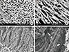
SEM observations of the surface of the nHA-coated silk scaffolds revealed a combination of porous spongy morphology and lamellar structures. In cross-sections, the scaffolds were observed to exhibit a highly interconnected porous structure comprising circularly shaped pores (Fig. 2A). Longitudinal sections showed an internal structure with lamellae mainly oriented parallel to the scaffold surface (Fig. 2B). SEM was used for assessing the results of the cell culture procedure. Fig. 2C and D show that cells were well spread out and attached to the surfaces of the scaffolds. Further, the formation of an ECM was observed. Such a response indicates good cytocompatibility and close interaction of the silk nanofibers with human PDLCs and DPCs.
Clinical findings
The scaffold-grafted defect sites showed minor exposure of scaffolds, and with the exception of three surgical wounds, were well closed upon stitch removal. All defect sites healed uneventfully with minimal signs of inflammation and without any clinical signs of postoperative complications including abnormal bleeding or infection following the surgery. After death, all scaffolds were retrieved with the surrounding soft and hard tissues intact.
Histologic findings
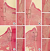
There was no significant difference in the apical extension of the junctional epithelium between the experimental groups and the control group. In the control group, the base of the defect showed slight bone formation along the root surface, whereas sites that received scaffolds showed minimal bone formation into the grafted area (Fig. 3A-D). The newly formed cementum was rarely observed, particularly at the experimental sites. Functionally oriented collagen fibers were found attached to the outer surfaces of denuded roots. In these areas of the experimental sites, collagen fibers originating from the space of the silk scaffolds were arranged obliquely or attached in parallel to the denuded root surface. The observed structures of the new attachments in this area did not differ significantly among the experimental groups (Fig. 3a-h).
The nHA-coated silk scaffolds occupied the spaces of the periodontal defect and were well maintained at all of the experimental sites. The tissues that had formed around all scaffolds resembled connective tissue rather than a bone-like tissue. After 8 weeks all experimental defects were surrounded by a thin fibrous capsule, which sporadically contained inflammatory cells. The upper surface of the silk scaffolds was covered with a periosteum-like connective tissue, with no infiltration by inflammatory cells (Fig. 3B-D).
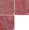
Most of the spaces within the silk scaffolds were filled with a loose connective-tissue matrix that contained a loose collagen fiber network and large numbers of fibroblasts. Cells and blood vessels were evident in all scaffolds loaded with PDLCs or DPCs. A small number of inflammatory cells were seen within the scaffolds. Blood vessels were occasionally observed at the periphery of the silk scaffolds but not in the central regions. These histologic characteristics in this area were observed in both the PDLC and the DPC groups. No hard-tissue formation was observed (Fig. 4).
DISCUSSION
In this study we developed nHA-coated silk scaffolds and cultured PDLCs or DPCs onto them so as to investigate their in vivo effect on periodontal regeneration. One of the aims of the study was to characterize the efficacy of nHA-coated silk scaffolds for one-wall intrabony periodontal defects. A previous study showed that a critical factor in determining the treatment outcomes is the number of bone walls remaining in intrabony periodontal defects; further, the regenerative potential of a three-wall defect is generally high, whereas that of a one-wall defect is low [21]. In addition, a one-wall intrabony defect has been shown to be a reliable model for evaluating regenerative periodontal therapies [24].
Wound healing and tissue regeneration were analyzed in this study after an 8-week healing period, which is sufficient to observe the initial healing process [21]. Many studies have used an 8-week healing interval to evaluate periodontal repair [25,26], and Choi et al. [27] reported no differences in bone regeneration by BMP between 8- and 24-week intervals.
The nHA-coated silk scaffolds in the present study did not cause any significant harmful side effects such as abnormal bleeding or infection following the surgery during the 8-week monitoring period in all the dogs. We prepared the silk scaffolds processed by extracting the sericin. To improve osteogenic outcomes and the structural rigidity, we combined these biodegradable polymers with an HA ceramic-based material. Such composite materials permit the fabrication of highly porous structures that allow cellular infiltration when culturing stem cells inside such scaffolds [28]. The histologic results obtained in the present study indicated that a well-maintained grafted volume was present at all experimental sites for 8 weeks. Further, the examination of the cell-seeded constructs by using SEM provided visual confirmation of the favorable characteristics of nHA-coated silk scaffolds for tissue engineering. The scaffolds exhibited a firm connective porous structure in cross section, and after the PDLCs and DPCs were seeded onto the scaffolds and cultured for 3 weeks, the attachment of the well-spread cells and the formation of ECM were observed. These results demonstrate that the nHA-coated silk-scaffolds architecture is suitable for the seeding and growth of PDLCs and DPCs.
We cultured PDLCs and DPCs in 3D scaffolds for 3 weeks. It is thought that the cell morphology resulting from differentiation in a 3D culture may be similar to that of in vivo differentiation because the microenvironment of a 3D culture resembles that of in vivo conditions [20]. Moreover, 3D cultures have recently been shown to be biologically more appropriate than 2D cultures for mimicking organogenesis in vitro [29,30]. This is consistent with a previous study finding that the osteoblastic differentiation efficiency was higher in a 3D culture than in a 2D culture [31]. Naito et al. [20] recently suggested that 3D culturing in a collagen hydrogel provides the advantage of the differentiation of mesenchymal stem cells into osteoblasts that have a similar phenotype to that of in vivo cells.
Fibrous capsules were more prominent than the regeneration of hard tissues such as cementum and bone in the scaffolds considered in this study. The minimal formation of hard tissue at the experimental sites may be attributable to the slow biodegradation process of a silk scaffold. Previous studies have found silk to be a biodegradable protein due to its susceptibility to proteolytic enzymes [32]; however, in most cases this process takes more than 60 days to complete [33]. Silks degrade very slowly, and thus, these systems maintain their open porous features longer, which will promote increased growth of cells and new tissue formation [34]. However, the rate of degradation must approximately match (or be less than) the rate of tissue growth. Achieving this balance assures appropriate physiological and mechanical compatibility during the integration of the host and the implanted tissue in vivo [33]; a mismatch in these rates may result in fibrous capsules interfering with the ingrowth of mineralized tissues into the scaffolds.
In addition to investigating silk scaffolds in general, we also attempted to compare their efficacies for the periodontal regeneration of two types of cells: PDLCs and DPCs. Previous analyses demonstrated that PDLCs and DPCs represent heterogeneous cell populations and can differentiate into various cell types. Seo et al. [35] reported that human PDL contains a population of multipotent postnatal stem cells that can be isolated and expanded in vitro, providing a unique reservoir of stem cells from an accessible tissue resource. A previous animal study found that PDLCs cultured in vitro could be successfully retransplanted into periodontal defects for periodontal regeneration [36]. Iwata et al. [37] transplanted three-layer PDLC sheets supported with polyglycolic acid onto dental root surfaces of three-wall periodontal defects and successfully demonstrated that PDLCs exhibit diverse differentiation properties, in terms of being able to regenerate periodontal tissues comprising hard and soft tissues [38]. Dental pulp tissue has also been regarded as a promising source of stem cells for research into bone defect repair and dental tissue engineering [39]. DPCs can promote the formation of reparative dentin by differentiating into odontoblasts and then producing a mineralized matrix. Wei et al. [17] isolated DPCs with stem-cell characteristics and confirmed their odontoblast-like differentiation potential.
While no study has compared the periodontal regenerative efficacies of PDLCs and DPCs, Park et al. [40] compared the regenerative potential of PDL stem cells (PDLSCs), periapical follicular stem cells, and dental pulp stem cells (DPSCs). They found that the extent of regeneration seen in the defects at 8 weeks after implantation did not differ significantly between those that received DPSCs and control defects that received no stem cells. These results suggest that DPSCs could play a role in bone and dentin regeneration; however, PDLSCs have a greater potential to form the bone, cementum, and PDL-like structures, and to enhance the overall periodontal regeneration. In contrast, there was no remarkable difference in periodontal regeneration among the PDLC group, DPC group, or even the NS group in our histologic results. This could be attributed to the fact that cells encounter difficulties when directly meeting a denuded root surface, particularly because of the prominent fibrous capsules around the silk scaffolds.
nHA-coated silk scaffolds were tested in this study, and it was found that these silk scaffolds have favorable biocompatibility and sufficient structural rigidity for periodontal regeneration. Although seeding and culturing of dental-derived cells onto these scaffolds were achieved, further investigations into how to control the degradation rate of the scaffold and how to effectively release cells that have been cultured on the scaffolds toward the denuded root surface are needed.
In conclusion, nHA-coated silk scaffolds could be useful biomaterials for periodontal regeneration. The in vivo effects of PDLCs and DPCs on different scaffold materials should be further studied in order to assess their ability to be applied in clinical periodontal regeneration.

 XML Download