

 PDF
PDF ePub
ePub Citation
 Print
Print


INTRODUCTION
Implant placement has become a widespread dental procedure to restore the edentulous jaw with functional defects. However, in many cases, insufficient vertical bone height of the residual ridge and poor bone quality give rise to difficulties in implant placement in the maxillary posterior area. This is partially due to the rapid progression of alveolar bone resorption and pneumatization of the maxillary sinus after tooth extraction. To overcome such anatomical and physiological problems, a sinus lift procedure, which was composed of a maxillary sinus membrane elevation step and bone graft step, was developed and has been applied widely in clinics. For maxillary sinus membrane elevation, either the lateral approach or the crestal approach is used depending on the bone height of the residual ridge.
When the crestal approach, which is known as the osteotome technique, was introduced, the crestal approach had the limelight among clinicians due to its many advantages in comparison with the lateral approach. First of all, the crestal approach is less aggressive than the lateral approach. Second, the crestal approach is a relatively simpler procedure and requires less time for wound healing. Nonetheless, the crestal approach has several drawbacks, in that the osteotome technique depends heavily on the skill of the clinician and causes ringing in the head of the patient due to malleting and maxillary sinus membrane perforation during malleting. Moreover, the osteotome technique gives rise to complications such as headache and vertigo after the sinus lift procedure [1-3]. Various surgical procedures and devices have been developed to overcome the shortcomings of the osteotome technique. Among these surgical procedures and devices, devices using hydraulic pressure for sinus membrane elevation have demonstrated a low risk of sinus membrane perforation as well as ease of application [4-10].
Recently, companies in the Republic of Korea have developed devices for the sinus lift procedure by the crestal approach using a special drilling system and hydraulic pressure. Although several newly developed sinus lift devices have been used widely and successfully, only a few reports on the devices developed in Korea have been published. Evaluating clinicians' opinions on the available sinus lift devices is undoubtedly important for the further development of safer and more user-friendly sinus lift devices.
The purpose of this study was to assess dentists' subjective satisfaction with the crestal approach sinus (CAS) kit (Osstem Implant Co., Busan, Korea), a newly developed device for maxillary sinus membrane elevation by the crestal approach using a special drilling system and hydraulic pressure. This paper also summarizes the sinus membrane perforation rate after the sinus lift procedure with the CAS kit.
MATERIALS AND METHODS
This study was carried out with the approval of Seoul National University Bundang Hospital Institutional Review Board (B-1205-156-302). The 30 dental clinicians who had experiences of dental implant placement after the sinus lift procedure with the CAS kit from June 2010 to May 2012 were included in this study.
The questionnaire was sent to the respondents and returned (Supplementary material). The questionnaire was composed of two main parts. The first part was related to the sinus membrane perforation rate. The second part was related to the dentists' subjective satisfaction with the CAS kit. For comparison with other sinus lift devices, the dentists were asked what sinus lift devices they preferred. Additional questions solicited users' opinions and advice on hydraulic membrane elevation for the development of safer and more user-friendly devices.
Features of the CAS kit
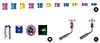
The CAS kit contains two different types of drills. The twist drill is used for the initial drilling and can be connected with a stopper. Stoppers of various lengths are provided, ranging from 2.0 mm to 12.0 mm (Fig. 1A). The twist drill can drill down to about 2.0 mm from the sinus floor, and its recommended speed is 1,000-1,500 rpm. The other type of special drill is named the CAS drill. Since the CAS drill tip is in conical form, a conical hole in the bone is formed after drilling. The CAS drill allows the dentist to elevate the sinus membrane safely. In addition, since the CAS drill makes the lateral side round, it can be used safely for maxillary sinuses in various forms. The CAS drill provides the additional function of collecting autogenous bone, and its recommended speed is 400-800 rpm (400-600 rpm recommended for beginners) (Fig. 1B). The depth gauge can measure the residual bone height and check membrane elevation. It can also be connected with a stopper (Fig. 1C).
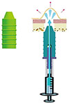
The hydraulic lifter is connected with a 1.0-mL syringe filled with saline solution. In the case of a single implant, saline solution of 0.2-0.3 mL elevates the membrane by about 3.0 mm (Fig. 2).
For the bone graft procedure, the bone carrier, condenser, and spreader are used The bone carrier has two different diameters: 3.5 mm and 3.9 mm. It is filled with particulate bone (Fig. 3A). The bone condenser fills a drilling hole with bone graft material to elevate the sinus membrane (Fig. 3B). The bone spreader spreads the bone graft material to the lateral part of the maxillary sinus to elevate the membrane. Using it at a low speed of 30 rpm is recommended (Fig. 3C).
Surgical procedure
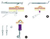
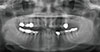
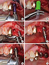
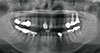
According to the usual procedure, the oral cavity was disinfected and local anesthesia was performed. Afterward, an incision was made along the alveolar crest, and the flap was elevated. Following the manufacturer's guideline strictly, the twist drill with a diameter of 2.0 mm was used to drill down to as much as 2.0 mm from the residual bone. For safe drilling, the twist drill was connected with the stopper before drilling was begun. The diameter of the drill was increased in consecutive order with the stopper still connected, considering the diameter of the implant to be placed. Maintaining a drilling speed of 800 rpm was recommended. Before performing the final drilling, the drill was connected with a stopper of the same height as the premeasured height of residual bone and the maxillary sinus membrane was elevated. The depth gauge was inserted to check the penetration through the maxillary sinus membrane. The hydraulic lifter was inserted into the drilled hole and 0.3 mL saline solution was injected slowly with the 1.0-mL syringe to elevate the maxillary sinus membrane. The bone carrier and the bone condenser were used to fill the hole with bone graft material. The speed of the bone spreader was maintained at 30 rpm to spread the material to the lateral part. The implant was placed using the self-tapping method, and the healing abutment was connected. Finally, the wound was sutured (Figs. 4, 5, 6).
RESULTS
Among the dentists from 30 dental clinics, 28 dentists answered the questionnaire. The CAS kit was used in a total of 924 implant cases combined with sinus membrane elevation, and sinus membrane perforation occurred in 38 implant cases (4.1%). Among the 28 respondents, only 13 dentists experienced sinus membrane perforation.
In response to the question on the preferred devices for sinus membrane elevation by the crestal approach other than the CAS kit, 26 dentists (92.9%) reported preferring an osteotome (Table 1).
General satisfaction with the CAS kit compared with the preferred devices
Among a total of 28 dentists who responded to the survey, 26 dentists (92.9%) reported that they were satisfied or very satisfied with the CAS kit. One dentist was dissatisfied with the CAS kit, pointing out that it was less efficient in sinus membrane elevation in comparison with other devices (Table 2).
Satisfaction with the cutting performance of the CAS drill
Among the 28 respondents, 23 dentists (82.1%) were satisfied or very satisfied with the cutting performance, with no respondent expressing dissatisfaction (Table 2).
Satisfaction with the stopper, depth gauge, bone carrier, bone condenser, and bone spreader
With regard to the use of the stopper, 17 dentists (60.7%) answered that there was no interference by adjacent teeth. On the other hand, 39% of the respondents replied that such interference caused problems during surgery. As for the depth gauge, 22 dentists (78.6%) answered that the depth gauge was useful in easily checking the sinus membrane. In terms of the bone carrier, bone condenser, and bone spreader, 15 dentists (53.6%) were satisfied with their functions, whereas 12 dentists (42.9%) did not find them to be different from those of other devices. One dentist was dissatisfied, pointing out that the bone carrier broke down frequently, and that filling the hole with the bone graft material was difficult (Table 2).
The advantages of the CAS kit
At least 24 dentists (85.7%) cited the safety, cutting performance, and user-friendliness of the CAS drill as the main advantage of the CAS kit. Only two dentists considered the function of autogenous bone collection of the CAS drill to be an advantage (Table 3).
Survey on the use of the hydraulic lifter for sinus membrane elevation with the CAS kit
Among the 28 dentists, only 21 dentists (75.0%) used the hydraulic lifter routinely. Seven dentists (25.0%) tended to skip the sinus elevation procedure using the hydraulic lifter. Ten dentists had used the hydraulic lifter 10 times or less. Only 2 dentists had used the hydraulic lifter more than 30 times (Table 4). Among those who used it, 15 dentists (71.4%) were satisfied or very satisfied with the hydraulic lifter for sinus membrane elevation, whereas 4 dentists (19.0%) did not sense any difference from other devices. On the other hand, 2 dentists (9.5%) expressed dissatisfaction, pointing out that they could not monitor the hydraulic pressure or sinus membrane elevation during the injection of saline solution, and that they were concerned about the risk of sinus membrane perforation (Table 2).
DISCUSSION
Since the osteotome technique was proposed by Summers [11] in 1994, it has been applied widely with the advantages that it requires a less complicated procedure and a shorter healing time than the conventional lateral approach. In addition, the osteotome technique was found to compact cancellous bone in the low-bone density area while elevating the sinus membrane so that bone quality could be improved. However, if used improperly, the osteotome technique may cause compression necrosis or fracture of cortical bone, and consequently, patients may suffer from headache or damage of the inner ear after a sinus lift procedure. In addition, because of the limited view of the surgical field during the entire procedure, the osteotome technique thoroughly depended on the dentists' skills and senses [12-15]. Because the osteotome technique may perforate the maxillary sinus membrane or form an excessive bony cavity at the implant placement area, there is a risk of instability of the implant in the initial stage as well as postoperative complications. Lalo et al. [5] proposed a device for reducing the sinus membrane perforation by an osteotome and drilling with a stopper, whereas Tilotta et al. [7] reported on a surgical procedure using an osteotome equipped with a trephine bur and stopper. The perforation rate in sinus membrane elevation using the osteotome technique was reported to be 0-21.4% (mean, 3.8%), and the 3-year survival rate of the implants placed in the sinus lift area was reportedly 87.4-96.0% (mean, 92.8%) [16]. Kolhatkar et al. [13] and Tetsch et al. [17] reported a 97.0-97.1% success rate of the implant placed in the sinus lift area with a crestal approach. Nkenke et al. [18] suggested that the sinus membrane elevation be limited on average to 3.0±0.8 mm using the osteotome technique to prevent perforation. As another limitation of the osteotome technique, at least 5.0 mm residual bone height is recommended to fix the implant properly in the initial stage. On the sinus membrane elevation and implant placement in relation to the residual bone height, a clinical guideline recommending that the lateral approach be used with delayed implant placement was suggested for less than 4.0 mm of residual bone height, the lateral approach and simultaneous implant placement for 4.0-6.0 mm, and the crestal approach for more than 5.0-6.0 mm [19]. The residual bone height is the most important factor for the success of a sinus bone graft. It is the present author's opinion that the sinus membrane can be elevated safely through the crestal approach with a bone height averaging 3 mm.
To overcome the shortcomings of the osteotome technique, various devices and surgical procedures have been developed. Kfir et al. [8] and Soltan and Smiler [20] introduced a minimally invasive method of maxillary sinus membrane elevation with balloon insertion into the hole and inflation after drilling down to the maxillary sinus floor and reported good clinical outcomes with the advantages of a low risk of sinus membrane perforation and short surgery time. Hu et al. [21] performed sinus membrane elevation using a water balloon with a balloon inflation volume of 0.67±0.17 mL and an elevation of 10.9±2.06 mm in radiological assessment and reported 2 sinus membrane perforation cases. Many researchers have introduced hydraulic sinus lift procedures. Piezoelastic internal sinus irrigation, water from a high-speed handpiece, metronidazole, and normal saline were used as fluid for hydraulic pressure, which places equal pressure on all surfaces, eliminating "point sources" of pressure and gently elevating the sinus membrane equally at all points of attachment. It was reported that hydraulic sinus lift procedures could reduce the sinus membrane perforation rate significantly [9,10,22-25].
Various minimally invasive sinus lift devices on the market could be grouped according to the drilling speed. High-speed drilling is applied when using the sinus crestal approach (SCA, NeoBiotech, Seoul, Korea), dentium advanced sinus kit (Dentium, Suwon, Korea), Samuel Lee's internal sinus graft system (Megagen, Daegue, Korea), and Santa system (Dentis, Daegue, Korea). On the other hand, low-speed drilling is recommended for the Hatch reamer (Sinustech America, Calabasas, CA, USA), cowellmedi sinus lift kit (Cowellmedi Co., Busan, Korea), disc-up sinus reamer (Dentimate Co., Seoul, Korea), sinu-lift system (Innovative Implant Technology, Aventura, FL, USA), bone compression kit (MIS, Tel-Aviv, Israel), and sinus master (Mr. Curette Tech., Seongnam, Korea). The Dr. Cosci drill (Dentech Co., Tokyo, Japan) and sinus lift drill (SSI, Seongnam, Korea) are devices that allow for both high- and low-speed drilling. Kang and Lee [26] and Cho et al. [3] reported that sinus membrane elevation using the Hatch reamer showed a very high success rate with rapid sinus membrane elevation and reduced the sinus membrane perforation rate. Lee and Kim [27] reported that quick and safe sinus membrane elevation could be possible using the SCA kit, a high-speed drill with a special blade, even at the septum area, reducing the risk of sinus membrane perforation.
In this study, the result of the survey on dentists' general satisfaction with the CAS kit showed that 92.9% of dentists were satisfied or very satisfied with it compared with their preferred devices. Other than the CAS kit, the preferred device was reported to be the conventional osteotome. This was partly due to the dentists' familiarity with the osteotome because the osteotome has been in use for a long time.
The 85.7% of the respondents cited the safety, cutting performance, and ease of use of the CAS drill as the strengths of the CAS kit. The CAS drill in the kit was designed to form a conical bone hole and to elevate the membrane safely with good bone cutting performance using either low- or high-speed drilling. On the other hand, only 57.1% of the dentists were satisfied with the bone carrier and bone condenser of the CAS kit, while the rest found no difference from their preferred devices.
The CAS kit was originally designed to elevate the maxillary sinus membrane safely using hydraulic pressure. However, only 75.0% of dentists routinely used the hydraulic lifter for hydraulic sinus membrane elevation. The sinus membrane perforation rate reported by the respondents was 4.1% in this study. Although the sinus membrane perforation rate was not high, the dentists requested a safer and more convenient procedure for additional reduction of the perforation rate. Thus, for the easy application of hydraulic pressure, it is necessary to develop a more user-friendly hydraulic lifter.
In conclusion, most of the dentists we surveyed were generally satisfied with the CAS kit, and the cutting performance and safety of the drill component was reported to be a strength of the CAS kit. Although hydraulic sinus membrane elevation was reported to be safe and convenient, it seemed that the hydraulic lifter in the CAS kit was not a very user-friendly component. The respondents to the survey desired further developments or modifications of sinus lift devices to make them safer and more user-friendly.


 XML Download