Journal List > J Periodontal Implant Sci > v.43(1) > 1082393

Bayarchimeg, Namgoong, Kim, Kim, Kim, Kim, Seol, Lee, Ku, Rhyu, Lee, and Koo: Evaluation of the correlation between insertion torque and primary stability of dental implants using a block bone test

Abstract

Purpose

Implant stability at the time of surgery is crucial for the long-term success of dental implants. Primary stability is considered of paramount importance to achieve osseointegration. The purpose of the present study was to investigate the correlation between the insertion torque and primary stability of dental implants using artificial bone blocks with different bone densities and compositions to mimic different circumstances that are encountered in routine daily clinical settings.

Methods

In order to validate the objectives, various sized holes were made in bone blocks with different bone densities (#10, #20, #30, #40, and #50) using a surgical drill and insertion torque together with implant stability quotient (ISQ) values that were measured using the Osstell Mentor. The experimental groups under evaluation were subdivided into 5 subgroups according to the circumstances.

Results

In group 1, the mean insertion torque and ISQ values increased as the density of the bone blocks increased. For group 2, the mean insertion torque values decreased as the final drill size expanded, but this was not the case for the ISQ values. The mean insertion torque values in group 3 increased with the thickness of the cortical bone, and the same was true for the ISQ values. For group 4, the mean insertion torque values increased as the cancellous bone density increased, but the correlation with the ISQ values was weak. Finally, in group 5, the mean insertion torque decreased as the final drill size increased, but the correlation with the ISQ value was weak.

Conclusions

Within the limitations of the study, it was concluded that primary stability does not simply depend on the insertion torque, but also on the bone quality.

INTRODUCTION

Implant stability at the time of surgery is crucial for the long-term success of dental implants. Primary stability is considered of paramount importance to achieve osteointegration [1]. Primary implant stability can be defined as a function of local bone quality and quantity, the geometry of an implant, the placement and surgical technique used, and the precise fit in the bone. Thus, primary implant stability is considered a significant parameter in achieving osseointegration, and the orchestration of the elements mentioned is crucial for the long-term success of the implant [2,3].
Two main factors that influence primary stability of an implant during placement are the amount of bone-implant contact and the role of compressive stresses at the implant-tissue interface. Such stresses may be beneficial for enhancing the primary stability of an implant, but they can reach a sufficiently high level to result in necrosis and local ischemia of the bone at the implant-tissue interface [4-6]. In the same respect, secondary stability can also be determined by the bone tissue response to the surgical trauma and the implant surface. The response is ultimately bone formation and remodeling at the implant interface leading to increased fixation and stability of the implant, although bone resorption resulting in implant failure can also occur during the initial healing period. In the literature, it is clear that surface roughness may result in more bone at the implant interface as well as a higher resistance to torque. Poor fixation may lead to micromovements during implant healing, which can potentially cause fibrous encapsulation and are associated with higher failure rates. Shorter healing periods are usually needed for implants with good primary stability. On the other hand, implants with poor stability need longer healing periods to achieve sufficient gain in secondary stability. This suggests the possibility of determining the length of the healing period on an individual basis, making implant treatment safer, more effective, and less time-consuming in some cases [7].
Generally, clinicians evaluate primary stability using the percussion test or using their own perception during the insertion process. However, the lack of precision has motivated the development of different methods to objectively evaluate primary stability; in particular, peak insertion torque (IT) and resonance frequency analysis (RFA) are the most used globally. Clinically, RFA values or implant stability quotient (ISQ) values have been correlated with changes in implant stability during osseous healing. Thus, IT and ISQ values are thought to have a positive correlation [8,9]. However, the formula of higher IT torque translating into higher primary stability may not always be true because the quantity and quality of bone varies significantly among patients. Therefore, the purpose of the present study was to investigate the correlation between IT and primary stability of dental implants using artificial bone blocks with different bone densities and compositions to mimic different circumstances that are encountered in routine daily clinical settings.

MATERIALS AND METHODS

Bone specimens

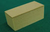
The bone block of solid rigid polyurethane foam (Sawbones, Vashon, WA, USA) with various bone densities (cancellous bone: #10, #20, #30; cortical bone: #50; and homogeneous bone: #10, #20, #30, #40) were used in the present study (Fig. 1). Because the mean bone mineral density was 0.31 g/cm3 for the posterior maxilla and 0.55 g/cm3 for the anterior maxilla, polyurethane foam blocks with a bone density of 0.48 g/cm3 were chosen. Short fiber-filled epoxy sheets were used as a substitute for cortical bone. Because the mean cortical thickness for the mandible was 2.22±0.47 mm and the mean cortical bone thickness for the maxilla was 1.49±0.34 mm, the sheets with a corresponding thickness were selected [10]. The following five different cortical thicknesses were used: blocks without a cortical layer (only homogeneous bone), blocks with a 0.5 mm cortical thickness, blocks with a 1.0 mm cortical thickness, blocks with a 1.5 mm cortical thickness, and blocks with a 2.0 mm cortical thickness.

Experimental design

The experimental group under evaluation was subdivided into 5 subgroups according to the objectives. In group 1, the correlation between IT and implant stability according to the bone density was evaluated. Implants were placed in homogeneous bone blocks with different bone densities (#10, #20, #30, and #40) following osteotomy preparation with a final drill diameter of Ø3.6 mm. An increase in numerical size of the bone blocks represented an increase in the bone density. Measurement of the IT and ISQ values were repeated 20 times for each bone density. In group 2, the correlation between the IT and implant stability according to the size of the final drill diameter used was evaluated. Homogeneous bone blocks with a density of #20 were used.
The final drill diameters under evaluation were Ø2.7 mm, Ø3.0 mm, Ø3.3 mm, and Ø3.6 mm. The remaining process was identical to that for group 1. The objective of group 3 was to evaluate the correlation between the IT and implant stability according to the thickness of the cortical bone. Using bone blocks with a cortical density of #50 and cancellous density of #20, the cortical bone was manipulated to have a thickness of 0.5 mm, 1.0 mm, 1.5 mm and 2.0 mm. Again, the final drill diameter was Ø3.6 mm and identical steps were performed.
The objective of group 4 was to evaluate the correlation between the IT and implant stability according to the cancellous bone density by controlling the cortical bone thickness to 1.5 mm and using block bones with a uniform cortical density of #50. Only the density of the cancellous bone (#10, #20, and #30) was different in the bone blocks for group 4.
In group 5, the correlation between the IT and implant stability according to the size of the final drill diameter in the bone blocks with a uniform cortical thickness of 1.5 mm was evaluated. The density of the cortical bone (#50) and the cancellous bone (#20) was controlled. The final drill diameters were Ø3.0 mm, Ø3.3 mm, Ø3.6 mm, and Ø3.8 mm.

Osteotomy preparation and fixture installation

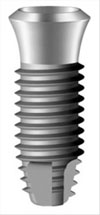
All of the osteotomies were prepared with a gentle surgical technique using a surgical drill at a rotational speed of 800 rpm with opious external cooling. The drill was fixed in a standard drilling set equipped for constant drilling. After drilling, the implant fixtures (Osstem Implant Co., Seoul, Korea) with a length of 11.5 mm and a diameter of 4.1 mm were placed in the prepared osteotomies. The diameter of the final drill was chosen and assigned according to the test protocol. The following diameters were used: Ø2.7 mm, Ø3.0 mm, Ø3.3 mm, Ø3.6 mm, and Ø3.8 mm (Fig. 2).

IT and RF measurements

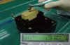
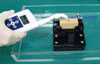
During installation, the peak IT was measured for all of the implants (Fig. 3). Following the final seating of the fixtures, the stability of each implant was measured in ISQ units using the Osstell Mentor (Osstell, Göteborg, Sweden). The RF values were represented in the ISQ on a scale from 1 to 100 and were averaged for each implant (Fig. 4). Each measurement was performed up to 20 times [11-15].

Statistical analysis

The IT and ISQ values were summarized using means and standard deviations. One-way analysis of variance was used to compare the mean IT and ISQ values. The Pearson's correlation coefficient was used to evaluate the correlation between the IT and the ISQ at implant placement. A P-value of <0.05 was considered statistically significant. Statistical analysis was performed using the IBM SPSS ver. 20.0 (IBM Co., Armonk, NY, USA) [16-22].

RESULTS

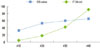
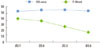
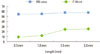
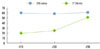
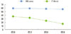
In group 1, the mean IT and ISQ value increased as the density of bone blocks increased, respectively (P=0.00 and P=0.00). The correlation between the two parameters was evaluated using Pearson's correlation coefficient, and the results suggested a positive correlation (CQ=0.82, P=0.00), which was statistically significant (Table 1, Fig. 5). For group 2, the mean IT values decreased as the final drill size expanded, but this was not the case for the ISQ values. The mean ISQ values showed limited change as the drill size increased. The correlation coefficient between the IT and ISQ value was -0.07 with a P-value of 0.77, but it was considered non-significant (Table 2, Fig. 6). The mean IT values in group 3 increased according to the thickness of the cortical bone, and the same was true for the ISQ values as well. It was also shown that the two parameters had a stronger linear relationship (CQ=0.84) with the values reaching statistical significance (P=0.00) (Table 3, Fig. 7). For group 4, the mean IT values increased as the cancellous bone density increased (P=0.00), but the correlation with the ISQ values was weak (CQ=0.45) and nonsignificant (P=0.09) (Table 4, Fig. 8). Finally, in group 5 the mean IT decreased according to the final drill size (P=0.00), but the correlation with the ISQ value was weak (CQ=0.57) and was non-significant (Table 5, Fig. 9).

DISCUSSION

The aim of the present study was to investigate the correlation between the IT and primary stability of dental implants through the IT test, and RFA analysis was performed using artificial bone blocks that simulated different circumstances that can ioccur in clinical practice. In general, primary implant stability is considered the most important factor in a successful implant treatment, and the distinct ranges of implant primary stability have been distinguished by the resonance frequency method [23-27]. Thus, RFA was used instead of the Periotest as a method to measure implant stability.
The results of the present study showed that the IT and initial stability increased according to the increase in the bone density, resulting in a strong positive correlation. In other words, the initial stability was shown to be highly dependent on the bone density. The IT also increased according to the thickness of the cortical bone, and a slight increase was observed for initial stability. This shows that the volume of high dense cortical bone affects the initial stability and it corroborates a recent study in which the same artificial bone model was used. In that study, an increase in the mean IT values was observed when the bone blocks with only trabecular bone (without a cortical layer) were compared to the groups with a cortical layer of 1 mm, 1 to 2 mm, and 2.5 mm [10].
The results from group 4 did not deviate much from group 2, which may suggest that the density of cancellous bone may have an impact on IT, but may have a limited effect on primary stability. This may indicate that the thickness of cortical bone or the cortical outer layer functions as the primary determinant for primary stability. This was also reflected in the results of groups 2, 4, and 5, where the correlation between the two parameters was weak. Therefore, the only factors that showed a positive correlation between the IT and the ISQ value were the bone density and thickness of the cortical bone.
In testing the impact of the final drill size, the IT decreased as the drill size expanded, but the initial stability showed limited change. This showed that initial stability cannot be acquired by simply reducing the diameter of the final drill in attempts to increase the IT. Biologic and anatomical consequences such as the thickness of cortical bone seem to be significant factors affecting primary stability, and estimation of bone density and the optimal selection of drill size are important.

Figures and Tables

Figure 1
Photographic presentation of the bone block with various bone densities that were used in the present study.
jpis-43-30-g001
Figure 2
Photographic presentation of the fixture type used in the present study (SS II fixture, Osstem Implant Co.).
jpis-43-30-g002
Figure 3
Torque measurement at fixture placement.
jpis-43-30-g003
Figure 4
Measurement of the implant stability quotient values using the Ostell Mentor (Osstell).
jpis-43-30-g004
Figure 5
A diagram presenting the correlation between the insertion torque (IT) and implant stability quotient (ISQ) values according to the various bone densities tested in the present study. The insertion torque increased according to the bone density, and the same was observed for the ISQ. The two variables appeared to have a strong positive correlation, which was statistically significant (correlation coefficient=0.82).
jpis-43-30-g005
Figure 6
Diagram illustrating the relationship between the insertion torque and implant stability quotient (ISQ) according to the various drill sizes under evaluation. The insertion torque (IT) decreased when the correlation with the ISQ value was weak and nonsignificant (correlation coefficient=0.07).
jpis-43-30-g006
Figure 7
The insertion torque (IT) increased according to the thickness of the cortical bone, as did the implant stability quotient (ISQ) value. The two variables had a stronger positive correlation (correlation coefficient=0.84) with statistically significant values.
jpis-43-30-g007
Figure 8
Insertion torque (IT) increased according to the bone density, while the correlation with the implant stability quotient (ISQ) value was weak and non-significant (correlation coefficient=0.45).
jpis-43-30-g008
Figure 9
Insertion torque (IT) decreased according to the size of the final drill, but the correlation (correlation coefficient=0.57) with the implant stability quotient (ISQ) value was weak and nonsignificant.
jpis-43-30-g009
Table 1
Correlation between the insertion torque and implant stability according to the bone density.
jpis-43-30-i001

SD: standard deviation, ISQ: implant stability quotient, ANOVA: analysis of variance.

a)ANOVA.

Table 2
Correlation between the insertion torque and implant stability according to the final drill diameter.
jpis-43-30-i002

SD: standard deviation, ISQ: implant stability quotient, ANOVA: analysis of variance.

a)ANOVA.

Table 3
Correlation between the insertion torque and implant stability according to the thickness of the cortical bone.
jpis-43-30-i003

SD: standard deviation, ISQ: implant stability quotient, ANOVA: analysis of variance.

a)ANOVA.

Table 4
Correlation between the insertion torque and implant stability according to the cancellous bone density with cortical bone.
jpis-43-30-i004

SD: standard deviation, ISQ: implant stability quotient, ANOVA: analysis of variance.

a)ANOVA

Table 5
Correlation between the insertion torque and implant stability according to the final drill diameter with cortical bone.
jpis-43-30-i005

SD: standard deviation, ISQ: implant stability quotient, ANOVA: analysis of variance.

a)ANOVA

ACKNOWLEDGEMENTS

Our special thanks to Osstem Implant Co. (Seoul, Korea) for the technical support to conduct this study.

Notes

Authors Byung Kook Kim and Myung Duk Kim are researchers employed under Osstem Implant Co. (Seoul, Korea).

References

1. Degidi M, Daprile G, Piattelli A, Iezzi G. Development of a new implant primary stability parameter: insertion torque revisited. Clin Implant Dent Relat Res. 2011; 10. 18. [Epub]. http://dx.doi.org/10.1111/j.1708-8208.2011.00392.x.
crossref
2. Dilek O, Tezulas E, Dincel M. Required minimum primary stability and torque values for immediate loading of mini dental implants: an experimental study in nonviable bovine femoral bone. Oral Surg Oral Med Oral Pathol Oral Radiol Endod. 2008; 105:e20–e27.
crossref
3. Stacchi C, Vercellotti T, Torelli L, Furlan F, Di Lenarda R. Changes in implant stability using different site preparation techniques: twist drills versus piezosurgery: a single-blinded, randomized, controlled clinical trial. Clin Implant Dent Relat Res. 2011; 04. 19. [Epub]. http://dx.doi.org/10.1111/j.1708-8208.2011.00341.x.
crossref
4. Nedir R, Bischof M, Szmukler-Moncler S, Bernard JP, Samson J. Predicting osseointegration by means of implant primary stability. Clin Oral Implants Res. 2004; 15:520–528.
crossref
5. Isoda K, Ayukawa Y, Tsukiyama Y, Sogo M, Matsushita Y, Koyano K. Relationship between the bone density estimated by cone-beam computed tomography and the primary stability of dental implants. Clin Oral Implants Res. 2012; 23:832–836.
crossref
6. Bilhan H, Geckili O, Mumcu E, Bozdag E, Sunbuloglu E, Kutay O. Influence of surgical technique, implant shape and diameter on the primary stability in cancellous bone. J Oral Rehabil. 2010; 37:900–907.
crossref
7. Esposito M, Hirsch JM, Lekholm U, Thomsen P. Biological factors contributing to failures of osseointegrated oral implants. (II). Etiopathogenesis. Eur J Oral Sci. 1998; 106:721–764.
crossref
8. Degidi M, Daprile G, Piattelli A. Primary stability determination by means of insertion torque and RFA in a sample of 4,135 implants. Clin Implant Dent Relat Res. 2010; 09. 17. [Epub]. http://dx.doi.org/10.1111/j.1708-8208.2010.00302.x.
crossref
9. Degidi M, Perrotti V, Strocchi R, Piattelli A, Iezzi G. Is insertion torque correlated to bone-implant contact percentage in the early healing period? A histological and histomorphometrical evaluation of 17 human-retrieved dental implants. Clin Oral Implants Res. 2009; 20:778–781.
crossref
10. Tabassum A, Meijer GJ, Wolke JG, Jansen JA. Influence of surgical technique and surface roughness on the primary stability of an implant in artificial bone with different cortical thickness: a laboratory study. Clin Oral Implants Res. 2010; 21:213–220.
crossref
11. Turkyilmaz I. A comparison between insertion torque and resonance frequency in the assessment of torque capacity and primary stability of Brånemark system implants. J Oral Rehabil. 2006; 33:754–759.
crossref
12. Kahraman S, Bal BT, Asar NV, Turkyilmaz I, Tozum TF. Clinical study on the insertion torque and wireless resonance frequency analysis in the assessment of torque capacity and stability of self-tapping dental implants. J Oral Rehabil. 2009; 36:755–761.
crossref
13. Roze J, Babu S, Saffarzadeh A, Gayet-Delacroix M, Hoornaert A, Layrolle P. Correlating implant stability to bone structure. Clin Oral Implants Res. 2009; 20:1140–1145.
crossref
14. Huang HM, Lee SY, Yeh CY, Lin CT. Resonance frequency assessment of dental implant stability with various bone qualities: a numerical approach. Clin Oral Implants Res. 2002; 13:65–74.
crossref
15. Hong J, Lim YJ, Park SO. Quantitative biomechanical analysis of the influence of the cortical bone and implant length on primary stability. Clin Oral Implants Res. 2012; 23:1193–1197.
crossref
16. Dos Santos MV, Elias CN, Cavalcanti Lima JH. The effects of superficial roughness and design on the primary stability of dental implants. Clin Implant Dent Relat Res. 2011; 13:215–223.
crossref
17. Bischof M, Nedir R, Szmukler-Moncler S, Bernard JP, Samson J. Implant stability measurement of delayed and immediately loaded implants during healing. Clin Oral Implants Res. 2004; 15:529–539.
crossref
18. Trisi P, De Benedittis S, Perfetti G, Berardi D. Primary stability, insertion torque and bone density of cylindric implant ad modum Branemark: is there a relationship? An in vitro study. Clin Oral Implants Res. 2011; 22:567–570.
crossref
19. Moon SH, Um HS, Lee JK, Chang BS, Lee MK. The effect of implant shape and bone preparation on primary stability. J Periodontal Implant Sci. 2010; 40:239–243.
crossref
20. Hsu JT, Fuh LJ, Tu MG, Li YF, Chen KT, Huang HL. The effects of cortical bone thickness and trabecular bone strength on noninvasive measures of the implant primary stability using synthetic bone models. Clin Implant Dent Relat Res. 2011; 05. 20. [Epub]. http://dx.doi.org/10.1111/j.1708-8208.2011.00349.x.
crossref
21. Merheb J, Van Assche N, Coucke W, Jacobs R, Naert I, Quirynen M. Relationship between cortical bone thickness or computerized tomography-derived bone density values and implant stability. Clin Oral Implants Res. 2010; 21:612–617.
crossref
22. Khayat PG, Arnal HM, Tourbah BI, Sennerby L. Clinical outcome of dental implants placed with high insertion torques (up to 176 Ncm). Clin Implant Dent Relat Res. 2011; 05. 20. [Epub]. http://dx.doi.org/10.1111/j.1708-8208.2011.00351.x.
23. Molly L. Bone density and primary stability in implant therapy. Clin Oral Implants Res. 2006; 17:Suppl 2. 124–135.
crossref
24. Sim CP, Lang NP. Factors influencing resonance frequency analysis assessed by Osstell mentor during implant tissue integration: I. Instrument positioning, bone structure, implant length. Clin Oral Implants Res. 2010; 21:598–604.
crossref
25. Martinez H, Davarpanah M, Missika P, Celletti R, Lazzara R. Optimal implant stabilization in low density bone. Clin Oral Implants Res. 2001; 12:423–432.
crossref
26. Katsoulis J, Avrampou M, Spycher C, Stipic M, Enkling N, Mericske-Stern R. Comparison of implant stability by means of resonance frequency analysis for flapless and conventionally inserted implants. Clin Implant Dent Relat Res. 2012; 14:915–923.
crossref
27. Ostman PO, Hellman M, Sennerby L. Direct implant loading in the edentulous maxilla using a bone density-adapted surgical protocol and primary implant stability criteria for inclusion. Clin Implant Dent Relat Res. 2005; 7:Suppl 1. S60–S69.
crossref
TOOLS
ORCID iDs

Tae-Il Kim
https://orcid.org/http://orcid.org/0000-0003-4087-8021

Yang Jo Seol
https://orcid.org/http://orcid.org/0000-0002-2076-5452

Young Ku
https://orcid.org/http://orcid.org/0000-0001-5682-0604

Similar articles