

 PDF
PDF ePub
ePub Citation
 Print
Print


INTRODUCTION
Aggressive periodontitis is characterized by a rapid loss of periodontal attachment and alveolar bone. It commonly affects young adults [1-3]. As opposed to chronic periodontitis, the amount of biofilm and calculus accumulation in aggressive periodontitis is inconsistent with the severity and rate of progression of the periodontal destruction. These infections are subdivided into localized and generalized forms, according to the extent of the periodontal destruction [4]. The stringent age requirement used previously for diagnosis of early-onset periodontitis is no longer considered to be essential [5]. Even though there have been attempts to analyze aggressive periodontitis biochemically and microbiologically, there is no specific way to screen for the disease. Currently, early detection depends primarily on clinical and radiographic examination [1,5].
Many reports have discussed host susceptibility factors, including family aggregation, single nucleotide polymorphisms, polymorphonuclear neutrophils, antibodies to bacteria, smoking, stress, a local contributing factor (root morphology), and herpes virus infections [6]. Anatomic variations in teeth, such as cervical enamel projections, enamel pearls, intermediate bifurcation ridges, and root grooves have been regarded as etiologic cofactors in this destructive periodontal disease process [7]. It has been shown that molars are more vulnerable to attachment loss and are more frequently extracted [8-10]. Anatomical variations in molar root form may favor plaque retention in these teeth and may contribute to an unfavorable crown-root ratio, resulting in increased susceptibility to loosening when they are subjected to heavy occlusal force [11]. Meng et al. [6] indicated that root shape abnormalities can be a susceptibility factor in the development of aggressive periodontitis and suggested root shape classification. Kim [12] reported that the ratio of root abnormalities was 1.76 times higher in aggressive periodontitis patients than in normal patients.
The aim of this study was to explore root shape abnormalities based on Meng classification, to investigate the influence of root form abnormalities on periodontal attachment loss and to gather basic data to assist in the diagnosis and treatment of aggressive periodontitis.
MATERIALS AND METHODS
Subjects
From January 2010 to June 2012, a survey was conducted of all 3,284 periodontitis patients who visited the Department of Periodontology, Daejeon Dental Hospital, Wonkwang University School of Dentistry. To qualify for inclusion in the survey, patients were required to display clinical and radiographic signs of aggressive periodontitis (based on the criteria of the American Academy of Periodontology International Classification of 1999) (Table 1). The patients were also required to be less than 35 years old at the time of the survey. Patients aged 35-40 years old who had suffered from periodontal disease since age twenty were also included. Of the 3,284 patients surveyed, 66 patients were selected as subjects for this study. The average age was 34.32 (±4.04). The numbers of males and females were 50 (2.2%) and 16 (1.6%), respectively, (P>0.05) (Table 2). Of the 66 subjects, 37 (56.1%) were smokers. To participate in the study, each subject was required to have a family history of destructive periodontitis back at least one generation. All of the subjects were thoroughly informed about the procedure and gave written consent for inclusion in the study. This study was approved by the Institutional Review Board of Wonkwang University Dental Hospital (IRB No. WKD IRB 20110201).
Clinical examinations
Before clinical periodontal assessment, complete medical and dental histories were taken. The workup included clinical assessment and examination by a single examiner, including the probing depths, gingival recession (data not shown) at six points per tooth, and Löe's plaque index [13]. The probing pocket depth was measured from the free gingiva to the pocket base using a periodontal probe (PW, Hu-Friedy Manufacturing Co., Chicago, IL, USA). We measured the gingival recession from the cemento-enamel junction to the free gingiva with the same tool. We expressed periodontal attachment loss, defined as the length from the cemento-enamel junction to the depth of the pocket base, as the sum of the depth of the pocket and the gingival recession.
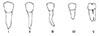
We classified roots according to the system published by Meng et al. [6]: cone root (type I), slender root (type II), curved root (type III), poor crown-root ratio (type IV), and syncretic root (type V) (Table 3, Fig. 1). We included the first and second premolars and the first and second molars of each arch in this root shape analysis study.
Tooth mortality was defined as the value obtained by adding the numbers of hopeless and missing teeth. The evaluation criterion for the hopeless category was an insufficient attachment level to maintain health, convenience, and function [14,15]. Following the clinical examination, full-mouth periapical radiographs were taken and analyzed for bone level, root deformity, or missing teeth using PiView STAR software (Infinitt Co., Seoul, Korea).
Statistical analysis
Data management procedures employed SPSS ver. 14.0. (SPSS Inc., Chicago, IL, USA). The tooth mortality and root form distribution were analyzed using the chi-square test. Periodontal probing depth and clinical attachment loss by root form was analyzed using one-way analysis of variance with Tukey or Dunnett T3 post hoc test.
RESULTS
There was no significant difference in prevalence of disease among smokers and nonsmokers (P>0.05) (Table 4). The plaque index averaged 1.7 (data was not shown).
The probing pocket depth was the deepest at the maxillary first molar and at the mandibular second molar (Table 5). The periodontal attachment loss was the highest at the first molar in both the maxilla and mandible (Table 6). The average number of missing teeth was 1.35 per subject. The tooth mortality was the highest among the maxillary first molars and the mandibular second molars (Table 7).
The results of root shape deformity analysis of bicuspids and molars showed type V deformity to be the highest in the second maxillary and mandibular molars. Type IV was the most common in the first mandibular molar (P<0.05). All five types of root shape deformity were seen in the maxillary second premolar only (Table 8). In a comparison of frequency of root form abnormalities, the maxillary incidence (16.7%) was higher than the mandibular incidence (10.9%), showing a significant difference (P=0.008) (Table 9). We analyzed the periodontal attachment loss of each tooth according to root form. In the second maxillary and mandibular molars, the type V root anomaly correlated with the highest periodontal attachment loss, 6.2 mm and 6.0 mm, respectively, among the deformity types, but not to a significant extent. There were no significant differences in attachment loss between the different deformities detected in the first and second premolars and first molars (Table 10). In the comparison between root shape abnormality and periodontal attachment loss regardless of tooth type, the type V root shape was associated with the highest periodontal attachment loss at 6.09±2.11 (P=0.01) (Table 11).
DISCUSSION
Although we did our best to identify aggressive periodontitis patients based on clinical and radiographic evidence, we could not confirm the subjects of this study to be aggressive periodontitis patients. Classification of periodontal disease and the criteria for distinction between the two major forms of periodontitis are listed in the Consensus Reports of the 1999 International Workshop. However, distinguishing between the two forms in clinical practice remains problematic. For example, we looked for the primary criteria of rapid attachment and bone loss to diagnose aggressive periodontitis. However, any evaluation of disease progression requires some inference of the condition's severity with respect to patient age. According to current classification principles, age should no longer constitute a primary determinant of diagnosis. Familial aggregation is also largely unhelpful [16]. A recent systematic review of the literature concluded that there is no evidence that the subgingival etiological factors in chronic periodontitis are substantially different from those of corresponding lesions in aggressive periodontitis [17]. Instead, it is generally accepted that the variation in susceptibility to periodontitis and types of disease are largely due to host factors [18]. In treating patients with aggressive periodontitis, therefore, early and aggressive treatment measures are more important than an investigation of etiology.
A review of the published literature suggests that the prevalence of aggressive periodontitis may vary significantly among countries and ethnicities. Albandar and Tinoco [19] reported that the prevalence of aggressive periodontitis was 10% in African-Americans, 5% in Hispanics, and 1.3% in white United States adolescents between 13 and 17 years of age. Low prevalence rates ranging between 0.1% and 0.2% have been reported in Europe [20]. In Japan, Kowashi [21] reported a 0.47% prevalence of aggressive periodontal disease in subjects between 19 and 28 years of age. The data from China showed the prevalence of aggressive periodontitis to vary between 0.12% and 0.47% in different areas of the country [22]. Of the surveyed initial 3,284 patients, 66 patients were provisionally assumed to have aggressive periodontitis (2.01% prevalence) with a mean age of 34.32, which was somewhat higher than that found in previous clinical studies [23]. It seems that this is because the subjects were patients who presented to the hospital rather than youths without symptoms. Considering an asymptomatic progression period and delay in patients visiting the hospital, we applied less strict criteria for age, especially since age is not a critical diagnostic criterion for aggressive periodontitis.
The gender ratio was male:female 3.13:1, but this was not a statistically significant difference. Studies are contradictory as to the predilection of periodontal disease according to gender. Differences in periodontal disease risk by sex have not been clearly demonstrated [24]. Baer [25] estimated a female:male ratio of about 3:1. Hormand and Frandsen [26] concluded that the disease affects females more often than males with a ratio of 2.5:1. Albandar and Rams [27] denied the existence of a relationship between aggressive periodontitis and gender. More studies are required to determine the gender ratio in aggressive periodontitis prevalence in Koreans.
In the present study, marked attachment loss over 5 mm was observed at the first molars. This result is consistent with our previous study [23]. It is assumed that the first molars might be affected earlier by periodontitis. Aggressive periodontitis developed as a localized form may progress into a generalized form with the involvement of more teeth with advancing age.
The average number of missing teeth was 1.35 at the first visit, but if we include the teeth diagnosed as hopeless, it doubled to 2.71. The most frequently missing teeth in the current study were the maxillary first molars (21.21%) and the mandibular second molars (15.15%). In the evaluation of tooth mortality, a high attachment loss of molars seems to lead to high tooth loss. We can assume that the high percentage of molar tooth loss might be because the molars had additional susceptibility factors such as occlusal interference, hygienic difficulty, and abnormal root form. Further epidemiologic studies of larger populations, comparing them with normal and chronic periodontitis subjects, are needed to assess the exact prevalence and pattern of aggressive periodontitis in Korea.
The shape, length, and spread of molar roots are important factors in tooth prognosis, as they can affect the anchorage and stability of molars. Molar root fusion is one of the most common anomalies in root morphological development [8,11]. Root fusion (type V) was the commonest anomaly in both the maxilla and mandible, and showed the highest prevalence in the second molars in this study. Ross and Evanchik [11] reported that a relatively high proportion of Europeans (70%) had one or more maxillary molars and 54% had one or more mandibular molars with fused roots. In the studies of Choi et al. [28] and Ryu et al. [29], root fusion demonstrated higher prevalence in the maxilla than mandible, and was the highest in the second maxillary molar. When we analyzed the degree of periodontal tissue destruction according to root shape, the second molars with root fusion (type V) had a greater risk of increased probing depth and periodontal attachment loss. In the correlation of root fusion and local inflammation, Choi et al. [28] reported a significance increase in clinical index-probing depth, gingival index, and tooth mobility in both the maxilla and mandible. Likewise, Hou and Tsai [8,30] described the correlation between root fusion and periodontal disease in a Chinese population. Negative characteristics of molars with root fusion, such as an unfavorable crown-to-root ratio, short root length, and a taper-shaped root, may offer less resistance to heavy occlusal loads and/or torque forces. This evidence suggests that molar root fusion may ultimately accelerate periodontal tissue destruction. However, Hou and Tsai's study was not conducted in patients with aggressive periodontitis. Given the similar results of the present study, we can cautiously infer that a fused root can be a contributing factor in disease progression of aggressive periodontitis as well as of chronic periodontitis.
In summary, periodontal attachment loss was the highest at the first molar in both the maxilla and mandible. Root fusion (type V) was the most prevalent among root abnormality types in both the maxilla and mandible, demonstrating the highest prevalence in the second molars. In the comparison between root shape abnormality and periodontal attachment loss regardless of tooth type, the type V root shape was associated with the highest attachment loss. Studies on the impact of root shape in patients with aggressive and chronic periodontitis are rare. Considering the small population and limited design of this study, definitive conclusions cannot be drawn. We suggest more large-scale, methodologically sophisticated studies that include normal controls and chronic periodontitis patients to clarify whether root form abnormalities are a potential risk factor for aggressive periodontitis.


 XML Download