

 PDF
PDF ePub
ePub Citation
 Print
Print


INTRODUCTION
The aim of nonsurgical periodontal debridement is to reduce plaque and calculus and create a relatively smooth root surface in order to achieve new attachment. Scaling and root planning are fundamental for periodontal treatment and hand/ultrasonic instrumentation was the only means for this purpose for years [1]. However, controversy remains between researchers who believe that manual instrumentation may lead to excessive root surface removal and others who report harmful effects of using ultrasonic scalers [2,3]. Among various determining factors for new attachment achievement, root surface smoothness following instrumentation and its effect on cell response have been discussed [4]. There are two methods for testing the above-mentioned controversies: by using a profilometer to assess root surface smoothness, and through quantitative and qualitative evaluation of surface ultrastructure by scanning electron microscopy [5].
Laser irradiation as an adjunct to traditional scaling and root planning has become popular recently and some studies suggest that lasers may improve root surface compatibility and facilitate adhesion of fibroblasts [6]. Researchers have demonstrated that although root surface roughness decreases following treatment with an erbium-doped yttrium aluminium garnet (Er:YAG) laser with an intensity of 120 mJ/pulse compared to manual instrumentation, some degree of unevenness and roughness remains on the root surface [7]. Therefore, some investigators have suggested the use of less powerful dental lasers to reduce mechanical and chemical side effects like carbonization.
During the past two decades, the application of lasers to dentistry has greatly increased for various purposes such as alleviation of dentinal hypersensitivity [8], increasing tissue healing following nonsurgical periodontal therapy [9], subgingival scaling and root planning [10], treatment and regeneration of osseous defects [11], photodynamic therapy [12], elimination of the inner epithelial wall of periodontal pockets [13], and tissue regeneration by stimulating the periodontal ligament's fibroblasts [14]. Nevertheless, it is crucially important to ensure the safety of laser treatment.
Quantitative assessment of root surfaces following laser treatment has been reported in the literature; however, the protocols used for treating the samples have differed from those used in routine clinical practice. A meta-analysis showed that erbium lasers would be better used as an adjunct rather than monotherapy for scaling [15]. The aim of the present study was to measure the root surface roughness of teeth with periodontitis following conventional instrumentation with and without Er:YAG laser irradiation.
MATERIALS AND METHODS
Sixty single-rooted maxillary and mandibular teeth with more than 5 mm attachment loss, extracted due to periodontal disease, were collected. The teeth had no history of root caries, fracture, or root canal therapy. The entire crown and apices of the roots were cut off using a diamond bur and water coolant. The specimens were mounted in an acrylic resin block in order to have an accessible plain root surface. The root surface roughness of all of the samples was determined at baseline with a profilometer (MahrSurf M300+RD18C system, Mahr GmbH, Göttingen, Germany) with a stylus tip radius of 2.5 µm. After primary evaluation and setting a baseline, the samples were divided into four groups.
Group 1
Fifteen roots were root planned using a #7.8 manual curette (Nordent Manufacturing Inc., Elk Grove Village, IL, USA). The treatment continued until reaching a smooth surface in probing by the operator.
Group 2
Fifteen roots were prepared with an ultrasonic scaler (Juya Electronic Co., Tehran, Iran) with the scaler intensity knob set to medium position and high water irrigation with 0-20° tip angulation using the same protocol.
Group 3
Fifteen roots were treated with hand instruments followed by Er:YAG laser irradiation (Smart 1240D plus laser, DEKA, Florence, Italy) at 10 Hz frequency with an intensity of 50 mJ and power of 50 W in "very short pulse" mode, 50-50% air-water coolant and a swiping motion for 45 seconds, and a maximum tip angulation of 20°.
Group 4
Fifteen roots were prepared with an ultrasonic scaler and subsequently treated with an Er:YAG laser according to the same protocol.
All of the procedures were carried out by an expert periodontist (R.A.) with 93% reproducibility based on the intraclass correlation coefficient index. The samples were sent to a laboratory and root surface roughness was measured by a blind operator with a profilometer under controlled laboratory conditions at 25℃ temperature and 41% humidity while keeping the probe at right angles to the surface during scanning.
RESULTS
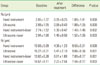
Baseline analysis found no significant differences among the groups in terms of surface roughness (Ra) or surface distortion (Rz) (P>0.2). Statistical tests revealed significant differences in each group before and after treatment. The highest changes were observed in groups 1 and 2 with a P-value of 0.000, while the smallest change was reported in group 3 (1.40±1.45, P=0.002). However, no statistically significant differences were observed among the four groups with P-values of 0.966 and 0.629 for Ra and Rz, respectively (Table 1).
In group1 (hand instrumentation), the mean surface roughness before and after intervention was 2.85±1.37 and 0.95±0.25, respectively which revealed root planning with a manual curette to be the most effective method for reduction of surface roughness. If we consider Rz to be the marker of surface distortion, group 2 (ultrasonic instrumentation) showed the highest changes in this respect. The average roughness in this group was 15.27±8.31 and 5.41±2.18 preintervention and postintervention, respectively (P=0.001). In terms of Ra, the smallest changes happened in group 3 (7.00±6.17) and the least significant changes occurred in samples treated with an ultrasonic instrument followed by laser irradiation (P=0.003).
The situation was a bit different for the other variable (Rmax). The lowest and the highest values for Rmax were reported after hand and ultrasonic+laser instrumentation. However, ultrasonic and hand instruments per se caused the most and least changes in Rmax (16.86±14.58 and 14.91±10.16, respectively). Similar to the results for Ra and Rz, the changes in Rmax before and after treatment were significant for all groups but the differences among the four groups were not statistically meaningful (P=0.973).
DISCUSSION
A significant number of studies have been seeking an ideal method for achieving a root surface with optimal conditions for cell response. Cell response as a biological term in nonsurgical periodontal therapy means new attachment with repopulation of gingival fibroblasts on the exposed root surfaces, although the physical characteristics of root surfaces after the procedure is another field of interest. The efficacy of conventional scaling and root planning with various periodontal hand and rotary instruments was examined by using a profilometer [16]. Some authors have used root roughness as the main criteria to explain biocompatibility as well as the cell response of different treatments [17]. A morphological analysis and roughness measurement done by Bolortuya et al. [18] showed that dentin surfaces treated with Er:YAG laser irradiation were rougher and subsequently exhibited the greatest number of attached fibroblasts among all experimental and control groups after 12 hours and 24 hours.
The effects of a laser on calculus removal and root planning have been the subject of interest for numerous research studies in the literature [19-23]. In comparison with hand/ultrasonic instrumentation, Er:YAG and erbium, chromium: yttrium, scandium, gallium, garnet (Er,Cr:YSGG) lasers can remove subgingival calculus with no adverse effects on tooth structures [24]. The results of several studies have suggested that use of an Er:YAG laser for scaling and root planning is more suitable than other types of lasers; however, controversy still exists on this subject [25].
In the present study, we evaluated the root surface roughness of teeth with three different variables (Ra, Rz, and Rmax) following four different protocols to determine ultrastructural changes. All four protocols (hand instrumentation, ultrasonic instrumentation, hand plus laser, and ultrasonic plus laser) reduced surface roughness, which would have, in turn, increased the probability of fibroblast adhesion and proliferation and have played a key role in periodontal healing [26]. The results of the current study revealed that minimal roughness resulted from hand and ultrasonic instrumentation (groups 1 and 2), while maximum roughness was achieved following ultrasonic instrumentation plus laser irradiation. However, the difference in this respect was not statistically significant.
Comparison of manual and ultrasonic instrumentation indicates that hand instrumentation produces a smoother surface than ultrasonic instrumentation, but this difference was not statistically significant. This result is comparable with previously published data [27-30].
Studies have shown that different lasers have various occluding effects on dentinal tubules. A recent in vitro study reported that the mean diameter of the dentinal tubule entrance after the application of Er,Cr:YSGG, 810-nm diode, CO2, and neodymium-doped:YAG lasers were 1.73, 3.27, 2.10, and 1.64 microns, respectively, compared with 3.52 microns before laser irradiation. However, dentinal tubule dimensions may be more important for root coverage procedures or desensitization rather than nonsurgical periodontal treatment on root surfaces where the exposed surface is mainly covered with cementum [31].
In conclusion, the Er:YAG laser as an adjunctive therapy to traditional scaling and root planning can decrease root surface roughness. However, the observed changes were not significantly different from those of traditional instrumentation.
 XML Download