

 PDF
PDF ePub
ePub Citation
 Print
Print


INTRODUCTION
The presence of the interdental papilla between two implants is an important factor for esthetics. The shape of the interdental papilla is determined by the contact relationships between the teeth, the width of the proximal tooth surfaces, and the position of the cemento-enamel junction [1].
The presence of the interdental papilla is determined by the alveolar bone height, the size of the interproximal space, the biotype of the gingiva, and the size of the contact area [2-7]. The bone loss around the implant, in particular, affects the height of the interproximal papilla between the implant and implant or implant and tooth [3,6,8].
In a single implant placement, the interdental papilla adjacent to the implant is well preserved and leads to a favorable esthetic result. The height of the interdental papilla is determined by the restoration environment [5]. The papilla adjacent to a tooth has a more favorable vertical soft tissue condition than the papilla adjacent to an implant. The papilla between implants has a 3 mm proximity limitation and the tip of the interdental papilla is only 3.5 mm in height, which is 2.5 mm less than that between the teeth.
In the cases of one stage implant surgery and the second surgery of two-stage implant surgery, the creation of interdental papilla is attempted using a soft tissue surgery method such as the Palacci technique, PK flap design, multiple Z-plasty, or connective tissue graft [5,9-11].
In the case of a lack of keratinized gingiva, interdental bone is often uncovered and exposed. Exposed bone will be covered by secondary healing. There are few clinical data on the difference of bone level change between exposed and unexposed bone. If exposed bone level is the same level of unexposed bone, clinician reduced the effort to cover the interdental bone using soft tissue surgery.
The purpose of this study was to evaluate the soft tissue and bone changes around two adjacent implants in one-stage implant surgery.
MATERIALS AND METHODS
Eleven subjects (7 males, 4 females), who ranged in age from 39 to 79 years (average, 53.6±7.59 years) were included from among the patients of the Department of Periodontology, the Institute of Oral Health Science, Samsung Medical Center (2006-07-012). Patients who needed 2 adjacent implants placed in premolar and molar area were recruited.
Exclusion criteria included heavy smoking, pregnancy or lactation, or suffering from a serious chronic medical condition (e.g., diabetes mellitus, kidney or liver disease). This study was performed in accordance with the requirements of the Samsung Medical Center Institutional Review Board. All patients signed informed consent forms.
The implants (Branemark MK III groovy) were placed according to the manufacturer's protocol. Briefly, a lingually positioned crestal incision was performed. Only buccal side mucoperiosteal flap was raised and two implants were placed with the platform at the level of the alveolar crest. The implant stability was evaluated using insertion torque and by feel during drilling. In case of good stability, we do one stage surgery. So we connect healing abutment and suture. If implant stability is not good, we do 2 stage surgery. We connect coverscrew and suture for full coverage of implant. The tooth side of the implant was fully covered with gingiva. However, the interproximal bone between the 2 implants could not be covered with gingiva and was exposed for the secondary healing procedure.
After surgery, an alginate impression was taken to record the gingival shape and radiographs were taken to evaluate implant placement. An alginate impression was taken again 4 weeks later. After 12 weeks, another alginate impression was taken and a radiograph was taken to evaluate alveolar bone change using extension cone paralleling for parallel technique.
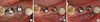
Using a master cast, the gingival height was measured using digital caliper (Digimatic caliper, Mitutoyo Co., Kawasaki, Japan). Gingival height was measured at 2 points: the distal side of the mesial implant (MiDs) and the mesial side of the distal implant (DiMs). The two measuring points are described in Fig. 1A.
On the radiograph, measurement was performed at the mesial and distal sides of both implants (mesial side of the mesial implant [MiMs]; MiDs; DiMs; distal side of the mesial implant) (Fig. 2). The alveolar bone level was measured using a method described previously [12]. In brief, the digitized images were analyzed using the Scion Image ver. 4.3 (Scion Co., Frederick, MD, USA). The distance from the fixture platform to the first bone contact was measured. The measurements were calibrated using the distance of the implant thread pitch.
Statistical analyses
Statistical analysis was performed with the help of an affiliated biostatistician. The results were analyzed using SAS ver. 8.2 (SAS Institute Inc., Cary, NC, USA). The changes in the gingival height and bone loss were tested using the Wilcoxon signed rank test with Bonferroni's correction or a paired t-test with Bonferroni's correction. The P-value was corrected by Bonferroni's method due to the multiple endpoint test. The significance level of both tests was P<0.05.
RESULTS
Eleven patients completed the study and no post-surgical complications were observed. The exposed bone had been covered with gingiva when patient visited 1 week after surgery.
Soft tissue change
The change of gingival levels of both implants was measured. Immediately after surgery, the MiMs was fully covered by gingiva and no bone exposure was found. However, the MiDs and DiMs were not fully covered by gingiva and alveolar bone was exposed. Soft tissue coverage was complete in all areas at 4 weeks. During the first 4 weeks, the thickness of soft tissue increased more than during the next 8 weeks. The gingival height of the MiDs and the DiMs increased to 1 mm. The gingival height of DiMs was 0.91 mm and 1.05 mm for 4 weeks and 12 weeks, respectively (P<0.05). The gingival height of the MiDs had also increased by 0.91 mm and 1.13 mm at 4 weeks and 12 weeks, respectively. However, the increases were not statistically significant (Table 1).
Alveolar bone level around the implants
The mean alveolar bone levels around the implants are presented in Table 2. The distance of two implants was 3.90±1.0 mm. All the alveolar bone levels of the 2 implants were reduced after a 12-week healing period.
No statistical difference was shown between baseline and 12 weeks in any of the 4 measurement sites around the implants. The alveolar bone loss of the MiMs increased from 1.28 to 1.46 mm during the 12 weeks (P<0.05). The change in the alveolar bone on the other sides of the implants did not show any statistical significance.
DISCUSSION
The original Branemark implant protocol was two-stage surgery. The implant was covered with soft tissue after the first surgery and protected from external force, infection, and the downward growth of the epithelium [13]. During this period, the implant achieved osseointegration. However, the International Team for Implantology group designed a one-stage implant and proposed one-stage surgery that requires no second surgery. Nowadays, the type of surgery is chosen according to the surgeon's preference, patient factors, alveolar bone condition, and implant stability.
To achieve an ideal esthetic and functional result, many soft tissue and hard tissue management techniques have been developed. It is well known that bone loss can be demonstrated around implants after implant placement [14]. This initial bone loss could be affected by surgical trauma, overload, peri-implantitis, microgaps, implant type, and disturbance of the biologic width [15,16].
Peri-implant bone loss could not been seen until the second surgery in two-stage surgery. After the second surgery, bacterial contamination of the gap between the implant and the healing abutment leads to peri-implant bone loss. Bone loss will progress until the biologic width has formed [15].
In the dog experiment by Berglundh and Lindhe [17] on the dimension of the transmucosal attachment, peri-implant bone resorption was observed in thin mucosa to establish space for the biologic width. However, the exposed alveolar bone area did not show any difference from the unexposed bone area in this study. During the 12-week healing period, a very small amount of alveolar bone loss was observed. The mean width of the exposed area was 2.90 mm at the baseline. This area was fully covered by a blood clot after treatment. This small area surrounded by gingival tissue is a good place for containing the blood clot. Early coverage of the exposed bone by a blood clot might reduce the alveolar bone loss.
The width of the alveolar ridge was wide enough to preserve the buccal and lingual sides of the bone because all implants were placed in the premolar and molar region. Five implants out of all 22 implants were placed in the molar area.
The short observation period of this study was not long enough for the biologic width to be formed by resorption of alveolar bone. Peri-implant bone loss will progress until the biologic width has created and stabilized. The peri-implant bone loss will progress both apically and horizontally [18]. Thus, future study designs should be based on an observation period longer than 3 months in order to accommodate the complete development of the biological width.
The limitations of this study were the small number of subjects and the lack of a control condition. This study showed that the alveolar bone level and gingival height around 2 adjacent implants in exposed bone areas did not differ from unexposed bone areas. Although this result showed that denudation of the gingiva did not affect alveolar bone loss, coverage of exposed bone using various techniques such as Palacci's technique is recommended. The coverage of exposed bone helps the papilla regeneration and improves the esthetic results.


 XML Download