

 PDF
PDF ePub
ePub Citation
 Print
Print


INTRODUCTION
Intentional replantation has been defined as "the act of deliberately removing a tooth and following some procedures, returning it to its original socket." [1]. Recent interest has focused on intentional replantation as an alternative for restoring an original tooth instead of replacing it with a prosthesis or implant. Some studies dealing with immediate replantation have shown successful results with intentional replantation for periodontally involved teeth. Lu [2] replanted a periodontally involved and endodontically mistreated mandibular first premolar and reported that the replanted tooth remained functional and asymptomatic for 32 months. Demiralp et al. [3] suggested that intentional replantation can be an alternative approach to extraction in cases where advanced periodontal destruction is present and no other treatments could be considered.
Although the above studies [2,3] have shown successful results with intentional replantation specifically for periodontally involved teeth, some complications should be considered. Common complications are root resorption and ankylosis that leads to a gradual resorption of the dental hard tissues and their replacement by bone [4,5]. To avoid postoperative complications, clinical and experimental studies have shown that a healthy periodontal status is of critical importance. With this understanding, two aspects could be considered. One is the extracted root surface and the other is the periodontium of the extraction socket.
Most studies have suggested not to touch the root surfaces of the tooth to be replanted if its periodontal ligament attachment is intact and sound [2,6-9]. However, our concern was the replantation of periodontally involved teeth that had deeper periodontal pockets, advanced bone loss, and a diseased periodontal ligament with necrotic cementum. These factors lead to the above-mentioned complications. Lindskog et al. [10] reported root surface resorption with or without accompanying ankylosis when the necrotic periodontal ligament was not removed prior to replantation of teeth. They suggested that denuding the root surface chemically prior to replantation of the teeth without a vital periodontal ligament would prevent resorption. Mahajan and Sidhu [11] also reported that the removal of the periodontal ligament raised the success rate of tooth replantation. This is thought to be due to either the effective elimination of a necrotic periodontal ligament and/or microorganisms prior to replantation.
Kratchman [6] suggested not touching the socket walls and rinsing with sterile saline only if necessary, because touching the walls of the socket might increase the risk of ankylosis. Furthermore, recent studies have shown that granulation tissues contained mesenchymal stem cells that were of help to healing of the socket. On the other hand, most clinicians perform curettage on the socket before replantation of periodontally involved teeth. It is generally agreed that the removal of granulation tissue raises the success rate of replantation [8,12]. In the absence of inflammation, "delayed replantation" could also be considered. Delayed replantation is to replant an extracted tooth after some time passes while a treatment is performed. This may decrease the complication rate by replantation after resolution of inflammation. The period between preparation of the recipient bed and replantation of the tooth also allows for improvement of the nutrition and preservation of the vitality of the cells of the remaining periodontal ligament and the root cementum. Although Nethander et al. [13] failed to show a difference between the immediate and delayed methods in terms of frequency of various types of root resorption, the teeth used in that study were not periodontally involved teeth. However, for periodontally involved teeth, delayed replantation may be more suitable. Nevertheless, no study has reported the optimum replantation timing during the healing process of an extraction socket for delayed replantation of periodontally involved teeth.
For that reason, the aims of the present study were to observe the healing process of the extraction socket of periodontally involved teeth and determine the point of resolution of inflammation of the periodontium so that its results serve as a reference for the ideal timing for delayed replantation.
MATERIALS AND METHODS
Study design
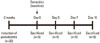
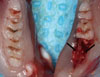
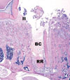
Twenty-eight six-week-old Sprague-Dawley rats were used for the present study. The animal experiment was conducted in accordance with the guidelines approved by the Animal Ethics Committee of the Yonsei University College of Dentistry, Seoul, Korea. All animals were randomly divided into two groups: a control group (n=8) and test group (n=20). In all of the rats of the test group, periodontitis was induced with ligature placement on the mandibular first left molars. Two weeks later, the mandibular first left molars of all of the rats in the control and test groups were extracted. The rats in each group were sequentially sacrificed on day 0, 3, 7, and 10 after extraction, with n=2 in the control group and n=5 in the test group on each day (Fig. 1). Mandibles were retrieved for histological and immunohistochemical analysis (Fig. 2). Neutral buffered formalin (10%) was used to fix the dissected block sections for 10 days.
Method
Male Sprague-Dawley rats (280 to 330 g) were obtained from a company (Orient Bio, Seongnam, Korea). Food and water were provided ad libitum. The animals were maintained in a temperature-controlled room (22℃) on a 12-hour light-dark cycle. In the test group, the rats were anesthetized with a 1:2 mixture of tiletamine/zolazepam (zoletil) and ketamine (-1.5 µL anesthetic per gram of body weight). A sterilized nylon thread ligature was placed around the cervix of the mandibular left first molar and knotted mesially. This induction of periodontitis was performed according to protocols previously described [14]. In the control group, the rats were kept without any intervention. Two weeks later, the mandibular first left molars of all of the rats in the control and test groups were extracted. Body weight and food intake were measured daily during the experimental period.
Evaluation method
Histological evaluation
After rinsing the block in sterile water, the sections were decalcified in 5% formic acid for 2 weeks, dehydrated in a graded ethanol series, and embedded in paraffin. Step-serial sections, 5-µm thick, were cut in the apico-coronal vertical plane. The central-most section was selected based on the position of the adjacent roots. Inflammatory infiltrates, new bone formation, and epithelium proliferation in the sections were observed under a microscope after being stained with hematoxylin-eosin.
Immunohistochemical evaluation
After deparaffinization and rehydration, the antigen was retrieved using a microwave-based technique in citrate buffer (pH 9.0) for 30 minutes. The endogenous peroxidase was blocked with 3% hydrogen peroxide for 5 minutes. The sections were incubated for 30 minutes at room temperature with primary antibodies: interleukin-6 (IL-6) (SC-1,265, Santa Cruz Biotechnology Inc., Santa Cruz, CA, USA) diluted 1:100, and TNF-a (ab6671, Abcam, Cambridge, UK) diluted 1:100. After washing three times, immunodetection was performed using a commercially available kit (EnVision Detection System, Dako, Kyoto, Japan) according to the manufacturer's instructions. Slides were then counterstained with hematoxylin.
RESULTS
Histological analysis
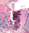
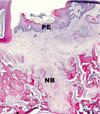
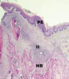
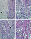
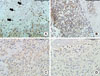
In the control group, all extraction sockets have shown uneventful and general healing processes. At day 0, a blood clot from the remaining periodontium and the gingival tissue filled the extraction socket (Fig. 3). At 3 days, the contracted clot was replaced by granulation tissue composed of fibroblasts and endothelial cells. Epithelium began to proliferate to cover the extraction socket. At 7 days, a woven bone and trabeculae pattern started to appear, beginning at the apical half of the socket. At 10 days, much more, and more mature, newly formed bone was found. Epithelium fully covered the extraction socket (Fig. 4). Fig. 5 shows histologic views of the extraction sockets at each day after tooth extraction in the control group.
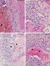
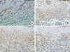
In the test group, the extraction socket was also filled with a blood clot at day 0. The blood clot mainly consisted of erythrocytes and fibrin strands with various leukocytes. At the coronal portion of the socket, abundant infiltration of inflammatory cells was observed in the remaining periodontium until 3 days (Fig. 6). At 7 days, the clot was replaced by granulation tissue and osteoblastic activity was observed, beginning along the alveolar wall. From 7 days, infiltration of inflammatory cells decreased gradually and only a little was found in the coronal half of the socket at 10 days. At 10 days, newly formed bone including the capillary-like vessels became more obvious in the apex of the socket histologically and epithelium had proliferated from the sides of the wound to cover the wound surface (Fig. 7). Fig. 8 shows histologic views of the extraction sockets at each day after tooth extraction in the test group.
Immunohistochemical analysis
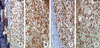
The immunoreactivities of the cytokines examined were consistently localized in the cytoplasm of the cells, showing a dark brown-colored reaction (Fig. 9A).
Figs. 9-12 show the immunohistochemical reactivities and Tables 1 and 2 show subjectively determined staining intensities of the tested cytokines in the control and test groups, respectively. For comparison of the immunoreactivities of the individual cytokines, the labeling intensity of the cells was scored on the following scale of 0 to ++++ using subjective criteria, similar to that described previously [15-17]: 0, no staining; +, weak staining (minimally detectable); ++, moderate staining; +++, strong staining; and ++++, very strong staining (dark brown, almost black).
In the control group, little interleukin-6 (IL-6) was observed at day 0 and was stained more strongly between 3 and 7 days after extraction (Fig. 9B and C); at day 10 after extraction, little staining was observed, as on day 0 (Fig. 9D). Although it was somewhat weaker at 0 and 10 days, similarly weak staining intensities were shown throughout the healing period in the case of tumor necrosis factor-α (TNF-α) (Fig. 10).
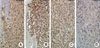
In the test group, IL-6 was stained very strongly on days 3 and 7 after extraction (Fig. 11B and C); at day 10 after extraction, little staining was observed (Fig. 11D). TNF-α staining was more intense at 3 days after extraction and gradually weakened at later points in time (Fig. 12).
DISCUSSION
Periodontal disease is mainly caused by interactions between bacteria of the dental plaque and components of the cellular and humoral host immune response, including cytokines and biological mediators released by activated immunocompetent cells [18,19]. Each cell type in the inflammatory infiltrates (neutrophils, T- and B-lymphocytes, plasma cells, and macrophages) plays a specific role in the progression of periodontal disease [19]. On the other hand, it has been suggested that periodontal disease will be resolved if plaque, inflammatory infiltrates, and cytokines disappear. Therefore, the present experiment was designed to histologically and immunohistochemically analyze the healing of sockets after extraction of periodontally involved teeth in rats and to determine the optimum timing of resolution of inflammation for delayed replantation. In all of the rats, healing was generally uneventful.
In histological analysis, the extraction sockets of the test group were filled with blood clots and the periodontium of the coronal portion demonstrated large numbers of inflammatory infiltrates on day 0. Until the first 3 days after tooth extraction, inflammatory infiltrates were found abundantly in the remaining periodontium and decreased at later points in time. At 7 days, the proliferative granulation tissue was found in the extraction socket. Granulation tissue originates from the fibroblasts and the endothelial cells in the remaining periodontium [20]. Moreover, the fibroblasts in the granulation tissue are suggested to differentiate into osteoblasts, thus forming new bone during socket healing. The extraction socket was observed to undergo the healing process and bone formation in the socket became obvious histologically beginning at 7 days after extraction (Fig. 8). Compared with the control group and a previous study [21] that demonstrated the socket healing process after sound tooth extraction, we found that socket healing after periodontally involved tooth extraction showed a delay of 2 to 4 days. It was assumed that inflammation could have caused this delayed socket healing.
In immunohistochemical analysis, we quantified the cytokines of IL-6 and TNF-α at the furcation area of the extraction socket because IL-6 and TNF-α play an important role in inflammation.
In response to Porphyromonas gingivalis oral gavage, mice with genetically deleted IL-6 had decreased bone loss compared to wild-type mice, suggesting that the production of IL-6, which is proinflammatory, contributed to bone resorption [22]. Borch et al. [23] also suggested that an exaggerated production of IL-6 occurs in generalized aggressive periodontitis. However, according to several studies, IL-6 did not seem to disappear instantly as the source of inflammation disappeared. A recent randomized-controlled clinical trial confirmed that intensive periodontal treatment resulted in a temporary increase of serum levels of IL-6 at 6 months after the treatment [24]. Lopez Carriches et al. [25] have found that levels of IL-6 were higher after surgical extraction of the lower third molars and remained high until the seventh day after. Miyawaki et al. [26] have proven that the level of IL-6 in plasma increases after different operations (cystectomy, benign tumor extirpation, etc.). Cruickshank et al. [27] studied the response of IL-6 in patients who had undergone different types of operations (minor surgery, cholecystectomy, hip surgery, colorectal surgery, and major vascular surgery), finding that levels of IL-6 were related to the duration of surgery. The authors concluded that IL-6 is a sensitive and early marker of tissue damage. Results of the present experiment also showed a similar correspondence with theirs. In both the control and test group, IL-6 was stained strongly at 3 and 7 days after extraction; at 10 days after extraction, it had decreased significantly. This finding suggests that IL-6 increased temporarily until 7 days through tissue damage of extraction and decreased at 10 days as inflammation was resolved. However, overall, IL-6 in the test group showed stronger staining than IL-6 in the control group due to induced inflammation.
TNF-α also plays a significant role in bone loss. Several studies have shown that TNF-α increased with a host response stimulated by dental plaque and bacterial products, raised osteoclastic activity, and consequently accelerated bone resorption and periodontal breakdown [28-30]. In other words, a decrease in TNF-α seems to reduce the host response, thereby reducing expression of the cytokines that stimulate bone resorption, which results in less net bone loss. In a study by Liu et al. [31] that examined the potential impact of TNF-α, bone resorption was induced following the placement of ligatures around rat molars for 7 days. At 4 and 9 days following removal of the ligatures, new bone formation occurred in normal mice with bone resorption ceasing. In the present study, TNF-α of the control group showed similar weak staining from 0 to 7 days. On the other hand, TNF-α of the test group showed a peak at 3 days and the TNF-α decreased gradually at later points in time. At 10 days after extraction, TNF-α had almost faded out, which implied the resolution of inflammation.
In fact, this study using rats cannot be applied to a human model directly, as there are differences in the healing process and time after extraction of periodontally involved teeth of rats and humans. The purpose of this study was not to reveal any clinical significance, but to serve as a reference for the ideal timing for future studies of delayed replantation following periodontally involved tooth extraction in rats.
The conclusions from this study are as follows. Within the limits of this study, it takes at least 10 days to resolve periodontal inflammation in the rat extraction socket. This can be a baseline for delayed replantation following periodontally involved tooth extraction in rats.
 XML Download