

 PDF
PDF ePub
ePub Citation
 Print
Print


INTRODUCTION
Various studies have been performed to regenerate or repair bone defects in the craniofacial region. In particular, one strategy among the methods that have been employed in bone tissue engineering is to utilize biomaterial as a scaffold for cell attachment. The scaffold material functions to provide 3-dimensional structure for cell attachment and cell delivery. This enables the biomaterial to provide cells needed for bone regeneration directly into bone defects. To acquire a suitable scaffold for cell attachment, it is important to investigate the cell-material interactions [1-4].
Calcium phosphate is one of the commonly used bone substitutes because of its high biocompatibility, osteoconductivity, and non-toxicity [5]. Among the calcium phosphates, hydroxyapatite (HA) and β-tricalcium phosphate (TCP) have been widely investigated and verified by in vitro and in vivo studies. HA has shown considerable osteogenic ability in the presence of bone marrow cells [6-8]. Also, it has been reported that TCP provides an excellent environment for bone regeneration despite its rapid degradation and weak mechanical properties [9-12]. Okumura et al. [6] demonstrated that the initial attachment of osteoblasts to HA was faster than to other materials and this might result in earlier osteogenesis on HA. Kon et al. [8] also reported that autologous bone marrow stromal cells transplanted on HA-based carriers led to extensive bone formation. Boo et al. [12] showed that TCP could play a role as a scaffold for bone formation and be useful for reconstructing bone.
High porosity and micro-architecture of deproteinized bovine bone mineral is like that of the human bone. It facilitates the invasion of blood vessels and cell attachment to the bovine bone [13,14]. It was reported that anorganic bovine bone was highly biocompatible and osteoconductive [15].
Fibronectin (FN) is one of the extracellular matrix glycoproteins, which play various roles in bone formation. It has been known that FN plays an important role in promoting cell attchment, cell spreading, and cell differentiation. Many attempts have been made to improve cell attchment by means of FN [1,16,17]. It was shown that FN coated biomaterials may be able to promote initial cell attachment.
The aim of this study is to determine whether these materials have potential as scaffolding material to support cell attachment. After seeding bone marrow stromal cells onto the biomaterials, we investigated their initial attachment in vitro.
MATERIALS AND METHODS
Cell isolation and culture
Rat bone marrow derived stromal cells were obtained according to the methods described previously by Maniatopoulos et al. [18]. Briefly, femora and tibiae were aseptically dissected from Sprague Dawley rats (130 to 150 g). The epiphyses were cut away and the bone marrow was flushed out by α-MEM medium (Gibco, Grand Island, NY, USA), which was expelled from a 10 mL syringe. The bone marrow cells were cultured in standard culture medium at 37℃ in 5% CO2. The culture medium contained α-MEM supplement with 10% fetal bovine serum (Gibco), 100 U/mL penicillin and 100 µg/mL streptomycin (Gibco). Hematopoietic cells and other unattached cells were washed away after 3 days. The culture medium was replaced twice a week. Near confluency, bone marrow stromal cells had been passaged and expanded until passage 4 or 5. Subsequently, they were seeded on the biomaterials.
Biomaterials
The tested biomaterials were divided into six groups. The first group was deproteinized bovine bone mineral (Bio-Oss, Geistlich Biomaterials, Wolhusen, Switzerland) as natural bone. In addition, the deproteinized bovine bone mineral (DBBM) was also studied after being coated with 100 µM human plasma fibronectin (FN; Gibco). The third material was synthetic HA, which is developed at Dentium Co. in Korea. Additionally, HA was investigated after coating it with 100 µM FN or β-TCP. Coating DBBM or HA with FN was performed by storing the materials overnight with 100 µM FN at 4℃. In the case of HA coated with β-TCP (HA/TCP), the material (Osteon, Dentium Co., Seoul, Korea) was obtained from Dentium Co. and consisted of 70% HA and 30% β-TCP. The sixth material was pure β-TCP particles (Cerasorb, Curasan AG, Kleinostheim, Germany), which were used after crushing and passing them through sieves. These biomaterials had particle sizes of 250 to 1,000 µm.
Confocal laser scanning microscopy
The bone marrow stromal cells were cultured on the biomaterials at a cell density of 5×10⁵ cells/well on a 48-well plate. 0.25 g of a given biomaterial was used per well. After 6, 12, and 24 hours of cell seeding, actin filaments, vinculin, and nuclei were stained with rhodamine-labelled phalloidin (Molecular Probes, Eugene, OR, USA), anti-vinculin antibody (Sigma-Aldrich Co., St. Louis, MO, USA), and DAPI (Molecular Probes), respectively, according to the manufacturer's protocol. In brief, cells were washed with phosphate buffered saline (PBS) and fixed in 10% neutral buffered formalin for 20 minutes and subsequently permeabilized with 0.1% Triton X-100 solution. The samples were incubated with 1% bovine serum albumin for 60 minutes. The vinculin was stained with mouse anti-vinculin antibody for 1 hour. Then the actin cytoskeleton was stained using rhodamine-labelled phalloidin for 20 minutes, followed by incubation with DAPI for 5 minutes. The samples were observed in confocal laser scanning microscopy (Olympus-FV300, Olympus, Tokyo, Japan).
Scanning electron microscopy
The bone marrow stromal cells were seeded on each biomaterial at a density of 5×10⁵ cells/well on a 48-well culture plate. 0.25 g of a biomaterial was used per well. After 1 and 7 days, the morphology of the cells was observed using a scanning electron microscope (S-4700, Hitach, Tokyo, Japan). The biomaterials were rinsed twice with PBS and fixed with 2.5% glutaraldehyde at 4℃ for 20 minutes. Each sample was fixed with 1% OsO4 at 4℃ for 20 minutes. The samples were dehydrated by ethanol in increasing concentrations of 70 to 100% at 4℃, and were also dehydrated with 1,1,1,3,3,3-hexamethyldisilazane (Sigma-Aldrich Co.) for 20 minutes. The specimens were sputter-coated with gold and observed in the scanning electron microscope.
RESULTS
Confocal laser scanning microscopy
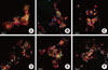
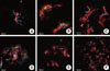
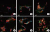
At 6 hours after cell seeding, the cells, which adhered on all the materials but HA/FN, were round or oval. On HA coated with FN (HA/FN), the cells seemed to be elongated (Fig. 1). After 12 hours, the cells on DBBM still exhibited round or oval shapes. On the other materials, the cells had spread more widely at 12 hours compared to the cells after 6 hours. Notably, actin filaments were widely distributed in cells adhered on HA and HA/FN. Cells on HA and HA/FN overlapped one another. Although cells on HA/TCP and TCP showed pronounced cell extensions for anchoring to the surface, they did not seem to overlap one another (Fig. 2). On DBBM and DBBM/FN, the cells after 24 hours showed similar shapes to those after 12 hours. The cells did not seem to spread extensively on DBBM. In addition, it was difficult to detect the cells adhered on DBBM and DBBM/FN. The cells on the other biomaterials displayed more extensive spreading and showed well-defined actin cytoskeletons after 24 hours (Fig. 3).
Scanning electron microscopy
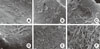
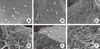
The morphological features of cells on each biomaterial were observed with scanning electron microscopy at day 1 and 7, respectively. After 1 day of incubation, even if some cells adhered flatly on the surface of DBBM and DBBM/FN, many cells were observed to have a circular morphology. The cells on HA and HA/FN appeared to be flattened out throughout the surface and were elongated. They seemed to be in close attachment to each other. In the case of HA/TCP, it was observed that β-TCP particles were coated on the HA surfaces and several layers of cells adhered to these surfaces. It was found cell density on HA/TCP and TCP were lower than it was on HA and HA/FN (Fig. 4). After 7 days, it was impossible to find well-spread cells on DBBM and DBBM/FN. In contrast, cells on the other biomaterials adhered close together. Cells adhered to HA and HA/FN developed lamellipodia and were widely spread all over the material. Some cells on HA and HA/FN formed multicellular layers. On HA/TCP and TCP, the cells showed a flat and polygonal shape. They had irregularly branched cytoplasm (Fig. 5).
DISCUSSION
In this study, we investigated rat bone marrow stromal cell responses to various bone substitutes. When biomaterials supporting bone formation are developed, attention should be paid to the cell-material interactions, such as cell attachment, proliferation, and differentiation. Particularly, cell attachment on biomaterial is the first step in cell-material interactions. Depending on the degree of cell attachment, the capacity to proliferate and differentiate may be determined [19].
The results of this study demonstrated that the degree of attachment was different according to bone substitutes. At day 7, the cells on DBBM showed round shapes and the isolation of individual cells was observed (Fig. 5). This may mean the initiation of apoptosis [20]. The cells grown on DBBM/FN also had similar shapes to those on DBBM. These results for Bio-Oss are in agreement with other in vitro studies [5,20-24]. Many studies have demonstrated that the viability of osteoblasts grown on Bio-Oss decreased over time [5,22]. However, some in vitro experiments [14,25,26] and clinical results [13,15,27,28] reported that DBBM produced a favorable cell response. These conflicting results about DBBM could stem from different culture conditions (e.g., seeding density, cultivation duration) among the studies [23]. Another study, which reported on cell viability with DBBM, demonstrated that the surface properties of hydroxyapatite changed when it was in contact with blood proteins and extracellular matrix components [22]. Additionally, oxygen and nutrients in vivo would provide support for cell proliferation by developing the vascularization. However, the in vitro environment was usually set up under static culture conditions [20]. These factors may result in the differences among the studies' results.
Fibronectin (FN) is an extracellular matrix protein that promotes cell adhesion. The binding of FN and other adhesion proteins to cell surface receptors enhances cell spreading, focal contact formation, and strength of adhesion [19,29]. In this study, 6 hours after cell seeding, the cells on HA/FN showed actin filaments, unlike those on HA (Fig. 1). At 12 hours, cells on DBBM/FN seemed to express actin filaments unlike those on DBBM. Grzesik and Robey [16] found that FN that contains the integrin-binding Arginine-Glycine-Aspartate sequence promoted bone cell attachment after 24 hours of incubation. In our study, at the initial phase, FN seemed to have a favorable effect on cell adhesion. Nevertheless, when the attachment tendencies were analyzed subsequently, the effect of FN seemed to be lost. After 12 hours, cells with or without fibronectin seemed to display no differences in cell morphology. The FN-coating method for biomaterials might have an effect on these results. To coat the biomaterials with FN, they were dried in FN solution overnight. Deduced from the results mentioned above, it is possible that FN, which attached to the surfaces of biomaterials, would be released over time. Therefore, the amount of FN on a surface might decrease, and this could reduce its effect on cell attachment. This is supported by previous studies [1,17]. Van den Dolder et al. [1] also explained that the surface concentration of FN might decrease continuously because of the competitive adsorption- desorption process of serum proteins. This could diminish the beneficial effects on the seeded cells. Another possible cause, which decreased the effectiveness of FN, is that we used whole FN to treat the biomaterials with FN. According to Grzesik et al. [17], the application of whole glycoproteins has limitations for various reasons, such as the opposite effect of multiple domains of the same factor, low availability of material, and loss of activity.
In the case of HA/TCP and TCP, cells seem to spread less extensively than those on pure HA in the initial phase (Fig. 2). HA/TCP, an alloplastic material, was covered with TCP particles on HA surfaces. Due to this surface characteristic of HA/TCP, a similar phenomenon was observed on TCP and HA/TCP. As previously mentioned, cell attachment on biomaterial is the first stage for cell proliferation and differentiation. Therefore, these results have implications for generating the proliferation of cells on biomaterials. Suzuki et al. [30] reported that osteoblast-like cells on pure HA exhibited higher growth rates than those on other ceramic plates including β-TCP. HA in the culture medium tend to have high pH on the thin surface layer of the ceramic carrier and to accelerate calcinations by trapping Ca ions from the medium, which stimulates the growth of osteoblast-like cells on HA.
Within the limitations of this study, we can conclude that while DBBM does not support cell attachment, HA provides a more favorable environment for cell attachment.
 XML Download