

 PDF
PDF ePub
ePub Citation
 Print
Print


INTRODUCTION
Dental implants are accepted as a valuable option for the restoration of missing teeth and various edentulous sites [1-3]. Clinicians often face esthetic challenges when restoring implants in anterior regions [4-6]. Especially when the restoration is in the maxillary anterior esthetic zone, the creation of a natural appearance is very important [6].
The provisional crown is a key factor in the success of soft tissue and interproximal papilla shaping [7]. The provisional crown should not induce extensive pressure on the gingiva, which could lead to recession [5,7]. Furthermore, the crown should mimic the natural color and shape of the symmetric tooth.
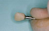
A fixed provisional crown is more comfortable and less vulnerable to fracture or loss than a temporary removable denture. On the other hand, the soft tissue shaping can be accomplished by a composite layering technique on fixed provisional crowns (Fig. 1) [8]. Provisionalisation of abutments in the anterior regions may also be necessary when the final crowns are not delivered at the time the abutments are placed. This may be because of a need to alter the aesthetics and occlusion before completing the treatment [8]. Major implant companies have introduced provisional abutments for these purposes. Nevertheless, these parts bring additional costs and make this treatment modality more expensive [9]. This article describes a practical method for provisionalisation of permanent metal abutments of implants placed in the maxillary anterior regions in several cases, as well as cases where these permanent metal abutments have been used instead of tooth colored zirconia abutments where excellent esthetic results could be obtained.
CASE DESCRIPTION
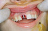
Patients with one missing anterior tooth who presented to the Department of Prosthodontics at a University Clinic for prosthetic treatment were included in this case series.
Information was given to each patient regarding alternative treatment options. This treatment was not offered to heavy smokers, since smoking could be unfavorable for later pink esthetics. Additionally, patients without a minimum of 1 mm of bone thickness surrounding the entire implant surface and patients with thin biotypes were excluded from the treatment group. In the end, twenty-two patients (9 males, 13 females; mean age, 36,72 years) with one missing anterior tooth fulfilling the above mentioned criteria were treated with this method.
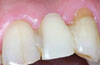
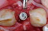
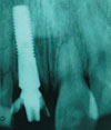
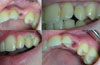
An artificial tooth (Optodent, Bayer, Leverkusen, Germany) was positioned in the edentulous place by attaching it to the neighboring teeth with visible-light-cured dental restorative composite material (Herculite XRV, Kerr, Orange, CA, USA) (Fig. 2). A polyvinylsiloxane impression was made for provisional crown fabrication (Brecision, bredent GmbH & Co.KG, Senden, Germany). After placement of the dental implant (in total 14 Astra-Tech AB, Mölndal, Sweden and 8 Straumann Holding AG, Basel, Switzerland) in a 3-dimensionally correct position as a substitute for the missing anterior tooth (Fig. 3), an abutment (Straumann Holding AG or Direct Abutment, Astra-Tech AB) was mounted (Fig. 1), upon which a provisional crown was fabricated chair-side using the polyvinylsiloxane impression with an auto-polymerizing acrylic resin (Dentalon, Heraeus Kulzer GmbH & Co.KG, Hanau, Germany) (Fig. 4). The marginal fit was finished on a laboratory analogue (Fig. 5) and temporarily cemented (Temp Bond, Kerr) to the abutment. An important step at this point was to control the marginal adaptation of the crown radiographically (Fig. 6). In cases of insufficient gingival contour or papillae, the temporary crown was used for gingival reshaping by composite addition to the marginal part of the acrylic crown (Fig. 7).
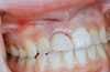
The patients were all satisfied with the final appearance and no complications occurred until the implants were loaded with permanent restorations. All implants were finally restored with cemented porcelain fused to metal crown restorations. Pink esthetics as well as the marginal bone levels were satisfactory in all cases during the follow-up period up to 60 months.
DISCUSSION
As progress in material and implant design continues noticeably over time, implant patients demand treatment protocols that take less time, require fewer surgeries, and have better esthetic outcomes [3,10]. One of the most frequently mentioned requests by patients is that some type of crown can be put on a dental implant right after its placement, especially in the anterior region. Following these expectations, most implant manufacturers have introduced a number of additional implant parts such as zirconia abutments for final restorations or tooth-colored plastic abutments for provisionalisation purposes [11]. Together with esthetics, these abutments also serve the important function of better shaping and maintaining the gum architecture around the tooth/implant. The provisional abutment is mainly used for the achievement of soft tissue modeling and shaping by the help of a temporary crown. The crown body is used to embody an ideal soft tissue contour of the gingiva as well as the interdental papillae, by adding composite or reducing the size [9].
The zirconia abutments on the market are a good solution especially in thin biotyped cases, where a grayish permeability of the soft tissue could distort the whole esthetic outcome. As is well known, however, the above-mentioned abutment types cause a rise in the global treatment cost for the patient [11].
Studies showing the long-term results of these two methods are scarce and the number of cases is limited [12,13]. In our experience, it seems that the use of temporary plastic abutments or zirconia abutments is not indispensable. For the achievement of esthetic results, several other factors seem to play a more important role. During surgery, dental implants should be positioned 3-dimensionally in the correct position and primary stability must be obtained for loading [2,12,13], both of which were adequately achieved in the cases presented here. Additionally, the socket walls where the implant will be placed must be intact to ensure later soft tissue esthetics [14]. Insufficient bone support and a thin biotype can often lead to undesirable consequences [15,16]. If the bony support is defective, a dehiscence is present, or a thin biotype is detected prior or during the surgery, a grafting procedure that will delay the loading time is indispensable. If the clinician is dealing with a thick biotyped gingiva, a metal abutment can perfectly fulfill the esthetic demands, as shown in the present cases that were followed up for several years. Another important risk factor is a history of aggressive periodontitis, especially combined with smoking [17]. These cases can show an unpredictable soft tissue recession, making a metal abutment visible. That is why patients with smoking habits were excluded from the present study. The use of definitive abutments instead of provisional abutments both reduced the costs and maintained the health of peri-implant tissues just like provisional abutments in the 3 months follow-up period until the final restoration was made.
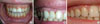
All patients in this article who had been restored by metal abutments showed a stable soft tissue response and excellent esthetic outcome (Fig. 8), indicating that in selected esthetic cases, costs can be reduced by the abdication of zirconia abutments for final restorations and temporary abutments for provisionalisation.
It should be noted that in case of a deep subgingival margin, which is often found in cases with thick gingiva, residual cement may cause infection. To avoid this complication screw-retained restorations could be used. However, several implant systems, as in this case series, offer a variety of abutment heights making prevention of deep subgingival margins possible.
Proper indications, good surgical technique, and the use of a prosthetic protocol are very important for the esthetic success of dental implant therapy, especially in the anterior regions. Nevertheless, there are also contraindications to the use of metal abutments in the esthetic zone. If even one among the patient-related factors of poor systemic health, a heavy smoking habit, poor oral hygiene, a thin biotype, or an infection in the extraction region is present, this treatment option should not be considered. Based on the presented case series, it can be concluded that the use of the definitive abutments for provisional restorations reduce costs and the same result can be obtained. The use of zirconia abutments could be relinquished in accurately selected cases for cost reduction as well.
 XML Download