

 PDF
PDF ePub
ePub Citation
 Print
Print


INTRODUCTION
Commercially pure titanium and titanium alloys have been used for several decades and their safety and efficacy as dental implant materials have been proven. However, their grayish color sometimes raises esthetic problems through the thin peri-implant mucosa. Tooth-colored materials, such as ceramics, can be a solution for these occasions. The demand for ceramic alternatives to titanium implants is not only based on esthetic considerations. Corrosion products of titanium have been found in inner organs and local lymph nodes [1,2]. Additionally, allergic reactions and sensitivities to titanium have been reported [3,4]. To resolve the allergy and esthetic problems caused by titanium implants, a ceramic implant was developed as a viable alternative. Aluminum oxide (alumina) was once used as an implant material. However, it was withdrawn from the market because of its insufficient physical properties. Ceramics are generally brittle and subject to premature failure, especially in repeated contact loading and in moist environments.
Recently, research has focused on another ceramic material, zirconium dioxide (zirconia), which has the potential for future use as a dental implant material. Tetragonal zirconia polycrystals, especially 3 mol% yttria-stabilized zirconia (3Y-TZP), serve as a metal substitute in substrates and possesses good physical characteristics [5,6].
In vitro and animal studies of zirconia have shown its good biocompatibility, high mechanical strength, and esthetic properties [7-9]. In particular, the role of the ZiO2 surface topography on the response of osteoblasts has been the focus of several in vitro studies in recent years [10-13]. Like titanium, the zirconia with rough surfaces had better initial cell responses than that with smooth surfaces. In addition to surface roughness, the coating of the surface with bioactive material such as bone morphogenetic protein-2 (BMP-2) to enhance osteogenesis around the titanium surface has been investigated. However, little information is available on the response of osteoblasts on zirconia to BMP-2.
The objective of this study was to investigate the effect of BMP-2 on the proliferation and differentiation of osteoblast-like MC3T3-E1 cells cultured on zirconia with rough surfaces and to compare this with the corresponding response in titanium.
MATERIALS AND METHODS
Sample preparation and surface analysis
Titanium discs were made out of commercially pure titanium grade 4 and zirconia discs were made out of yttrium-stabilized tetragonal poly-crystals (Y-TZP). All discs had a diameter of 12 mm and a thickness of 3 mm. The sandblasted/acidetched titanium discs were purchased (Warantec Co., Seoul, Korea). The zirconia discs were sandblasted with 125-µm Al2O3 powders at 3.5 bar for 1 minute. After surface treatment, all the discs were washed in distilled water, cleaned in an ultrasonic bath, and sterilized by EO-gas.
The surface topography of the discs was examined by confocal laser scanning microscopy (LSM 5 Pascal, Zeiss, Obercochen, Germany).
Osteoblastic cell culture
MC3T3-E1 cells are the non-transformed cell line established from newborn mouse calvaria. These cells exhibit the osteoblastic phenotype, as evidenced by the expression of ALPase activity [14], the synthesis of extracellular matrix (ECM) components such as osteocalcin and type-1 collagen [15], and their ability to mineralize the ECM.
MC3T3-E1 cells were purchased from Riken BioResource Center (Shiga, Japan). MC3T3-E1 cells were grown in alpha MEM medium (Gibco, Grand Island, NY, USA) supplemented with 10% fetal bovine serum (Gibco), 100 U/mL penicillin and 100 µg/mL streptomycin (Gibco) at 37℃ in a humidified atmosphere of 5% CO2. The culture medium was replaced twice a week. The cells were plated on discs of zirconia and titanium in 24-well tissue culture plates. One day after cell seeding, the commercially available demineralized bone matrix (DBM) gel (Rafugen, human demineralized bone matrix, Korea Bone Bank, Seoul, Korea) was added (25 mg/600 µL of culture medium) to the culture medium adjacent to the zirconia and titanium discs, and the DBM gel with BMP-2 (including BMP-2 of 50 µg/mL) (Korea Bone Bank) was added in the same manner. The discs were classified into 6 groups: zirconia discs with DBM gel, zirconia discs with the DBM gel including BMP-2, titanium discs with DBM gel, titanium discs with the DBM gel including BMP-2, zirconia discs without gel, and titanium discs without gel.
Release kinetics of BMP-2 from the DBM gel with BMP-2
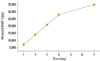
The kinetics of BMP-2 release from the DBM gel with BMP-2 was examined (Fig. 1). 50 mg of the DBM gel with BMP-2 was placed in a tube and 1 mL of phosphate buffered saline (pH 7.4) was added to the tube as a releasing medium. The tubes were placed in a shaking incubator at 37℃ and shaken at 15 rpm. The supernatants were decanted and replenished with fresh phosphate-buffered saline (PBS) at predetermined time intervals over a 1-week period. The concentrated BMP-2 released from the gel was assayed using a BMP-2 immunoassay kit (Quantikine BMP-2, R&D Systems Inc., Minneapolis, MN, USA).
Cell morphology
Cells were seeded onto the materials in 24-multiwell plates at a final density of 1×10⁴ cells/cm2. One day later, commercially available DBM gel alone and the DBM gel with BMP-2 were added to the culture medium (25 mg/600 µL culture medium/well). 24 hours after gel loading, the cells were fixed and stained with 4'-6-Diamidino-2-phenylindole for nuclei and phalloidin for actin filaments. The specimens were examined by Confocal laser scanning microscopy (Olympus-FV300, Olympus, Tokyo, Japan).
Colorimetric methyl tetrazole sulfate (MTS) assay
The cell viability was evaluated by measuring mitochondrial dehydrogenase activity with the MTS assay (Cell Titer 96 AQ Nonradioactive Cell Proliferation Assay, Promega Co., Madison, WI, USA) at 1, 4, and 7 days after gel loading. The assay measures the conversion of MTS into an aqueous soluble formazan product. 60 µL of reaction solution (in 600 µL culture medium per well) were added. The samples were incubated with the reagent for 3 hours in the CO2 incubator at 37℃ and the optical density was read at 490 nm by a spectrophotometric plate reader (THERMOmax, Molecular Devices Co., Sunnyvale, CA, USA).
Alkaline phosphatase (ALP) Activity
Seven days after gel loading, the alkaline phosphatase activity was measured according to the protocol of the ALP activity assay kit (AnaSpec Inc., San Jose, CA, USA). Briefly, the culture medium was removed. The cells were rinsed twice with PBS and lysed in lysis buffer with 1% Triton X-100 at 4℃ for 10 minutes. The lysates were harvested and clarified by centrifugation at 13,000 rpm at 4℃ for 10 minutes. The supernatants were incubated with 3.7 mM 4-nitrophenyl phosphate in 100 mM diethanolamine, pH 9.8, containing 0.1% Triton X-100 at 37℃ for 30 minutes. The reaction was stopped with 100 mM NaOH. The amount of released 4-nitrophenolate was determined photometrically at 405 nm. Alkaline phosphatase activity was expressed as the concentration of p-nitrophenyl phosphate (in nmole) transformed per microgram of protein. The protein concentration was measured with a commercially available kit (SMART microBCA kit; iNtRON Biotechnology, Seoul, Korea).
Real-time polymerase chain reaction (PCR)
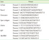
The osteoblastic differentiation of MC3T3-E1 cells was evaluated by real-time PCR examination of ALPase, bone sialoprotein, type I collagen, runt-related transcription factor 2 (Runx-2), osteocalcin, and osterix. The cells were plated at a density of 5×10⁴ cells/cm2 on Ti and Zr discs and cultured for 4 and 7 days after gel loading.
The total cellular RNA was isolated using Easy-BLUE Total RNA Extraction kits (iNtRON Biotechnology) according to the manufacturer's protocol. The first-strand of single-strand cDNA was synthesized from 1µg of RNA and used as a PCR template with a SuperScript First-Strand cDNA Synthesis Kit (Invitrogen, Carlsbad, CA, USA). Quantitative real-time PCR (QRT-PCR) was carried out using the Applied Biosystems 7500 Real-time PCR system (Applied Biosystems, Foster City, CA, USA) with a reaction mixture containing SYBR Premix Ex Taq II (Takara Bio Co., Shiga, Japan), cDNA, and PCR forward and reverse primers. Oligonucleotide primers for ALPase, Runx-2, osteocalcin, type I collagen, osterix, BSP, and 18s rRNA were purchased from Macrogen (Seoul, Korea) (Table 1). The thermal cycling conditions were 95℃ for 15 seconds, 95℃ for 15 seconds, 60℃ for 15 seconds, and 72℃ for 33 seconds. The relative gene expression of the samples of interest was compared with that of the titanium discs, with each sample being normalized to 18s rRNA to correct for differences in RNA quality and quantity.
RESULTS
Surface topography of the substrates
The surface roughness Ra (arithmetical mean deviation of the profile) of the titanium discs had an average of 1.04 µm, and that of the zirconia discs had an average of 1.34 µm. The roughness of the titanium discs treated by sandblasting and acid-etching did not differ from that of the sandblasted zirconia discs.
Cellular attachment and morphology
The morphology of adherent cells on the different surfaces was visualized by fluorescence staining of cytoskeleton actin filaments. 24 hours after gel loading (48 hours after cell seeding), confocal laser scanning microscopy images of MC3T3-E1 cells cultured on the different samples showed that the cells adhered properly on both zirconia and titanium surfaces (Fig. 2). Cells on both surfaces appeared to be well distributed and in contact with each other via cellular extensions. The MC3T3-E1 cells cultured on both zirconia and titanium surfaces showed well-organized actin fibers. The cells cultured with DBM gel or BMP-2 gel showed more well-organized actin fibers and cellular adhesions than did the cells cultured on the discs alone. However, there was no difference in cell morphology or adhesion between the cells on the zirconia and titanium surfaces.
Cellular proliferation
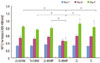
To examine the proliferation of MC3T3-E1 cells, we carried out a measurement of MTS activity at 1, 4, and 7 days after gel loading. Fig. 3 shows the optical density of formazan produced by the cells on the zirconia and titanium disc after 1, 4, and 7 days. The cells on the zirconia disc proliferated continuously for 7 days, when they were cultured with DBM gel or BMP-2 gel. However, cell proliferation on the titanium disc increased for 4 days and then decreased until day 7, when they were cultured with BMP-2 gel, and their proliferation was significantly lower than that on the titanium discs with DBM gel and the zirconia discs with and without gel (P<0.05).
Cell proliferation on the titanium disc cultured with DBM gel increased continuously for 7 days, like that on the zirconia disc. At day 1 and day 4, no statistically significant differences were observed between the cells cultured on the zirconia and titanium discs. There were no statistically significant differences in cell proliferation among the specimens cultured with and without DBM gel or BMP-2 gel at day 1 and day 4.
Cellular differentiation
ALPase activity
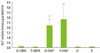
Early differentiation was measured by the ALPase activity at day 7 after gel loading (Fig. 4). Cells on the zirconia and titanium disc with BMP-2 gel exhibited significantly higher.
ALPase activity than those on the zirconia/titanium with DBM gel or without gel. However, ALPase activity of cells cultured on the zirconia disc with BMP-2 gel was not significantly different than that of cells cultured on titanium discs with BMP-2 gel (2.23±0.60 nmoL/µg protein/m on zirconia, 2.82±1.13 nmoL/µg protein/m on titanium). Therefore, BMP-2 stimulated the differentiation of MC3T3-E1 cells. However, DBM gel did not stimulate the differentiation of cells.
Osteoblastic gene expression
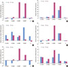
The effect of BMP-2 on the differentiation of cells on zirconia and titanium and the difference in the cell response on zirconia and titanium with BMP-2 were investigated by quantitative real-time RT-PCR. The gene expression of the osteogenic marker genes ALPase, osteocalcin, Runx-2, osterix, type I collagen, and BSP were analyzed at 4 days and 7 days after gel loading (Fig. 5).
The relative gene expression level was calculated by the 2-ΔΔCt formula and that may be interpreted as 'the expression of the gene of interest relative to the internal control (18s rRNA) in treated samples compared with the untreated control'. In this study, the extent of gene expression of the samples of interest was compared with that of the titanium disc. In investigating ALPase gene expression, at day 7, that of the cells on the zirconia disc with BMP-2 increased very extensively, followed by the titanium disc with BMP-2. At day 4, ALPase gene expression of the cells without BMP-2 decreased, compared with those on the titanium disc. In BSP gene expression, cells on the titanium with BMP-2 showed a higher expression level than those on the zirconia with BMP-2 at day 4. However, by day 7, BSP gene expression on zirconia had increased and was higher than that of titanium cultured with BMP-2. Similar results were found for osteocalcin, osterix, and Runx-2 gene expression.
DISCUSSION
In this study, the response of MC3T3-E1 cells to BMP-2 on zirconia was compared with that of such cells on titanium. On the basis of previous studies on the surface roughness of zirconia [11-13], the surface of zirconia was treated by sandblasting to obtain a moderately rough (Ra, 1.34 µm) finish. The sandblasted/etched titanium used in this study also had a moderately rough surface (Ra, 1.04 µm). Albrektsson and Wennerberg [16,17] reported that the roughness value in the moderate range (Sa between 1 and 2 µm) was optimal for the surface roughness.
Twenty-four hours after gel loading, confocal laser scanning microscopy images of the MC3T3-E1 cells showed that the cells adhered properly on both the zirconia and titanium surfaces. Cells cultured with DBM gel or BMP-2 gel showed very well-organized actin fibers and a much flatter appearance than those cultured on discs only. It has been suggested that cell morphology with a fully spreading shape and a regular cytoskeleton enables better cell proliferation and differentiation [18].
In this study, there was no difference in cell morphology and adhesion between the zirconia and titanium. In concord with this study, Yamashita et al. [11] demonstrated that the expression of actin filaments was similar on both zirconia and titanium. However, Hempel et al. [13] showed that, within 24 hours, osteblasts on titanium failed to express cellular extensions and retained smaller size compared with cells on zirconia.
The cell proliferation kinetics on the zirconia was similar to those on titanium, in agreement with other studies [11,19]. DBM gel or BMP-2 gel did not influence cell proliferation, but stimulated cell differentiation. At day 7, ALP activity and relative ALP mRNA expression was significantly greater in the cells cultured on the zirconia and titanium discs with BMP-2 compared to those on titanium alone.
In particular, the proliferation of cells on titanium with BMP-2 decreased significantly compared with the other groups, but ALP activity and ALP mRNA expression increased at 7 days. On the other hand, the proliferation capacity, ALP activity, and ALP mRNA expression of cells on the zirconia with BMP-2 increased until day 7. Cells on the zirconia, responding to BMP-2, proliferated longer and differentiated more actively than those on titanium with BMP-2.
In the osteogenic gene expression with ALPase, BSP, osteocalcin, Runx-2, and osterix, cells on zirconia with BMP-2 showed a higher expression level than those on titanium with BMP-2 at day 7. On the other hand, at day 4 the gene expression in cells on the titanium with BMP-2 was higher than that on zirconia with BMP-2. At the early stage, the gene expression in cells on titanium was higher in response to BMP-2, but at the later stage, the cells on zirconia showed a stronger response.
In conclusion, the data in this study demonstrate that osteoblastic cell attachment and proliferation of zirconia were comparable to those of titanium. With the stimulation of BMP-2, zirconia has a more pronounced effect on the proliferation and differentiation of the osteoblastic cells compared with titanium.
Further studies are necessary to address the bone-tissue response of zirconia implant in BMP-2 in animals.

 XML Download