

 PDF
PDF ePub
ePub Citation
 Print
Print


INTRODUCTION
Stem cells are undifferentiated cells characterized by the ability at the single cell level to both self-renew and differentiate to produce mature progeny cells [1,2]. The embryonic stem cells and the adult stem cells are two major categories of stem cells. Adult stem cells are undifferentiated cells found in specialized tissues and organs of adults. Compared to the embryonic stem cells, adult stem cells that exist in various organs of the body are easily accessible and less controversial in ethical terms [3,4].
The presence of adult stem cells in dental tissue has been formally proven. Seo et al. [5] reported that periodontal ligament stem cells (PDLSCs) expressed the mesenchymal stem-cell markers STRO-1 and CD146/MUC18. Up to now, 5 different human dental stem/progenitor cells have been described in the literature: dental pulp stem cells (DPSCs), stem cells from exfoliated deciduous teeth (SHED), periodontal ligament stem cells (PDLSCs), stem cells from apical papilla (SCAP), and dental follicle progenitor cells (DFPCs) [5-11]. These cells are easily accessible and, in contrast to bone-marrow-derived mesenchymal stem cells (BMSCs), are more intimately associated with dental tissues [3].
There are plenty of adult stem cells available for possible cell-based tissue engineering. Traditionally, BMSCs have been studied for bone regeneration. BMSCs are a population of multipotent, non-hematopoietic marrow-derived cells that are easily expanded in culture and differentiate into cells with an osteogenic phenotype [12,13]. Transplantation of BMSCs showed enhancement of periodontal tissue regeneration and bone formation [14,15]. Since dental stem cells were first identified, these cells have come under the spotlight in dental tissue engineering. Recently, numerous investigators have attempted to use these cells for dental tissue regeneration to assess their potential in pre-clinical applications [16,17]. Consequently, dental tissues are clinically accessible in routine clinical practice like tooth extraction, possibly providing a readily available source of stem cells, and these dental stem cells may be an ideal source for clinical periodontal regenerative therapy [5,16,18].
However, little is known about the characteristics of dental stem cells. In the case of PDLSCs, the PDL is composed of heterogeneous cell populations and thus far no highly purified PDLSCs clone has yet been established from human PDL tissue [18-20]. The challenge lies in the ability to identify a dental stem cell-specific marker that allows for the selection of a pure dental stem cell population [21].
The development of microarray methods for large-scale analysis of mRNA gene expression makes it possible to search systematically for key molecules [22,23]. After introduction of these genome-wide research techniques, there have been various attempts to describe and compare the gene expression patterns of more specialized adult stem cells for cell characterization [24-26].
The aim of this study was to compare the gene expression profile in mesenchymal stem cells derived from dental tissues and bone marrow for characterization of dental stem cells. In this study, the gene profiles from two individual PDLSCs and DPSCs samples were compared with BMSCs as a control.
MATERIALS AND METHODS
Subjects
Normal human extracted premolars were collected from one adult man (19 years old) and one adult woman (18 years old) at the Department of Dentistry, Asan Medical Center. The subjects were selected according to the following inclusion criteria: A person scheduled for tooth extraction for orthodontic reasons, general good health, absence of periodontal disease, and being a non smoker. Both of the subjects were thoroughly informed about the use of the extracted teeth and gave written consent for inclusion and genetic testing in the study. This study was approved by the Institutional Review Board of Asan Medical Center (No. 2010-0233).
Cell culture
The extracted premolars were placed into regular transfer medium and the PDL and the pulp tissue extracted and prepared as single cell suspensions as previously described [5,6].
Briefly, the PDL was gently separated from the root surface of the middle third of the extracted teeth. The pulp tissue was also separated after the tooth surfaces were cleaned and cut around the cementum-enamel junction by using a sterilized dental disk to reveal the pulp chamber. The PDL and pulp tissues obtained were digested in a solution of 3 mg/mL collagenase type I (Gibco, Grand Island, NY, USA) and 4 mg/mL dispase (Gibco) for 1 hour at 37℃. Single-cell suspensions were obtained by passing through a 70 µm stainer (BD Biosciences, Franklin Lakes, NJ, USA).
To identify the putative stem cell, single-cell suspensions (1×10⁴ cells) were seeded into 10 cm culture dishes with alpha-modification of Eagle's medium (Gibco) supplement with 15% fetal bovine serum (FBS) (Gibco), 100 mol/L ascorbic acid 2-phospate (Fluka Chemie GmbH, Buchs, Switzerland), 100 U/mL penicillin (Gibco), and 100 µg/mL streptomycin (Gibco) and incubated at 37℃ in 5% carbon dioxide. The cells of the colonies (aggregates of 50 or more cells) were collected and stored in liquid nitrogen until use.
BMSCs, processed from marrow aspirates of a normal human adult volunteer (female, 22 years old), were purchased from Lonza Walkersville Inc. (Walkersville, MD, USA).
In this study, cell passage 2 of the PDLSCs and DPSCs and passage 4 of the BMSCs were used for purification by a FACS Vantage cell sorter (FACS Vantage SE, Becton Dickinson, San Jose, CA, USA) and for further procedures.
Sorting of STRO-1+ cells
The sorting was carried out on a FACS Vantage Sorter using immunocytochemical staining of the early mesenchymal stem cell surface marker STRO-1 following previously reported methods [27].
Cells were incubated with a monoclonal anti-human STRO-1 primary antibody (R&D Systems Inc., Minneapolis, MN, USA) in 2% FBS/phosphate buffered saline (PBS) for 1 hour. After three washes with 2% FBS/PBS, the cells were incubated with fluorescein isothiocyanate-conjugated goat anti-mouse immunoglobulin M secondary antibody (BioFX Laboratories Inc., Owings Mills, MD, USA) for 30 minutes. Cells were washed and resuspended in regular growth medium. All the above procedures were performed in the dark at 4℃.
The expression profiles of STRO-1 on cells were examined and sorted by the FACS Vantage sorter. The sorting gates were set to sort the strong positive STRO-1 cells and exclude the weak positive ones. The sorted cells were collected in a tube containing growth medium. The cells were seeded into 10 cm culture dishes and incubated until they were just sub-confluent.
RNA isolation
The sorted and expanded cells were stored in RNAlater (Ambion Diagnostics Inc., Austin, TX, USA) for RNA extraction and preparation for the gene chip experiments. Total RNA was isolated applying a mirVana miRNA isolation kit (Ambion Diagnostics Inc.), according to the manufacturer's instructions. RNA concentration was quantified with a NanoDrop spectrophotometer (NanoDrop Technologies Inc., Wilmington, DE, USA), and the RNA integrity was evaluated using Agilent Bioanalyzer (Agilent Technologies, Santa Clara, CA, USA).
Microarray analysis
300 ng of total RNA was used for labeling. Probe synthesis from the total RNA samples, hybridization, detection, and scanning were performed according to standard protocols from Affymetrix, Inc. (Santa Clara, CA, USA).
Briefly, cDNA was synthesized from total RNA using the Whole Transcript (WT) sense target labeling and control reagents kit (Affymetrix, Inc.). Double-stranded cDNA was synthesized with random hexamers tagged with a T7 promoter sequence. The double-stranded cDNA was subsequently used as a template second cycle of cDNA synthesis, and random hexamers were used to prime reverse transcription of the cRNA from the first cycle to produce single-stranded DNA in the sense orientation. In order to reproducibly fragment the single-stranded DNA and improve the robustness of the assay, a novel approach was utilized where UTP was incorporated in the DNA during the second-cycle, first-strand reverse transcription reaction. This single-stranded DNA sample was then treated with a combinatoion of Uracil-DNA glycosylase & apurinic/apyrimidinic endonuclease 1 that specifically recognized the unnatural dUTP residues and broke the DNA strand. DNA was labeled by terminal deoxynucleotidyl transferases with the WT terminal labeling kit (Affymetrix, Inc.). The labeled cDNA was hybridized to the Human Gene 1.0 ST array (Affymetrix, Inc.) at 45℃ for 17 hours according to the Affymetrix standard protocol. After hybridization, the arrays were washed in a GeneChip Fluidics Station 450 with a non-stringent wash buffer at 25℃ followed by a stringent wash buffer at 50℃. After washing, the arrays were stained with a streptavidin-phycoerythrin complex. After staining, intensities were determined with a GeneChip scanner 3000 (Affymetrix, Inc.), controlled by GCOS Affymetrix software.
Data analysis
We performed three comparisons: BMSCs and PDLSCs, BMSCs and DPSCs, and PDLSCs and DPSCs. First, we fit a model that had a mean for each group, and tested whether the group means were different. The eBayes function in the R package Limma [28] was used to compute an empirical Bayes factor, pooling the variances from all the genes to estimate significance. After that, we had a model fit that estimated the log ratios between the two group samples for each of the three times. The fit also had an estimate of the Bayes factor and the log odds of differential expression for each gene. Estimated P-values using a number of multiple testing corrections were computed using Benjamini-Hochberg correction [29]. The adjusted P-value's cutoff is 0.001. We identified differentially expressed genes in each test. Each differentially expressed gene was analyzed using DAVID for analysis [30].
RESULTS
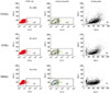
Isolation of mesenchymal stem cells using FACS Vantage Sorter
Isolation of STRO-1+ cells from each heterogeneous population was conducted using a FACS sorter machine. 0.96% and 1.38% STRO-1+ PDLSCs, 0.81% and 2.3% STRO-1+ DPSCs and 0.73% STRO-1+ BMSCs were successfully isolated using sorting gates strictly (Fig. 1). There were some differences in expression of STRO-1 between the patients and among the cell types.
Gene expression profile
We employed GeneChip analysis to expression levels of approximately 32,321 kinds of transcripts in 5 samples of BMSCs (n=1), PDLSCs (n=2), and DPSCs (n=2). Each sample was tested in triplicate to reduce technical errors. Altogether, 15 GeneChips were used to examine the gene expression profile of three different types of mesenchymal stem cells.
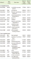
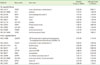
Differential expression was stated if a distinct transcript met the adjusted P-value's cutoff of 0.001. We identified 999 differentially expressed genes. In BMSCs, we identified 379 up-regulated transcripts, such as IGFBP5, KRT34, and DKK1, and 133 down-regulated transcripts, such as BEX1, GSTT1, GREM1, and NES. In PDLSCs, we identified 68 up-regulated transcripts, such as EBF2, PLXNC1, IL7R, and SATB2, and 64 down-regulated transcripts, such as TLR4, EDN1, and LMCD1. In DPSCs, we identified 218 up-regulated transcripts, such as BMP2, NES, COL18A1, EFHD1, and TGFB2, and 231 down-regulated transcripts, such as KRT34, GALNT5, and PENK. The most thoroughly up- and down-regulated genes in each cell types are listed in Tables 1, 2, 3.
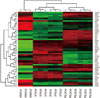
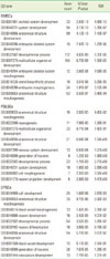
Additionally, heatmap, comparing the 53 genes most differentially expressed in PDLSCs, DPSCs, and BMSCs was shown (Fig. 2).
Gene Ontology (GO) Analysis
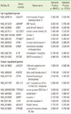
Major over-represented GO classes are listed in Table 4. In BMSCs, enrichment of gene productions were associated with the major GO classes of skeletal system, system, anatomical structure, and embryonic skeletal system development. In general, anatomical structure development and anatomical structure morphogenesis GO terms were over-represented in all three-different mesenchymal stem cells. In DPSCs, only GO terms related to blood vessels and neurons were over-represented.
DISCUSSION
In this study, we compared mesenchymal stem cells derived from dental tissues and bone marrow by employing genome-wide gene expression profiling and gene ontology analysis. The expression profiles of BMSCs, PDLSCs, and DPSCs were found to differ from those of each others. By GeneChip analysis, 999 genes were found to be definitely up- or down-regulated. In addition, GOstat analyses of regulated gene products provided over-represented GO classes. These data provide a first step for discovering key molecules related to the characteristics of dental stem cells.
The development of genome wide research techniques enables describing and comparing the gene expression patterns of different cells. Using these data, we can better understand the mechanisms governing the display of each cell's characteristics [25]. Recently, several microarray studies have been performed to identify new diagnostic markers and to investigate the origin of, as well as a genes characterizing progression of, various diseases including cancers [31-34].
In the dental field, a few studies about the characteristics and differentiation of dental cells were reported using genome-wide research techniques [24,35,36]. Furthermore, previous studies about dental stem cells only used heterogeneous dental stem cells. In this study, we used a FACS Vantage Sorter for isolation of STRO-1+ mesenchymal stem cells, and we first attempted to solve the problem of heterogeneity in dental stem cells for microarray studies.
The heterogeneity of the dental stem cell population remains one of the key obstacles to implementing optimal approaches to cell-based tissue engineering. Because dental stem cell-specific surface markers have not yet been identified, general mesenchymal stem cell markers like CD44, CD90, CD146, and STRO-1 have been used to identify stem cells from dental tissues [27,37]. Although the cells sorted using STRO-1 and CD146 markers have been used in some previous studies, no marker for dental stem cells has been established. In our study, we used the STRO-1 marker, which is the most common marker for mesenchymal stem cells. The subset of marrow cells that expresses the STRO-1 antigen is capable of differentiating into multiple mesenchymal lineages including hematopoiesis-supportive stromal cells with a vascular smooth muscle-like phenotype, adipocytes, osteoblasts, and chondrocytes was reported. Flow cytometry revealed that 1 to 5% of the PDL cells cultured using previously described methods [5] were STRO-1+ in human and animal models and the fluorescence values from the FACS data decreased with an increasing number of passages [16,27,37,38]. Comparably, 0.73-2.3% STRO-1+ cells were successfully isolated in this study by strict screening of strong positive cells.
Shi et al. [24] reported that insulin-like factor binding protein-7 (IGFBP7) was highly expressed in BMSCs and collagen type XVIII α1 (COL18A1) was highly expressed in DPSCs. In the present study, IGFBP5 was strongly up regulated in BMSCs and COL18A1 similarly in DPSCs. Representatively, interleukin 7 receptor (IL7R) was highly expressed in PDLSCs and transforming growth factor (TGFB2), while bone morphogenetic protein 2 (BMP2) was in DPSCs.
IL7R has been shown to play a critical role in V(D)J recombination during lymphocyte development. Several diseases are associated with IL7R including multiple sclerosis, rheumatoid arthritis, and juvenile idiopathic arthritis [39]. IL7R gene expression in PDLSCs may be a target molecule to study of chronic inflammatory disease like periodontitis. BMP2 is one of osteoinductive BMPs: it have been demonstrated to potently induce osteoblast differentiation in a variety of cell types [40]. The increased levels of BMP2 in DPSCs may suggest the possibility of using DPSCs as cell-sources for bone regeneration in tissue engineering.
Gene ontology is a tool for the systematic analysis of large-scale gene-expression data. GO analysis is based on the hypothesis that functionally related and differentially expressed genes should accumulate in the corresponding GO term. When the data of microarrys show a long list of significant genes, the soution can be found by pooling of genes into fuctional classes and GO database provides such a fuctional classificaton. According to GO analysis, anatomical structure development and anatomical structure morphogenesis GO terms were over-represented in all three different mesenchymal stem cells. However, GO terms related to blood vessels and neurons was over-represented in DPSCs, uniquely. These facts clearly show that the characteristic of dental stem cells is similar to that of BMSCs, and the dental stem cells also have other specific characteristics. Additional studies should be carried out to reveal the meaning of major over-represented GO terms in each cell.
In conclusion, our study demonstrated the genome-wide gene expression patterns of STRO-1+ mesenchymal stem cells derived from dental tissues and bone marrow. The difference of the expression profile of BMSCs, PDLSCs and DPSCs were showed and 999 candidate genes were found to be definite up- or down-regulated. A validation study of candidate gene expression is required using additional samples and additional analytical studies should be carried out to fully explore the functional role of the differentially expressed genes identified using microarray analysis. The study of the differences according to the presence of specific stem cell markers is also needed. Prospectively, the gene expression information of dental stem cells might provide significant insights into the development of dental tissues and the cellular differentiation processes. Dental stem cells could also be practicable tools for dental tissue engineering.
 XML Download