Journal List > J Periodontal Implant Sci > v.41(4) > 1082282

Cho, Park, Cha, Kim, Jung, Kim, Choi, and Kim: Dimensional change of the healed periosteum on surgically created defects

Abstract

Purpose

The final goal of regenerative periodontal therapy is to restore the structure and function of the periodontium destroyed or lost due to periodontitis. However, the role of periosteum in periodontal regeneration was relatively neglected while bone repair in the skeleton occurs as a result of a significant contribution from the periosteum. The aim of this study is to understand the histological characteristics of periosteum and compare the native periosteum with the repaired periosteum after elevating flap or after surgical intervention with flap elevation.

Methods

Buccal and lingual mucoperiosteal flaps were reflected to surgically create critical-size, "box-type" (4 mm width, 5 mm depth), one-wall, intrabony defects at the distal aspect of the 2nd and the mesial aspect of the 4th mandibular premolars in the right and left jaw quadrants. Animals were sacrificed after 24 weeks.

Results

The results from this study are as follows: 1) thickness of periosteum showed difference as follows (P<0.05): control group (0.45±0.22 mm)>flap-elevation group (0.36±0.07 mm)>defect formation group (0.26±0.03 mm), 2) thickness of gingival tissue showed difference as follows (P<0.05): defect formation group (3.15±0.40 mm)>flap-elevation group (2.02±0.25 mm)>control group (1.88±0.27 mm), 3) higher cellular activity was observed in defect formation group and flap-elevation groups than control group, 4) the number of blood vessles was higher in defect formation group than control group.

Conclusions

In conclusion, prolonged operation with increased surgical trauma seems to decrease the thickness of repaired periosteum and increase the thickness of gingiva. More blood vessles and high cellular activity were observed in defect formation group.

INTRODUCTION

Periodontitis is an infectious and common disease of the periodontal tissue that requires proper treatment to prevent alveolar bone loss or tooth loss. For decades, resective surgical therapy with or without osseous recontouring was regarded as the standard of treatment. Recently, attention has moved from resective surgeries to regenerative and reconstructive therapies. The final goal of regenerative periodontal therapy is to restore the structure and function of the periodontium destroyed or lost due to periodontitis. This includes the formation of a new connective tissue attachment, new cementum, and supporting bone [1]. However, the role of the periosteum in periodontal regeneration has been relatively neglected while bone repair in the skeleton occurs as a result of a significant contribution from the periosteum [2-5].
The periosteum is a specialized fibrovascular membrane covering all bone surfaces except for the surface of the joints. It is composed of an inner osteogenic layer and outer fibrous layer. The inner osteogenic layer contains mesenchymal stem cells (MSCs) that have the potential to differentiate into osteoblasts [6], chondroblasts [7-9], adipocytes [10], and myocytes [11]. The outer fibrous layer of dense collagen positioned next to the gingival connective tissue, contains fibroblasts and MSCs. Between the inner osteogenic and outer fibrous layers, a fibrovascular component harbors fibroblasts, endothelial progenitor cells to regenerate blood vessels, and nerves [12,13].
The mucoperiosteal flap is widely used for access in procedures such as flap surgery, mucogingival surgery, and regenerative periodontal treatment. In normal periodontium, the blood supply to the bone comes from three different paths: the periodontal ligaments, the connective tissue above the periosteum, and within the bone [14]. When soft tissue flaps are reflected, the blood supply from the soft tissue is removed, leaving poorly vascularized cortical bone exposed and resulting in bone resorption [15-17]. To prevent crestal bone resorption caused from elevation of a mucoperiosteal flap, a flapless implant surgical procedure was proposed as a predictable protocol [14,18]. Studies reported that the preservation of the periosteum maintained a better blood supply to the surgical site and helped to optimize the healing of the periimplant tissue, which implies the importance of the periosteum in the bone healing mechanism [19,20].
There have been various kinds of trials for application of the periosteum or periosteum-derived stem cells in periodontal regeneration therapy. The periosteum is composed of fibroblasts and an extracellular matrix, which was demonstrated to have regenerative cell populations [21]. The regenerative cells in the periosteum include chondroprogenitor and osteoprogenitor cells, which can form cartilage and bone under specific conditions. These cells play a crucial role in bone regeneration and wound healing of surgical bone defects or fractures. Also, the periosteum was shown to have the potential to stimulate bone formation when used as a graft material in animal and human studies [22-25]. A controlled clinical study was performed using human cultured periosteum sheets in combination with autologous platelet rich plasma and hydroxyapatite granules for periodontal regeneration therapy and significantly more favorable clinical improvement was observed [26]. Also, periosteal stem cells were shown to have the potential to regenerate osseous defects, especially when combined with a biocompatible scaffold [27,28]. Given these experimental results, the clinical application of cultured periosteal cells to repair bone defects was considered possible. However, little is known about the detailed cellular structure of native periosteum and repaired periosteum or the osteogenic mechanism of cultured periosteal cells of human periosteum.
The aim of this study is to understand the histological characteristics of alveolar periosteum and compare the dimensional difference between native periosteum and repaired periosteum after elevating a flap or after surgical intervention with flap elevation.

MATERIALS AND METHODS

Animal model

Nine male beagle dogs, with an approximate mean age of 15 months, and a weight of 15±1.2 kg, obtained from Samtako Company (an experimental animal breeder), Osan, Korea, were used. Upon receipt, a health examination of the animals was performed. The animals were examined for any abnormalities in posture or movement. Only healthy animals were included in the study. The animals were acclimatized for 1 week prior to experimentation. Animal selection and management, surgery protocol, and periodontal defect preparation followed a study protocol approved by the Institutional Animal Care and Use Committee, Yonsei University, Seoul, Korea.

Study design

Nine animals were divided into 2 experimental groups (group 1, flap only; group 2, defect formation) and 1 control group (control, native periodontium) with a 24-week healing period. While mucoperiosteal flaps were reflected and immediately repositioned in the flap only group, a 4×5 mm box type one-wall defect was formed and flaps were repositioned in the defect formation group.
They were housed in stainless steel cages with a floor area of 7,056 cm2 and a height of 60 cm labeled with cards identifying the study number, species/strain, sex, cage number, and animal ID. The cages were housed in a room air-conditioned with 10 to 20 air changes/hr. The temperature was 22±3℃ and the relative humidity 50 to 60%. The temperature and humidity were monitored daily. A light/dark cycle of 12 hours light/12 hours dark was applied.
The animals had ad libitum access to water and a pelleted laboratory diet with the exception of one week immediately postextraction and postsurgery when they were fed a canned soft dog food diet.

Surgical protocol

Food was withheld the night preceding surgery. The surgical procedure was performed under general anesthesia induced by intravenous injection of atropine (0.04 mg/kg; Kwangmyung Pharmaceutical Industrial Co., Seoul, Korea) and intramuscular injection of a combination of xylazine (Rompun, Bayer Korea Ltd, Seoul, Korea) and ketamine (Ketara, Yuhan, Seoul, Korea) followed by inhalation anesthesia (Gerolan, JW Pharmaceutical, Seoul, Korea). Routine dental infiltration anesthesia was used at the surgical sites.
The mandibular 1st and 3rd premolars were extracted to create an edentulous ridge prior to the experimental surgery and the extraction sites were allowed to heal for 2 months. The remaining dentition received oral prophylaxis in conjunction with the extractions.
The experimental surgery for group 1 included elevation of the buccal and lingual mucoperiosteal flaps to surgically create critically-sized, "box-type" (4 mm width, 5 mm depth), one-wall, intrabony defects at the distal aspect of the 2nd and the mesial aspect of the 4th mandibular premolars in the right and left jaw quadrants [29]. Following root planing, a reference notch was made with a round bur on the root surface at the base of the defect. The experimental surgery for group 2 included only elevation of the buccal and lingual mucoperiosteal flaps (Fig. 1).
The mucogingival flaps were advanced, adapted, and sutured using a resorbable suture material (Vicryl 5.0, Polyglactin 910, Ethicon Inc., Somerville, NJ, USA).

Postsurgical care

Postsurgical care included intramuscular administration of antibiotics (Cefazoline Sodium 20 mg/kg IV; Yuhan) and daily topical application of a 0.2% chlorhexidine solution (Hexamedin, Bukwang Pharm Co., Seoul, Korea) for infection control. Observations of the experimental sites with regard to gingival health, suture line closure, edema, and evidence of tissue necrosis or infection were made daily until suture removal, and at least twice weekly thereafter.

Euthanasia

The animals were euthanized at week 24 postsurgery using an overdose of pentobarbital (90 to 120 mg/kg IV). Block sections including the defect sites and surrounding alveolar bone and mucosal tissues were collected. The block specimens were rinsed in sterile saline and were immersed in 10% neutral buffered formalin at a volume 10 times that of the block section for 10 days.

Histological preparation

After rinsing in sterile water, the sections were decalcified in 5% formic acid for 14 days, trimmed, dehydrated in a graded ethanol series, and embedded in paraffin. Step-serial sections, 5-µm thick, were cut in a mesial-distal vertical plane, at approximately 80-µm intervals. The sections were stained using hematoxylin-eosin stains. The four most central sections of each defect site selected based on the width of the root canal were used for the histological and histometric analysis.

Histological analysis

One experienced masked examiner performed the histopathologic evaluation of the tissue specimens using incandescent microscopy (Multi-view microscope BH2, Olympus, Tokyo, Japan) and a PC-based image analysis system (Image-Pro Plus, Media Cybernetics, Silver Spring, MD, USA). Four central sections stained with hematoxylin/eosin were evaluated. The width of following parameters were measured at 1 mm, 2 mm, 3 mm and 4 mm from the foot surface and analyzed for the four central sections (Figs. 2 and 3):
  1. The inner osteogenic layer: This layer contains more cellular components and less fibrous tissue.

  2. The outer fibrous layer: This layer contains fewer cellular components and fibroblast and fibrous tissue outnumber other cellular components.

  3. The gingival tissue: It is composed of an epithelial layer and connective tissue layer.

Statistical analysis

Summary statistics (mean±SD) based on animal means for the experimental treatments were calculated using the four central sections from each defect, with defects being averaged for each site. Animal means were used to test for differences between experimental conditions using one-way analysis of variance and a post-hoc test. The level of significance was set at 5%.

RESULTS

Clinical observation

Surgical healings were uneventful. Despite the extent and size of the surgically involved areas, wound closure was successfully maintained throughout the experiment for all defects. During this study, no sign of infection or clinical complication was found.

Histological observation and measurement

There were no histologically observable pathological tissue reactions at the time of necropsy (Fig. 3). The measurement of gingival thickness and of the periosteum is shown in Table 1.
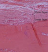
In the control group, the inner layer of the periosteum was firmly anchored to bone through fiber insertion, which is comprised of a direct continuation by the periosteal collagen fibers (Figs. 4 and 5). Osteoblasts were arranged in a layer adjacent to the bone surface and few activated osteoblasts were observed. Most of the osteoblasts were elongated, and basophilic characteristics of the cytoplasm were low. The cells over the osteoblast layer were small and spindle shaped osteogenic progenitor cells, which are similar to fibroblasts. Over these osteogenic progenitor cells, fibroblasts were layered in abundance and a very few blood vessels were observed throughout the periosteum (Figs. 3-5).
The outer layer of the periosteum, which is adjacent to the gingival connective tissue, is comprised of numerous dense collagen fibers, fibroblasts, and their progenitor cells. A few capillaries were observed. The thickness of the outer fibrous layer (0.21±0.05 mm) was similar to inner osteogenic layer (0.23±0.05 mm). The cellular density of the inner layer was several times higher than the outer layer.
In the flap only group, high cellular activity was observed inside the periosteum (Figs. 6 and 7). Osteoblasts were more cuboidal than in the control group and basophilic characteristics of the cytoplasm were greater, which means there was higher bone remodeling activity. In the outer layer, lots of fibroblasts and relatively dense collagen fibers were observed. The overall periosteal tissue seemed to be recovering from surgical trauma. Both the outer and inner layers were thinner than in the control group, but gingival thickness was greater than the control group even though there was no statistically significant difference (Table 1, Figs. 6-8).
In the defect formation group, the thickness of the outer and inner layers (0.14±0.04 mm, 0.12±0.05 mm each) was decreased and a statistical difference from the control and flap only group was noted (Table 1, Figs. 8 and 9). The thickness of the gingiva was the highest in the defect formation group and the scar-like tissue formation inside the gingival tissue was frequently observed. Numerous arterioles were observed between the outer layer and inner layer. Unlike the control group and flap only group, the activity of the osteoclasts was greatly increased and osteoblasts were observed more along the outline of the alveolar bone, which implies a high rate of bone remodeling and new bone formation (Figs. 9-11).

DISCUSSION

The periosteum is a specialized reactive layer composed of connective tissue covering the cortical bone and is a well vascularized osteogenic organ. Many studies have demonstrated the involvement of the periosteum in bone repair and remodeling of the jaw bone [2-5]. However, the importance of the periosteum in periodontal regeneration has been underappreciated so far. No potential conflict of interest relevant to this article was reported. Even though the original division of the periosteum into two anatomical layers was made in 1966, only in 1986 osteogenic cells of the cambium were clearly delineated from fibroblasts [30,31]. In periodontal regeneration therapy, the focus of evaluating the potency of regeneration was usually on the bone and cementum tissues, not on the periosteum. To understand the role of the periosteum in periodontal regeneration, it is assumed that the histological and histomorphometric difference between native periosteum and repaired periosteum after flap elevation or a surgical operation should be clarified first.
Many factors contribute to healing of periodontal defects. These include the shape of the defect, host factors, regenerative materials, and presence of growth factors and stem cells. However, the periosteum can also be an important factor for periodontal healing, which contributes as a source of stem cells as well as blood supply. MSCs in the periosteum are normally quiescent, but they are activated by injury, local infection, hypoxia, and surgical trauma such as the manipulation involved in releasing the periosteum [32]. There seem to be certain differences of histology among native and repaired periosteum according to the extent of surgical trauma applied, i.e. mucoperiosteal flap elevation, and defect formation.
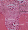
The results from this study show that elevation of a flap significantly reduced the thickness of the periosteum both in the outer and inner layers, and defect formation reduced the thickness of the periosteum even more. Flap elevation seems to be a direct cause of trauma, and the blood supply was severely compromised resulting in the moderate degree of tissue necrosis, which ultimately resulted a thinner width of repaired periosteum. Also, the extended time consumed in the additional operation and more traumatic procedures for defect formation might have caused much more trauma to the existing periosteum and resulted more necrosis of the periosteal tissue. The mechanism of surgical trauma is mainly based on the blood supply being interfered with. The elevation of the periosteum causes circulatory insufficiency through Volkmann's canals in the vascular plexus of the periosteum and periodontal ligament, and it could cause hypoxia of the periosteum and periodontal ligament [33]. Also, a series of morphological changes in the vascular plexus occur in the mucoperiosteal flap in the wound healing process after flap surgery [34]. Active angiogenesis, induced by elevation of the periosteum, increases blood flow and vascular permeability, which facilitates tissue repair responses in the series of steps of activation of osteoclasts, bone resorption, transition, and layered bone formation [33,35]. Therefore, the defect formation group showed the highest number of blood vessels in the periosteum and activated osteoblasts were dominantly observed along the alveolar bone (Fig. 10). The control group showed the lowest number of blood vessels and few activated osteoblasts, which implies that the bone remodeling process was relatively slow in the native periosteum compared to the flap only group or defect formation group (Fig. 11).
The thickness of the gingival tissue was greater in the flap only group than in the control group without statistical significance, and the defect formation group showed the greatest thickness of gingival tissue. It is assumed that the scar tissue was formed during the wound healing process from the surgical trauma and large amount of collagen fibers were produced in quantity creating the thickest gingival tissue in defect formation group followed by flap only group.
It is well known phenomenon that the osteogenic layer is immediately destroyed when periosteum is stripped from the adjacent bone surface, and blood supply over the underlying cortical bone is compromised. During this process, the cellular components show different distribution and activity degrees, and the total volume of periosteum changes according to the extent of surgical intervention, i.e, mucoperiosteal flap elevation or defect formation (Table 1, Fig. 12).
The conclusions from this study are as follows:
  1. The thickness of periosteum showed differences as follows (P<0.05): The control group (0.45±0.22 mm) had the greatest thickness followed by the flap only group (0.36±0.07 mm) and defect formation group (0.26±0.03 mm).

  2. Thickness of gingival tissue showed difference as follows (P<0.05): Defect formation group (3.15±0.40 mm) had the greatest thickness followed by flap only group (2.02±0.25 mm) and control group (1.88±0.27 mm).

  3. Higher cellular activity was observed in the defect formation group and flap only group than control group.

  4. The number of blood vessels was higher in one-wall group than pristine group.

In conclusion, prolonged operation with increased surgical trauma seems to decrease the thickness of the repaired periosteum and increase the thickness of gingiva. More blood vessels and high cellular activity were observed in the defect formation group.

Figures and Tables

Figure 1
Surgically created, critical-size, one-wall, intrabony, periodontal defect at the distal aspect of the mandibular 2nd and the mesial aspect of the mandibular 4th premolar teeth (A). Mucoperiosteal flaps are adapted and sutured for primary intention healing (B).
jpis-41-176-g001
Figure 2
Schematic illustration of the measurement parameters. CEJ: cemento-enamel junction.
jpis-41-176-g002
Figure 3
Measurement of point of control and defect formation group. Defect shape is illustrated by dotted line. Rt: root, Epi: epithelium, CT: connective tissue, PT: periosteum, AB: alveolar bone (H&E staining, ×20).
jpis-41-176-g003
Figure 4
Control group. The periosteum is divided into outer fibrous layer and inner osteogenic layer (H&E staining, ×200).
jpis-41-176-g004
Figure 5
Control group. Normal anatomy of periosteum divided into outer fibrous layer and inner osteogenic layer is shown (H&E staining, ×400).
jpis-41-176-g005
Figure 6
Flap only group. The periosteum shows two distinctive layers. A few numbers of capillaries were observed (H&E staining, ×200).
jpis-41-176-g006
Figure 7
Flap only group. Cuboidal osteoblasts were aligned along the alveolar bone and osteoclasts was observed resorbing the alveolar bone during the remodeling process (H&E staining, ×400).
jpis-41-176-g007
Figure 8
Defect formation group. Relatively large blood vessels were observed inside the periosteum (H&E staining, ×200).
jpis-41-176-g008
Figure 9
Defect formation group. Activated osteoblasts are aligned along the alveolar bone and relatively large blood vessels are located nearby (H&E staining, ×400).
jpis-41-176-g009
Figure 10
Histological photos focused on important anatomy, e.g., blood vessels, Sharpey's fibers, and activated osteoblasts from the control, flap elevation, and one-wall groups. (A-C); Caplillaries magnified ×100, (D-F); Sharpey's fibers magnified ×1000, (G-I); (H&E staining, ×200).
jpis-41-176-g010
Figure 11
Hihgher magnification of characteristic periodontal tissues. (A) Inactivated and elongated osteoblast (B) Activated osteoblast (C) Osteoclast actively absorbing alveolar bone with ruffled border (D) Fiber insertion into the alveolar bone (pristine group) (H&E staining, ×800).
jpis-41-176-g011
Figure 12
Measurement of gingival thickness and periosteum of the control group, flap only group, and defect formation group in mm. a)Statistically significant difference (P<0.05).
jpis-41-176-g012
Table 1
Measurement of gingival thickness and periosteum of control group, flap only group and defect formation group in mm (mean values and SD)
jpis-41-176-i001

a)Statistically significant difference (P<0.05).

ACKNOWLEDGEMENTS

This research was supported by Basic Science Research Program through the National Research Foundation of Korea (NRF) funded by the Ministry of Education, Science and Technology (R13-2003-013-04002-0).

Notes

No potential conflict of interest relevant to this article was reported.

References

1. Karring T, Nyman S, Gottlow J, Laurell L. Development of the biological concept of guided tissue regeneration--animal and human studies. Periodontol 2000. 1993. 1:26–35.
crossref
2. Nemcovsky CE, Artzi Z, Moses O. Rotated split palatal flap for soft tissue primary coverage over extraction sites with immediate implant placement. Description of the surgical procedure and clinical results. J Periodontol. 1999. 70:926–934.
crossref
3. Wang PD, Pitman DP, Jans HH. Ridge augmentation using a subepithelial connective tissue pedicle graft. Pract Periodontics Aesthet Dent. 1993. 5:47–51.
4. Nemcovsky CE, Moses O, Artzi Z, Gelernter I. Clinical coverage of dehiscence defects in immediate implant procedures: three surgical modalities to achieve primary soft tissue closure. Int J Oral Maxillofac Implants. 2000. 15:843–852.
5. Steiner GG, Kallet MP, Steiner DM, Roulet DN. The inverted periosteal graft. Compend Contin Educ Dent. 2007. 28:154–161.
6. Allen MR, Hock JM, Burr DB. Periosteum: biology, regulation, and response to osteoporosis therapies. Bone. 2004. 35:1003–1012.
crossref
7. Engdahl E, Ritsilä V, Uddströmer L. Growth potential of cranial suture bone autograft. II. An experimental microscopic investigation in young rabbits. Scand J Plast Reconstr Surg. 1978. 12:125–129.
crossref
8. O'Driscoll SW, Salter RB. The repair of major osteochondral defects in joint surfaces by neochondrogenesis with autogenous osteoperiosteal grafts stimulated by continuous passive motion. An experimental investigation in the rabbit. Clin Orthop Relat Res. 1986. (208):131–140.
9. O'Driscoll SW, Fitzsimmons JS. The importance of procedure specific training in harvesting periosteum for chondrogenesis. Clin Orthop Relat Res. 2000. (380):269–278.
10. Sakaguchi Y, Sekiya I, Yagishita K, Muneta T. Comparison of human stem cells derived from various mesenchymal tissues: superiority of synovium as a cell source. Arthritis Rheum. 2005. 52:2521–2529.
crossref
11. De Bari C, Dell'Accio F, Vanlauwe J, Eyckmans J, Khan IM, Archer CW, et al. Mesenchymal multipotency of adult human periosteal cells demonstrated by single-cell lineage analysis. Arthritis Rheum. 2006. 54:1209–1221.
crossref
12. Simpson AH. The blood supply of the periosteum. J Anat. 1985. 140(Pt 4):697–704.
crossref
13. Squier CA, Ghoneim S, Kremenak CR. Ultrastructure of the periosteum from membrane bone. J Anat. 1990. 171:233–239.
14. Campelo LD, Camara JR. Flapless implant surgery: a 10-year clinical retrospective analysis. Int J Oral Maxillofac Implants. 2002. 17:271–276.
15. Ramfjord SF, Costich ER. Healing after exposure of periosteum on the alveolar process. J Periodontol. 1968. 39:199–207.
crossref
16. Wood DL, Hoag PM, Donnenfeld OW, Rosenfeld LD. Alveolar crest reduction following full and partial thickness flaps. J Periodontol. 1972. 43:141–144.
crossref
17. Wilderman MN, Pennel BM, King K, Barron JM. Histogenesis of repair following osseous surgery. J Periodontol. 1970. 41:551–565.
crossref
18. Van der Zee E, Oosterveld P, Van Waas MA. Effect of GBR and fixture installation on gingiva and bone levels at adjacent teeth. Clin Oral Implants Res. 2004. 15:62–65.
crossref
19. Casap N, Tarazi E, Wexler A, Sonnenfeld U, Lustmann J. Intraoperative computerized navigation for flapless implant surgery and immediate loading in the edentulous mandible. Int J Oral Maxillofac Implants. 2005. 20:92–98.
crossref
20. Hahn J. Single-stage, immediate loading, and flapless surgery. J Oral Implantol. 2000. 26:193–198.
crossref
21. Breitbart AS, Grande DA, Kessler R, Ryaby JT, Fitzsimmons RJ, Grant RT. Tissue engineered bone repair of calvarial defects using cultured periosteal cells. Plast Reconstr Surg. 1998. 101:567–574.
crossref
22. Ritsilä V, Alhopuro S, Rintala A. Bone formation with free periosteum. An experimental study. Scand J Plast Reconstr Surg. 1972. 6:51–56.
23. Ritsilä V, Alhopuro S, Gylling U, Rintala A. The use of free periosteum for bone formation in congenital clefts of the maxilla. A preliminary report. Scand J Plast Reconstr Surg. 1972. 6:57–60.
crossref
24. Goldman HM, Smukler H. Controlled surgical stimulation of periosteum. J Periodontol. 1978. 49:518–522.
crossref
25. Cohen J, Lacroix P. Bone and cartilage formation by periosteum; assay of experimental autogenous grafts. J Bone Joint Surg Am. 1955. 37-A:717–730.
26. Yamamiya K, Okuda K, Kawase T, Hata K, Wolff LF, Yoshie H. Tissue-engineered cultured periosteum used with platelet-rich plasma and hydroxyapatite in treating human osseous defects. J Periodontol. 2008. 79:811–818.
crossref
27. Schantz JT, Hutmacher DW, Chim H, Ng KW, Lim TC, Teoh SH. Induction of ectopic bone formation by using human periosteal cells in combination with a novel scaffold technology. Cell Transplant. 2002. 11:125–138.
crossref
28. Sakata Y, Ueno T, Kagawa T, Kanou M, Fujii T, Yamachika E, et al. Osteogenic potential of cultured human periosteum-derived cells - a pilot study of human cell transplantation into a rat calvarial defect model. J Craniomaxillofac Surg. 2006. 34:461–465.
crossref
29. Kim CS, Choi SH, Chai JK, Cho KS, Moon IS, Wikesjö UM, et al. Periodontal repair in surgically created intrabony defects in dogs: influence of the number of bone walls on healing response. J Periodontol. 2004. 75:229–235.
crossref
30. Tang XM, Chai BF. Ultrastructural investigation of osteogenic cells. Chin Med J (Engl). 1986. 99:950–956.
31. Tonna EA. Response of the cellular phase of the skeleton to trauma. Periodontics. 1966. 4:105–114.
32. Simon TM, Van Sickle DC, Kunishima DH, Jackson DW. Cambium cell stimulation from surgical release of the periosteum. J Orthop Res. 2003. 21:470–480.
crossref
33. Nobuto T, Suwa F, Kono T, Taguchi Y, Takahashi T, Kanemura N, et al. Microvascular response in the periosteum following mucoperiosteal flap surgery in dogs: angiogenesis and bone resorption and formation. J Periodontol. 2005. 76:1346–1353.
crossref
34. Nobuto T, Yanagihara K, Teranishi Y, Minamibayashi S, Imai H, Yamaoka A. Periosteal microvasculature in the dog alveolar process. J Periodontol. 1989. 60:709–715.
crossref
35. Nobuto T, Imai H, Suwa F, Kono T, Suga H, Jyoshi K, et al. Microvascular response in the periodontal ligament following mucoperiosteal flap surgery. J Periodontol. 2003. 74:521–528.
crossref
TOOLS
ORCID iDs

Ui-Won Jung
https://orcid.org/http://orcid.org/0000-0001-6371-4172

Similar articles