

 PDF
PDF ePub
ePub Citation
 Print
Print


INTRODUCTION
Nitric oxide (NO), a free radical synthesized from L-arginine by NO synthases (NOS), can modulate various tissue and cell activities, including vasodilation, neurotransmission, immune responses, and death control [1,2]. NO is also involved in the regulation of bone metabolism through the several biological process [3]. Bone forming osteoblasts constitutively produce a small amount of NO which regulates proliferation and differentiation of osteoblasts [4,5]. Overproduction of NO, which is stimulated by inflammatory cytokines or mechanical stress of osteoblasts, may lead to cell damage and initiate and regulate apoptosis [4,6,7]. In inflammation-induced osteoporosis, elevated levels of NO have been shown to cause apoptosis and to increase bone-resorption by osteoclasts, resulting in decreased bone mineral density [8].
In the central nervous system, NO production is closely associated with N-methyl-D-aspartic acid (NMDA) receptors [9]. NMDA receptors, as glutamate receptors residing in presynaptic and postsynaptic neurons, have been known to control synaptic plasticity and memory formation in the central nervous system [10]. Upon stimulation of NMDA receptors by an external signal, Ca2+ enters the cell through the activated ionotropic NMDA receptor and then NO is produced by NOS which translocates to the membrane and is activated by Ca2+ [11]. An increase in NO induces excitotoxic cell death or apoptotic events in afflicted neurons.
Previous studies have revealed that glutamate NMDA receptors in osteoblasts may be involved in regulation of bone formation [12,13]. Recently, we have also reported that NMDA receptors are involved in periodontal ligament fibroblast (PDLF) differentiation but not their proliferation [14]. PDL cells form the connective tissue between the tooth cementum and alveolar bone to anchor teeth and maintain structural integrity. In addition, PDLFs have the potential to differentiate into cementoblasts and osteoblasts needed for cementum and alveolar bone formation, respectively [15-17].
Mechanical stress has been shown to promote an increase in NO production in cultured human PDL cells [18]. Constitutive NO production arising from chewing or other mechanical-stress of teeth may play several roles in periodontal tissues, such as serving to maintain structural integrity of PDL [19] and regulating PDL response to various stimuli like proinflammatory cytokines, mechanical strain, and sex hormones [6,20,21]. However, excessively produced NO may lead to cell damage in periodontal tissues similar to that in endothelial cells and osteoblasts [22,23]. For example, lipoplysaccharide (LPS)-induced nitrite synthesis in PDLFs and osteoblasts led to cell apoptosis and increased pro-apoptotic responses to cytokines [24,25]. This has been shown in the cell types such as human monocytes, endothelial cells, macrophages, and gingival fibroblasts, which produce inflammatory cytokines via stimuli of bacteria or bacterial components like LPS [26].
However, the function of NMDA receptor in NO-mediated PDLF cell damage has not been well characterized. We investigated the relationship between the effect of excessively produced NO using sodium nitroprusside (SNP) and NMDA receptor antagonist.
MATERIALS AND METHODS
Chemicals and antibodies
(+)-5-methyl-10, 11-dihydro-5H-dibenzo[a,d]cyclohepten-5, 10-imine hydrogen maleate (MK801) and SNP were purchased from Sigma Aldrich (St. Louis, MO, USA). 3-(4,5-dimethylthiazol-2-yl)-5-(3-carboxymethoxyphenyl)-2-(4-sulfophenyl)-2H-tetrazolium, inner salt (MTS) was purchased from Promega (Madison, WI, USA ). Cell culture reagents were obtained from Hyclone (Logan, UT, USA) and Gibco (Paisley, Scotland). Protease inhibitor cocktail was purchased from Roche (Rotkreuz, Switzerland). Primary antibodies of caspase-3, Bax, cytochrome c, c-Jun N-terminal kinase/stress-activated protein kinase (JNK/SARK), mitogen-activated protein kinases (p38) and anti-rabbit, anti-mouse second antibodies were obtained from Santa Cruz Biotechnology (Santa Cruz, CA, USA). Anti-phosphorylated extracellular signal-regulated kinases (pERK), ERK rabbit polyclonal antibodies were prepared by Cell Signaling Technology (Beverly, MA, USA). β-actin mouse monoclonal antibody was purchased from Sigma Aldrich.
Cell culture and chemical treatment
Human PDLFs were purchased from ScienCell (San Diego, CA, USA). They were cultured in α-MEM supplemented with 10% fetal bovine serum, 100 U/mL penicillin and 100 µg/mL streptomycin at 37℃ in a 5% CO2 humidified atmosphere. SNP was dissolved in phosphate buffered saline (PBS) buffer to 1 M as stock solution, stored frozen at -20℃ and protected from light for use in related experiments. PDLFs were treated with various concentrations (0, 0.5, 1, 1.5, 2, 2.5, 3, 3.5, and 4 mM) of SNP with or without 200 µM MK801 in culture media for 16 hours and PDLF morphologies were observed and photographed. All of the media were changed every 3 days.
Cell proliferation assay
After the cells were treated with SNP at the various concentrations for 16 hours, the cell medium was removed and then replaced by fresh medium containing MTS reagent. The cells were incubated at 37℃ in a 5% CO2 for 3 hours, and then the absorbance at 490 nm was measured on a microplate reader (Molecular Devices, Sunnyvale, CA, USA).
Western blot analysis
Human PDLFs were washed with PBS and collected in radioimmunoprecipitation assay buffer (150 mM sodium chloride, 1% Triton X-100, 1% sodium deoxycholate, 0.1% sodium dodecyl sulfate [SDS], 50 mM Tris-HCl, pH 7.5, and 2 mM ethylenediaminetetraacetic acid) with protease inhibitor cocktail (Roche, Rotkreuz, Switzerland). Equal amounts of proteins were separated by SDS-polyacrylamide gel electrophoresis and transferred to polyvinylidine fluoride membrane. The membrane was incubated with blocking buffer containing 5% skim milk in tris buffered saline for 2 hours. After blocking, the membranes were incubated for 2 hours with primary antibody, 1:1,000 anti-pERK, anti-ERK, anti-caspase-3 rabbit polyclonal antibodies, and anti-Bax, anti-cytochrome c, anti-β-actin mouse monoclonal antibodies, followed by incubation for 1 hour with 1:5,000 anti-rabbit and anti-mouse secondary antibodies, respectively. The membrane was detected with the aid of electrochemiluminescence detection reagents (Takara, Kyoto, Japan) using a fluorescence imaging system (Fuji Film, Tokyo, Japan).
Data analysis
A statistical software package (SPSS Inc., Chicago, IL, USA) was used to evaluate the data on cell proliferation. The differences for each value among the sample groups were compared using analysis of variance. The statistical significance of the results was analyzed using 95% confidence intervals.
RESULTS
Effect of NO and NMDA receptor antagonist on the viability in PDLFs
To determine the effect of SNP on the PDLFs, we first observed morphological changes. PDLF morphologies were observed and photographed following treatments of 0 to 4 mM SNP with or without 200 µM MK801, the NMDA receptor antagonist, for 16 hours. There was alteration of PDLF cell morphology in a SNP concentration-dependent manner. The 3 mM SNP treated PDLFs led to a dramatic reduction in the numbers of flat, spread, and spindle-shaped fibroblasts and loss of cell-cell contact, and increased cellular debris from apparently dead/dying cells (Fig. 1A). The PDLF morphologies were largely unaltered when they were cultured with 0 to 2 mM SNP. Interestingly, the PDLF morphology and viability were more sensitive to SNP in the presence of MK801, the blocker of NMDA receptors; the rounded and floating cells, and large loss of flat and spread PDLFs were observed at 2 mM SNP (Fig. 1A). We determined cell viability by MTS assay after SNP and MK801 exposure. Exposure of PDLFs to 0 and 0.5 mM SNP for 16 hours did not affect cell viability. In the 2.5 mM SNP treated PDLFs, significantly decreased optical density (A490) was determined by MTS assay, and in the presence of MK801, 1 mM SNP treated PDLFs showed similarly significant reduction compared to the untreated control (Fig. 1B), suggesting that PDLF viability is sensitive to NO and MK801.
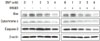
Effects of NO and NMDA antagonist on the expression of apoptotic proteins
Fig. 2 shows the effects of SNP on Bax, cytochrome c, and caspase-3 proteins. Administration of SNP in PDLFs increased Bax and cytochrome c production. However caspase-3 protein was decreased. Bax protein production was increased in 2 mM SNP treated PDLFs, and was highly expressed at 1 mM SNP treated cells in the presence of MK801. Cytochrome c protein represented the high expression in 2 mM SNP treated PDLFs. In MK801 treated PDLFs showed that cytochrome c was highly expressed at 1 mM SNP treatment. Caspase-3 protein was decreased in 3 mM SNP treated PDLFs and 2 mM SNP treated PDLFs in the presence of MK801. These results suggest that PDLFs reacted more sensitively in the presence of MK801 compared to the group treated with SNP
alone.
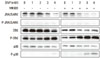
Effects of NO and NMDA antagonist on the expression of apoptotic proteins and MAPK pathway proteins
SNP treated PDLFs increased the phosphorylation of mitogen-activated protein kinase (MAPK) pathway proteins such as JNK/SARK, ERK, and p38 (Fig. 3). Western blot results revealed that phosphorylation of JNK/SARK and ERK proteins were induced from 3 mM SNP treated PDLFs. In the presence of MK801, increased phosphorylation of these proteins was shown in 2 mM SNP treated PDLFs. Phosphorylation of p38 was detected in 4 mM SNP treated PDLFs and revealed with 3 mM SNP in the presence of MK801. Notably, pERK was significantly increased in the presence of SNP and MK801.
DISCUSSION
Many of the pathogenic effects of bacteria in periodontitis involving alveolar bone destruction are mediated by bacterial products including endotoxin or LPS [27,28]. LPS can directly induce cell death or apoptosis in many cell types, and by stimulating NO production in macrophages, epithelial cells, and fibroblasts [29-31]. The production of high levels of NO in various cells is associated with autocytotoxicity, suppression of carcinogenesis, and inhibition of metastasis that result partially from induction of apoptosis [32].
In central neurons, NO is produced by elevated cytosolic Ca2+ which constitutively activates neuronal NOS (nNOS) via the activation of glutamate NMDA receptors [9]. There are two NOS subtypes: inducible NOS (iNOS) found in fibroblasts and macrophages and endothelial NOS (eNOS) in blood vessel walls [33]. PDL cells express both eNOS and iNOS, and NO products by eNOS, and iNOS modulates the function of PDL cells [19] and by occlusal stimuli in the rat PDL promotes PDL healing in transplanted teeth [34]. In this study we focused on the relation of NO and NMDA receptors in PDLFs.
NO released by SNP, an extracellular NO donor, has similar cytotoxic effects on PDLFs to induction of intracellular NO which is produced by NOS. A high level of NO induces oxidative stress in cells that may lead to apoptosis accompanied by changes in gene expression, and cell dysfunction and damage. As shown in Fig. 1, SNP treated PDLFs are associated with cell viability as evidenced by changes in their morphology into shrunken and round from spindle-shaped, and by the decrease in cell number and reduction in response to MTS assay. When PDLFs were treated with NMDA receptor antagonist, cell death of PDLFs was promoted at a lower concentration than only administration of SNP. In addition, our results showed that the oxidative stress by SNP-induced NO increased expression of apoptotic marker proteins such as Bax and cytochrome c. Interestingly, MK801 treatment of SNP stimulated PDLFs further enhanced the apoptotic responses of these cells in morphology and apoptosis marker protein expression than SNP treatment alone. Our results suggest that SNP, as a nitric donor, promotes apoptotic protein-induced PDLF cell death.
De novo synthesis of Bax protein by SNP has been shown to be proapoptotic in NO-induced osteoblast death [35], suggesting that increased Bax protein levels may be a critical trigger of apoptosis induced by oxidative stress in osteoblasts [36]. Cytochrome c is another well-known marker protein of apoptosis that may be released from mitochondria to the cytoplasm in osteoblasts under oxidative stress and associated depolarization of the mitochondrial membrane potential [37]. Decreased levels of caspase-3 expression in SNP treated PDLFs are also consistent with conversion and activation from the inactive form of caspase-3, which is specifically labeled as anti-caspase-3 antibody in the immunoblot analysis shown in Fig. 2. Activation of caspase-3 can cause the digestion of key cellular proteins and induce DNA fragmentation and cell apoptosis [38,39]. Possibly, similar proapoptotic alterations in Bax levels, cytochrome c-mediated activation of caspase-3 activity, and DNA fragmentation may have occurred in PDLFs treated with SNP in our study.
In most cells, NO-mediated apoptosis typically involves activation by phosphorylation of the JNK/SARK, ERK, and p38 groups of MAPKs [40]. We monitored the phosphorylation of ERK, p38, and JNK following treatment with SNP and SNP/MK801 to determine whether these kinases were involved in the signaling pathway of NO-mediated apoptosis in PDLFs. In NO induced PDLFs, phosphorylation of JNK/SARK, ERK and p38 was increased compared to the control PDLFs. In PDLFs treated with SNP and MK801, the phosphorylation of the MAPK panel occurred at lower concentrations of SNP that also produced morphologic and biochemical differences as shown in Fig. 1. Overall, these results indicate that in PDLFs, NO overproduction may induce an apoptosis signal via the MAPK pathway, and suggest that activated glutamate NMDA receptors in PDLFs may inhibit cell death and apoptosis, which are induced by conditions that stimulate excessive NO production, such as periodontal bacterial infections.

 XML Download