

 PDF
PDF ePub
ePub Citation
 Print
Print


INTRODUCTION
High levels of oral hygiene combined with active periodontal therapy and regular maintenance tooth cleaning can successfully manage periodontal disease [1]. Although supragingival plaque control alone is not as effective as professional subgingival tooth cleaning combined with a high level of supragingival plaque control, the role of individual supragingival plaque control is evident, as a community measure, in the management of periodontal disease [2].
Generally, the interproximal surface and the mandibular lingual surfaces seem to form the most plaque in undisturbed plaque accumulation experiments [3]. In subjects maintaining ordinary oral hygiene habits, the buccal surface of the maxillary second molars showed significantly more plaque accumulation compared to that of the first molars, possibly due to the difficulties of access for cleansing [4].
Despite the improvements made in the design of brushes, the average person removes only about 50% of plaque [5]. The effects of different types of toothbrushes have been the focus of many studies [6,7]. Different shapes and locations for the handle and bristles have been designed to increase plaque removing efficiency in hard-to-reach places [8].
The role of the single-tufted brush is to effectively remove the plaque in the hard-to-reach places, such as the buccal, oral, and distal sides of the molars [9]. However, no clinical comparison of plaque removal efficacy of the single-tufted brush and flat-trimmed brush could be found in the published literature. Thus, the purpose of the present study was to test the effectiveness of a single-tufted brush in the removal of plaque on the buccal and lingual surfaces of the molars compared with a flat-trimmed brush.
MATERIALS AND METHODS
The present study is a crossover and randomized clinical trial to compare the plaque removal efficacy of the single-tufted brush with the flat-trimmed brush. This study was approved by the Institutional Review Board of Gangnam Severance Hospital, Yonsei University (IRB-320110091). Patients were informed of the study procedures and all provided written informed consent.
Brush design
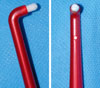
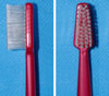
The single-tufted brush (ICUE, ICU, Anyang, Korea) has an angled handle, a brush diameter of 4 mm, and a rounded bristle tips (Fig. 1). The control brush (ICU403, ICU, Anyang, Korea) has a flat-trimmed brush, with a brush head measuring 28 mm in length and width tapered from 12 mm to 7 mm. The bristle tufts are positioned from 4 rows tapered to 2 rows, placed perpendicularly to the straight handle (Fig. 2).
Participants
This study was based on 26 male and 24 female, right-handed Korean patients aged 20 to 23 years (mean age, 22.3 years) who volunteered for an examination at the Department of Periodontics, Gangnam Severance Hospital, from September 2009 to December 2010. The main reason for visiting the Department of Periodontics was simple supragingival scaling. The exclusion criteria were as follows: subjects who had undergone prosthodontic or restorative therapy in the posterior molars, subjects who lost any molars, subjects who were taking any medicine known to affect the gingival dimensions, subjects with a probing depth >4 mm, subjects who had undergone any type of periodontal surgery including a soft tissue graft, subjects with a pre-brushing mean plaque index less than 1.8 [10].
Outline of the study
Each subject received a single-tufted brush and a flat-trimm-ed brush. Professional instruction and written brushing instructions were given. Prior to the examination, subjects were given a period of 3 weeks for familiarization with the brush [11]. After thorough supra-gingival scaling and polishing, all subjects were asked to abstain from oral hygiene procedures for 24 hours prior to the first experiment. Simple randomization for the treatment sequence was performed by coin tossing. Randomization and patient allocation were carried out with a hygienist who was not participating in the present experiment. Plaque scoring was done at the maxillary and mandibular molars after disclosing it with 1.4% erythrosine solution at six sites per tooth (mesiobuccal, midbuccal, distobuccal, mesiolingual [palatal], midlingual [palatal] and distolingual [palatal]), using the modification of the Quigley and Hein plaque index (PI) [12,13]. The PI was recorded pre- and post-tooth brushing, without the use of toothpaste. A washout period of at least 4 days was allowed between the test periods [14]. With dental prophylaxis, all the remaining plaque was professionally removed 24 hours prior to the second examination. Twenty-four hours of brushing abstinence was again performed. The PI was recorded pre- and post-tooth brushing after the subjects were given the second toothbrush in the cross-over sequence. The brushing time was 30 seconds per quadrant, for a total full-mouth brushing time of 2 minutes. All PI scoring was performed by a single blinded examiner. Adverse effects were monitored by the same examiner.
Statistical analysis
The average index score was determined for each individual. The plaque-reduction percentage was calculated by dividing the difference between the baseline and end PI by the baseline scores. Exploratory analysis on the plaque-reduction percentage was performed on individual regions (maxillary-buccal-marginal/maxillary-buccal-interproximal/mandibular-buccal-marginal/mandibular-buccal-interproximal) to determine the origin of possible differences, using a paired t-test [11]. P<0.05 was deemed significant.
RESULTS
One of the female subjects refused to continue participation in the experiment, due to the traumatic gingival abrasion caused by the single-tufted brush during the familiarization period. Thus, in total, 26 male and 23 female subjects completed the study. Among them, 5 subjects reported gingival trauma, probably due to inadequate handling of the single-tufted brush. The overall plaque scores and percentage reductions for the test and flat-trimmed brush are presented in Table 1. The single-tufted brush removed statistically significantly more plaque than the flat-trimmed brush.
The plaque reduction percentages for each of the tooth surfaces are listed in Table 2. The mesiobuccal (lingual) and distobuccal (lingual) PI are combined into the interproximal PI. The efficacy of plaque reduction was statistically different for the maxillary buccal interproximal/marginal and the mandibular lingual interproximal sites. However, the test and control brushes showed no statistical differences for the other sites.
DISCUSSION
Although single-use tooth brushing studies are routinely used for screening the efficacy of the test brush, the results could not provide definitive proof of superiority [14]. However, studies on the efficacy of plaque removal are still held using the single-use tooth brushing protocol [15]. The reasons for using the single-use tooth brushing protocol are its cost-effectiveness and ethical acceptability. In addition, no existing disease could worsen during the 1-day plaque accumulation [10]. Also, the present study was performed using a cross-over design, which is a valid model for assessing plaque removal efficacy [10]. The carry-over effects were minimized by the wash-out periods. However, the relatively short wash-out period might have increased the period effect.
The present study was mainly focused on plaque removal efficacy on the marginal/interproximal site of the posterior molars. However, during the study, the subjects complained that using the single-tufted brush on the occlusal surface and coronal of the posterior teeth was irksome. These drawbacks indicate that the single-tufted brush could only be used as a supplementary tool for the hard-to-reach sites. Also, some of the patients damaged the marginal gingival tissue due to improper usage of the single-tufted brush. Determining adequate stiffness of the single tufted brush is necessary in order to prevent unintentional damage to the periodontium.
The results indicate that the test brush was effective in removing the plaque on the relatively hard-to-reach sites. The buccal-proximal surfaces of the maxillary molar and lingual-proximal surface of the mandibular molars are known to accumulate more plaque than other sites [16]. In the present study, the most apparent difference in the plaque removal percentage was observed in the maxillary buccal marginal portion (about 44% difference). However, for other sites, even the sites which showed statistically effective removal of plaque, showed at most an 8% difference in the plaque removal percentage. The relatively small differences between the two brushes could be due to the fact that the base plaque was accumulated for only 24 hours. One-day accumulation of plaque could result in a relatively small amount of plaque accumulation, resulting in a less dramatic PI difference after brushing. However, due to potential ethical problems and subject cooperation, 24-hour oral hygiene abstinence was adopted. The data and methods of the present study per se could not allow any clinical conclusions about differences of this magnitude. The overall plaque reduction percentage differed by about 7%. The clinical effect, especially on the gingival status, of this degree of difference has not been investigated previously for the single-tufted brush. Thus, any further conclusions about the superiority of the single-tufted brush over the flat-trimmed brush should not be drawn. Moreover, the consensus has been that there is no one superior design of the manual toothbrush [5]. Nonetheless, a previous study revealed that an 8% difference in efficacy between a powered brush and a manual brush resulted in 22% less bleeding upon probing [17].
In conclusion, the single-tufted brush showed statistically significant plaque removal efficacy on the buccal side of the maxillary molars and the lingual interproximal side of the mandibular molars. However, the clinical relevance of the present results could not be determined. The clinical effect of the minor difference between the single-tufted brush and flat-trimmed brush should be the subject of a longitudinal study.


 XML Download