

 PDF
PDF ePub
ePub Citation
 Print
Print


INTRODUCTION
The placement of implants in the posterior maxilla is limited occasionally by insufficient bone volume as a result of alveolar atrophy or pneumatization of the maxillary sinus. This clinical problem can be resolved by sinus augmentation using surgical procedures such as onlay augmentation of the alveolar crest [1,2], Le Fort I osteotomy with an interpositional bone graft [3,4], lateral-approach sinus augmentation [5-7], or osteotome sinus augmentation [8-11].
In 1994, Summers introduced a less invasive sinus floor elevation procedure employing simultaneous grafting and the immediate placement of implants [8]. Using the Summers osteotome kit [8,9], which was specifically designed for this procedure, the pre-existing crestal bone is displaced toward the sinus floor as the osteotomes are inserted. Various graft materials and implants can be used in this surgical procedure. However, a minimum native bone height is required to get initial stability of the implant, and at least 5 mm of alveolar ridge height under the sinus is recommended for an implant that is 10 mm or longer [9]. Clinical case reports and studies on the bone-added osteotome sinus floor elevation (BAOSFE) procedure with simultaneous placement of implants show a relatively high survival rate in non-submerged sand blasted with large grit and acid etched (SLA) implants (94-98%) [10-15], but implant survival rates drop significantly when native bone height is 4 mm or less. Therefore, there are only a few clinical case reports involving sites with less than 4 mm of native bone height.
This report evaluates the clinical results of non-submerged SLA implants placed at the time of the BAOSFE procedure at sites where native bone height was less than 4 mm. Changes in graft height after the BAOSFE procedure were assessed radiographically for 5 years after the implant procedure.
MATERIALS AND METHODS
Patients
Four consecutive patients (2 women and 2 men, mean age of 61) with severe atrophy of the alveolar process in the posterior maxilla were treated at the Department of Periodontology, Yonsei University College of Dentistry. The patients showed no signs or symptoms of sinus or intraoral disease. All four patients underwent the BAOSFE procedure with simultaneous placement of a total of 7 Straumann SLA implants (Institut Straumann AG, Basel, Switzerland) (Table 1). The patients provided informed consent to participate in this clinical evaluation. The evaluation was approved by the Institutional Review Board at Yonsei University Dental Hospital (IRB No. 2-2009-0024).
Surgical techniques
All patients' medical histories were reviewed at an initial examination in order to rule out any local or systemic diseases that might contraindicate the surgical procedures. The patients received oral hygiene instructions and whole-mouth scaling prior to the surgery.
The BAOSFE procedure was performed using a Summers Osteotome kit, (3i Implant Innovations, Palm Beach Gardens, USA), as described by Summers [8,9]. Briefly, an incision was made under local anesthesia of lidocaine 2% with 1:80,000 epinephrine (Kwangmyung Pharmaceutical, Seoul, Korea) at the edentulous area to be treated. After the crestal incision was made, full-thickness buccal and palatal flaps were reflected. Site preparation was begun using the Summers #1 and #2 osteotomes. When the bone was too dense for hand instrumentation, 2-mm twist drilling was used to reach the cancellous bone, stopping 1 mm below the floor of the sinus. The preparation site was widened using #2 and #3 Summers osteotomes. Prepared bone graft material with beta-tricalcium phosphate, Cerasorb (Curasan AG, Kleinostheim, Germany), and demineralized freeze-dried bone, Dembone (Pacific Coast Tissue Bank, Los Angeles, USA), which acts as a shock absorber, was added to the preparation site with a carrier. Elevation of the maxillary sinus membrane was achieved using the #3 osteotome that was used previously to force the graft ahead of its tip to achieve the sinus floor up-fracture. At this stage, the integrity of the sinus membrane was confirmed by the Valsalva maneuver. Finally, the non-submerged Straumann SLA implants were place into the osteotomy site. Primary stability was achieved for all implants. Primary closure was achieved using monofilament suture material, Ethilon (Johnson & Johnson Int., Edinburgh, UK). All surgical procedures were performed by C. S. Kim.
Postoperatively, patients were instructed to rinse their mouth twice a day with a 0.12% chlorhexidine solution, Hexamedin (Bukwang Pharmaceutical Co., Seoul, Korea) for 2 weeks after surgery. Antibiotics were prescribed for 7 days, and sutures were removed after 10 days. After a mean healing period of 7 months, all patients were rehabilitated with fixed crowns or bridges.
Follow-up
After inserting the implants, the patients received follow-up care at 1 and 2 weeks, at 3, 6, and 9 months, and every 6 months thereafter. Clinical and radiological evaluations were performed using standardized radiographs according to the following schedule: prior to surgery, immediately after surgery, 6 months after surgery, and then every year after surgery up to 5 years.
Radiographic analysis of the grafted bone height
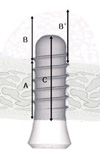
Using a scanner, HP scanjet 7400c (Hewlett Packard, Palo Alto, USA), the radiographs were digitalized. The digital image analysis program Image-Pro Plus (Media Cybernetics, Silver Spring, USA) was used for linear analysis of the radiographs. The magnification of each radiograph was corrected using the known actual length of the inserted implants so that an accurate graft height could be obtained. The radiographs from the same patient were blinded in terms of which time point they represented. Native bone height, grafted bone height, and implant height were measured on each radiograph as described in Fig. 1.
RESULTS
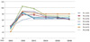
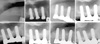
Radiographic examination showed that the sinus floor was elevated immediately after surgery in all patients. Table 1 shows the radiographic measurements for each patient. The mean native bone height was 3.4 mm. The average gain in the grafted bone height of the implants was 8.6 mm (range, 6.9-9.9 mm). The grafted bone area was easily distinguished from the sinus floor on the radiographs. Clinical and radiographic examination during the initial healing period showed normal healing in all patients. At 6 months, radiographic evaluation showed the maturation of the grafted bone, including increased density and sinus floor remodeling. Although the change in grafted bone height varied from patient to patient, there were marked differences in bone height immediately after the surgery versus 2 years after surgery. The mean reduction in grafted bone height, which was gradual, was 1.6 mm (85% of the mean total reduction) during the first 2 years. (Fig. 2) In contrast, subsequent grafted bone height reduction was minimal: After 2 years, the mean bone height was further reduced by 0.3 mm (15% of the mean total reduction). In case of patient no.4, significant radiographic remodeling of grafted bone occurred also during the first 2 years. And the mean reduction was minimal between 2 and 6 years (Fig. 3). Thus, the total mean reduction in the grafted bone height was 1.9 mm 5 years after surgery. All implants were functionally stable, and crestal bone remodeling was minimal.
DISCUSSION
This report evaluated the clinical results of non-submerged SLA implants placed simultaneously in sites with less than 4 mm of native bone height using the BAOSFE procedure. Using radiographs, this report also assessed changes in the grafted bone height during the long-term (5-year) healing period.
All implants were maintained successfully for over 5 years. The results suggested that simultaneous placement of non-submerged Straumann SLA implants using the BAOSFE procedure is a feasible treatment option for patients with atrophic posterior maxillas. However, radiographic reduction of the grafted bone height was observed, especially during the first 2 years of the healing period, although there was some variation among the patients. Therefore, patients must be chosen carefully and the clinicians should consider that some reduction will occur. There was some variation in results among patients, depending on the follow-up time, inclusion criteria, surgical and prosthetic techniques, and other factors; however, the BAOSFE procedure with simultaneous placement of an implant shows a predictable survival rate ranging from 95-100% [6,10,11]. The 1-step approach using the BAOSFE procedure has the advantage of being less invasive, and this technique can enhance the bone quality of the implant site from type III or IV to type II. Reducing the surgical and healing times can be achieved because coordinated consolidation of the graft around the implants during the healing period is expected. Moreover, little difference has been reported between the survival rate of implants placed at the time of grafting versus those placed after a delay [16]. Differences in implant design and surface characteristics may influence the survival rate of different types of implants [11]. The superiority of SLA surface implants in conjunction with the osteotome sinus floor elevation technique has been documented in many studies [17,18]. Regarding the extent of bone retention, some studies have reported that the SLA surface is superior to a machined-surface implant [19,20]. Moreover, the survival rate of SLA-surface implants in the sinusaugmented maxilla is markedly higher than that of the machined-surface implants [21].
The survival rate of implants is also influenced by the quality and quantity of the native bone [11,12,22]. In particular, the survival rate is clearly reduced when the native bone height in an implant site is 4 mm or less [11]: It is difficult to achieve primary stability of the implant, and there is a higher possibility that the Schneiderian membrane will tear [23]. However, this is somewhat controversial. Peleg et al. [24] evaluated the efficacy of augmentation of the maxillary sinus using a lateral approach with simultaneous placement of hydroxyapatite surface implants in patients with 3-5 mm of residual bone height. In 63 patients, all 160 implants were stable during the 2- to 4-year follow-up periods. Together with previous studies, these results show that using rough surface implants in the augmented sinus area results in a predictable prognosis. Therefore, a 1-step procedure involving both grafting of the maxillary sinus and simultaneous placement of rough surface implants might be a feasible treatment option for patients with as little as 5 mm of native bone height.
In this report, the height of the grafted bone was reduced markedly by an overall mean of 1.6 mm during the course of the short-term healing period, i.e. the first 2 years. During the long-term healing period, i.e. over 5 years, the height of the grafted bone was reduced by an overall mean of 1.9 mm. Dimensional changes in the height of augmented grafts in the sinus have been documented in clinical and radiographic studies [25,26]. At the Sinus consensus conference in 1996, there was a report on 100 patients and 145 sinus-grafting sites that were evaluated using panoramic radiographs over a 3-year period. All graft materials resulted in a radiographic reduction ranging from 0.79-2.09 mm. However, it was not determined whether this reduction in graft height occurred in the initial healing period or was part of an ongoing healing process. Hallman et al. analyzed 30 maxillary sinuses in 20 patients who were grafted with a mixture of autogenous bone and bovine hydroxyapatite, and reported that a small (<10%) but statistically significant dimensional reduction was observed 12 months after surgery and after 1 year of loading [27]. Other studies on the reduction of sinus grafts using X-rays have also been performed; most of these studies show agreement with the results of this report in that that shrinkage of the grafted materials and reduction in grafted bone height were observed during the initial healing period after the BAOSFE procedures were performed [28-30]. Hatano et al. [31] assessed long-term changes in the sinus-graft height after a maxillary sinus floor augmentation with simultaneous placement of implants. Those results showed that the graft height decreased during the first 2-3 years after augmentation, but all subsequent changes were minimal.
While small, this report suggests that simultaneous placement of non-submerged SLA implants using the BAOSFE procedure is a feasible treatment option for patients with severely atrophic posterior maxillas. However, the grafted bone height is reduced during the healing period, and clinicians should expect some radiographic reduction of the grafted bone height, especially in the first few years after the procedure.

 XML Download