

 PDF
PDF ePub
ePub Citation
 Print
Print


INTRODUCTION
Alveolar bone resorption in maxillary posterior edentulous region and advanced pneumatization of the maxillary sinus can result in a lack of bone support for dental implants, leaving only a thin wall of bone between the maxillary sinus and oral cavity. When this area does not offer adequate conditions for anchoring, long-term maintenance of dental implants is hard to achieve and a bone graft is required.
The placement and integration of dental implants in such patients requires augmentation of the maxillary sinus. The classic procedure for maxillary sinus augmentation was first introduced by Boyne and James [1]. The periosteal membrane of the sinus mucosa that adheres to the maxillary sinus floor has few elastic fibers. Thus, separation of this sinus membrane is relatively simple and has become a standard method with good results [2,3]. Numerous clinical studies have reported the clinical outcomes of placing implants in the augmented maxillary sinus. The insertion of dental implants in combination with maxillary sinus floor elevation is a predictable treatment method showing high implant survival rates and low incidences of surgical complications [4]. However, it sometimes leads to complications due to anatomical structure, infections, iatrogenic factors, or other unknown factors. Therefore, pre- and post-operative evaluation of the maxillary sinus is very important. However, there has been limited availability of data on this procedure based on computed tomography (CT).
This case report describes a 53-year-old male patient who received maxillary sinus augmentation and implant installation with a staged approach. Although the patient had no subjective symptoms and there was no abnormal radiolucency on the panoramic radiograph, incomplete bone formation in the central portion of the augmented sinus was found fortuitously in the CT scan.
CASE DESCRIPTION
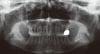
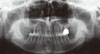
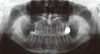
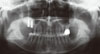
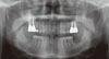
A 53-year-old male patient who was in good systemic condition visited the Department of Periodontology, Kyung Hee University School of Dentistry with the chief complaints of pain and gingival bleeding in the upper right 1st molar (#16) area. The upper right 2nd molar (#17) area was in an edentulous state (Fig. 1). Even though he was treated with non-surgical/surgical periodontal therapy, extraction of #16 was inevitable. The residual bone height was between 2 to 4 mm (Fig. 2). Augmentation of the maxillary sinus was scheduled to be conducted with a diagnostic stent 3 months after the extraction of #16, followed by placement of two dental implants 6 months after the augmentation of the maxillary sinus (Fig. 3). The maxillary sinus was clinically healthy. The sinus membrane was elevated from a lateral approach and the sinus was grafted with deproteinized bovine bone (DBB; Bio-Oss, Geistlich Pharma AG, Wolhusen, Switzerland) (Fig. 4). Prophylactic antibiotics (amoxicillin 500 mg, Chong Kun Dang Pharm., Seoul, Korea) were prescribed three times a day for 14 days and 0.12% chlorhexidine solution (Hexamedine, Bukwang Pharm., Seoul, Korea) was also prescribed twice a day for the first 2 weeks to prevent infection of the surgical wound. Healing was uneventful and there was no infection or other post-surgical complications during the healing period.
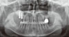
After a healing period of 6 months, a two stage implant surgery was planned (Fig. 5). Full thickness flap reflection under local anesthesia was followed by placement of two dental implants (Replace Select tapered HA, Nobel Biocare, Göteborg, Sweden) with a diameter of 4.3 mm and length of 13 mm in the site of #16i and #17i (Fig. 6). Both antibiotics and analgesics were administered for seven days, and mouth rinse with 0.12% chlorhexidine was also recommended for the following two weeks. No complications occurred after implant installation.
The second implant surgery was performed 6 months later (Fig. 7) and the implants were first loaded 2 months after the second surgery. Final prosthetic loading was initiated 13 months after the first implant surgery (Fig. 8).
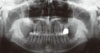
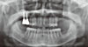
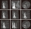
The implants placed in the augmented sinus were clinically healthy and the implant-supported restorations had been functioning successfully at 17 months after initial loading. Unexpectedly, the patient visited the dental clinic with the chief complaints of pain on biting in the upper right 2nd premolar (#15) since he had eaten hard food 3 days earlier. The #15 tooth was diagnosed as cracked and endodontic therapy was required. During endodontic therapy, a CT scan was taken to locate the buccal canal of the tooth. Peri-implant radiolucency in the apical portion of the implant placed in the augmented maxillary sinus was found by accident in the CT scan although a conventional (panoramic) radiograph revealed no signs of peri-implant radiolucency (Fig. 9). This was after a healing period of 32 months since sinus augmentation. The fortuitously discovered radiolucent portion can be described as incomplete bone formation or bone cavity in the augmented maxillary sinus. Nevertheless, the dental implants that were placed in the grafted sinus had been functioning well after prosthetic loading for more than 60 months and no enlargement of the bone cavity was found in follow-up radiographic views (Fig. 10). The patient has had no subjective symptoms such as discomfort or pain in the #16i and 17i area and has been receiving follow-up care on a regular basis.
DISCUSSION
This study presents the fortuitous discovery of incomplete bone formation (a bone cavity) in the central portion of the maxillary sinus after sinus augmentation using DBB.
Although sinus augmentation is very predictable [5] and complications caused by sinus graft are very rare, clinicians have observed various types of complications, such as perforation of the sinus membrane, excessive bleeding, infection of the grafted sinus, and failure of bone formation during and after sinus augmentation procedures [6-9]. Among these complications, incomplete bone formation of the grafted sinus is not common and the reason for this phenomenon has not yet been determined.
Several causes of incomplete bone formation can be suggested. First, the graft material used in augmentation was solely DBB, which is a cell-free grafting material with osteoconductive properties [10-13]. Cells possessing osteogenic potential are rich in residual host bone and elevated sinus membrane, whereas osteogenic potential of the graft is poor [1,14]. Therefore, new bone originates from the maxillary bone and progresses towards the augmented area [15,16]. The area of newly mineralized bone on the sinus floor is expected to be larger in the vicinity of the residual host bone [17]. Busenlechner et al. [16] reported the relative portion of newly formed bone after sinus augmentation with DBB in a minipig model study. In that study, with increasing distance from the host bone, the relative portion of newly formed bone declined from 38±13.3% at a 0-1 mm distance to 6.6±7% at a 4-5 mm distance. Fuerst et al. [14] and Roldan et al. [15] have also described the inhomogeneous distribution of bone and biomaterials within the augmented sinus in analysis by focusing on selected regions. The addition of autogenous bone, which contains bone-derived progenitor cells and osteoblasts, to bone substitutes is thought to enhance new bone formation [14]. In several animal model studies, e.g. in sheep [18] and in monkeys [19-21], the percentage of newly formed bone was increased by adding autogenous bone to bone substitutes. Experimental studies have shown that culture expanded autogenous bone-derived cells (ABC) added to cell-free grafting materials also enhanced the percentage of bone newly formed in critical-size defects of the rodent calvaria, and the dog and sheep mandible [22-25]. ABC possess an osteogenic potential and therefore increase bone formation in regions with a low number of bone-forming cells [23]. It can be speculated that supplementation of bovine bone mineral (BBM) with ABC may be recommended in cases where bony consolidation is complicated by the large volume of the grafting material or the poor regenerative potential of the host bone. If we had added autogenous bone to cell-free DBB during the sinus graft, more prominent bone formation could have been achieved.
Second, overpacking of graft material could restrict the blood supply. Rosenberg [26] and Garrett et al. [27] suggested packing the bone graft loosely. Loose packing of the graft can create better interparticulate spacing, which facilitates more rapid vascularization and more abundant bone formation. In contrast, tightly packed particles allow cellular and vascular access only to the outside layer of particles [28].
Third, Verdugo et al. [29] reported that bone contamination by specific pathogens could impair osteogenesis and induce greater bone loss in regenerative procedures. Similarly, Choukroun et al. [30] reported the accidental discovery of bubble-like lacunae in the grafted maxillary sinus when a sinus lift was performed with graft material without a 0.5% metronidazole solution. In that study, the formation of bubble-like lacunae within the graft was hypothesized to have resulted from anaerobic contamination after sinus grafting: the "septic theory." This "septic theory" suggests that grafted bone contamination by anaerobic bacteria could possibly induce problems with healing. They suggested the local use of a very small quantity of metronidazole (10 mg) as a sterile solution incorporated into the sinus bone graft.
Fourth, whether the healing period was long enough should also be considered. Hanisch et al. [31] reported that newly formed bone after sinus augmentation procedures using an allogenic-xenogenic bone graft in the grafted area at 12 months (20.7±8.3%) was significantly higher than at 6 months (8.1±3.0%), but it still remained lower than the volume of residual bone. In other words, the mineralization process of an allogenic-xenogenic sinus graft requires more than 12 months. According to Nkenke and Stelzle [32], healing periods after simultaneous implant placement ranged from 2 to 10 months. In staged approaches, healing periods for the graft material from 3 to 13 months were chosen. After implant placement, additional healing periods of up to 10 months were reported. In the present study, the healing period between sinus elevation and implant placement was 6 months, and the implant was loaded 8 months after the implant placement. Therefore, the healing period of the present case was long enough.
As the placement of endosseous implants has become the treatment of choice for restoring function and reconstructing edentulous areas, the number of patients having surgical complications is also on the rise. In this case report, we described one of these complications: the incomplete formation of maxillary sinus bone after sinus augmentation. As we have discussed above, it is possible that 1) osteoconductive graft material with poor osteogenic potential, 2) overpacking of graft material that restricts the blood supply, or 3) bone microbial contamination may cause the appearance of incomplete bone formation after sinus augmentation. Although the irregular appearance of the augmented maxillary sinus does not preclude implant placement and the success rate of implants placed in the subsinus area is very similar to that of implants placed in other regions [5,30,33], there is still lack of sound scientific data about whether the heterogeneity can be considered as a suitable condition for implant placement and survival. Further studies are needed to elucidate the mechanism of this unexpected result and care has to be taken to prevent it.

 XML Download