

 PDF
PDF ePub
ePub Citation
 Print
Print


INTRODUCTION
Mesenchymal stem cells (MSCs), which are nonhematopoietic multipotent stem cells, can differentiate into various types of progeny and are continuously self-renewing. MSCs suppress the activation of the immune reaction both in vivo and in vitro, known as "immunomodulation" or "immunosuppression." Several studies have demonstrated the immunomodulatory effects of MSCs on both allogenic and xenogenic immune cells [1,2]. This immunomodulatory capacity of MSCs has roused clinical interest in the medical field for areas such as hematopoietic stem cell transplantation [3]. However, the mechanisms involved in these activities are not yet completely understood.
In the field of dentistry, periodontal ligament stem cells (PDLSCs) have been considered to have therapeutic potential for the reconstruction of tissues destroyed by periodontal diseases. PDLSCs are highly proliferative and capable of regenerating cementum- and PDL-like tissues [4]. When stem cells from the root apical papilla and PDLSCs were transplanted into an animal model, a root/periodontal complex was generated [5,6]. Though the gold standard for periodontal tissue engineering is autologous PDLSCs, these cells may be unavailable in edentulous or medically compromised patients. In these cases, either allogenic or xenogenic PDLSCs can be an alternative source. However, allogenic or xenogenic cell transplantation still has the potential risk of a host immune response against the donor tissue.
The purpose of this study was to investigate the immunomodulatory effects of canine PDLSCs on allogenic and xenogenic immune cells, i.e., peripheral blood mononuclear cells (PBMCs).
MATERIALS AND METHODS
Animals
Five adult male beagle dogs (one year of age), weighting 10 to 12 kg, were used for the experiments. The animal research protocol was approved by the Institute of Laboratory Animal Resources, Seoul National University (SNU-100126-6).
Isolation and culture of canine PDLSCs and BMSCs
Two beagle dogs were purchased from Central Lab. Animal Inc. (Seoul, Korea) from which to isolate canine PDLSCs and bone marrow stem cells (BMSCs). These dogs were anesthetized with an intramuscular injection of 5 mg/kg xylazine (Rompun, Yuhan, Seoul, Korea) and 10 mg/kg ketamine (Ketamin, Yuhan, Seoul, Korea). The area around the iliac crest was depilated by an electric shaver and disinfected with betadine for bone marrow aspiration. Local anesthesia in the form of a 2% lidocaine solution (Lignospan, Septodont Lab., Saint-Maur-des-Fosses, France) was administered on the teeth to be extracted.
To collect the canine BMSCs, bone marrow aspirates of 1 mL were taken from the iliac crest of a beagle dog. A BMSC culture was performed in accordance with the technique described by Tsutsumi et al. [7]. Briefly, the cells, including erythrocytes, from bone marrow aspirates were seeded at 2×10⁸ cells in 100 mm tissue culture dishes and maintained in 10 mL of Dulbecco's modified Eagle's medium (Invitrogen, Carsbad, CA, USA) supplemented with 10% fetal bovine serum (Invitrogen, Carlsbad, CA, USA), 100 U/mL penicillin and 100 µg/mL streptomycin. Three days after seeding, the floating cells were removed. The medium was replaced every three days. Passage was performed when cells were approaching confluence. Cells were seeded at 5×10³ cells/cm2 and maintained in media containing 3 ng/mL FGF-2 (Invitrogen, Carlsbad, CA, USA) for two weeks. The cells were harvested prior to confluence by means of a sterile trypsin-EDTA (Invitrogen, Carlsbad, CA, USA) solution (0.5 g/L trypsin, 0.2 g/L EDTA in normal phosphate buffered saline, pH 7.4) and were stored in liquid nitrogen until use.
To obtain the canine PDLSCs, all mandibular premolars and first molars of a beagle dog were extracted bilaterally. The PDL was gently separated from the root surface of the middle thirds of the extracted mandibular premolars and first molars. Following separation, the PDLSC culture was performed in accordance with the technique described by Seo et al. [4]. The obtained PDL was digested in a solution of 3 mg/mL collagenase type I and 4 mg/mL dispase for 1 hour at 37℃. Single cell suspensions were obtained by filtration through a 70 µm strainer (BD Biosciences, Bedford, MA, USA). The cells were collected and stored in liquid nitrogen until use. In this study, cell passages 2-4 were used for the experiments.
Isolation of canine and human PBMCs
Canine whole blood was collected from beagle dogs that were purchased from Gaoyao Kangda Laboratory Animal Sciences & Technology Co. (Guangdong, China). Human whole blood was drawn from 10 healthy male volunteers (mean age 30) under a protocol approved by the Institutional Review Board, Seoul National University Dental Hospital (CRI09028).
PBMCs were acquired by means of gradient centrifugation separation. Fresh heparinized peripheral blood was diluted with an equal volume of balanced salt solution (Invitrogen, Carlsbad, CA, USA). The diluted blood was layered on Ficoll-Paque Plus (Amersham Biosciences, Wikströms, Sweden) and centrifuged at 400×g for 40 minutes. The buffy coat layer was separated and washed three times with HBSS (Invitrogen, Carlsbad, CA, USA). The deposited cells were resuspended in Roswell Park Memorial Institute (RPMI)-1640 medium (Invitrogen, Carlsbad, CA, USA) containing 10% fetal bovine serum, 100 U/mL penicillin, and 100 µg/mL streptomycin.
Mixed cell cultures
Mixed cell cultures were designed as follows. Canine PDLSCs and BMSCs were used as stimulators, while the allogenic canine and xenogenic human PBMCs were used as responders. All experiments were performed in 200 µL RPMI-1640 medium (Invitrogen, Carlsbad, CA, USA) using a 48-well flat bottom plate (Nunc, Roskilde, Denmark) and were run in triplicate.
Stimulator canine PDLSCs and BMSCs were inactivated by mitomycin C (15 µg/mL, 2 hours at 37℃; Sigma-Aldrich, St. Louis, MO, USA). These cells (1×10⁵) were co-cultured with canine and human PBMCs (1×10⁵) following mitogenic stimulation with 5 and 0.625 µg/mL phytohemagglutinin (PHA; Sigma-Aldrich, St. Louis, MO, USA), respectively. Optimal stimulant concentrations and culture times were determined via preliminary kinetic experiments in which four groups were designed: 1) canine PDLSCs+allogenic canine PBMCs, 2) canine PDLSCs+xenogenic human PBMCs, 3) canine BMSCs+allogenic canine PBMCs, 4) canine BMSCs+xenogenic human PBMCs. Canine and human PBMCs with mitogenic PHA stimulation were used as the negative control. Demecolcine (100 ng/mL; Colcemid, Sigma-Aldrich, St. Louis, MO, USA), a cell-cycle arresting agent, was applied as a positive control.
MTS assays for proliferation of PBMCs
After five days in culture, the proliferation of PBMCs was analyzed using an MTS (Promega, Madison, WI, USA) assay according to the manufacturer's protocol. The absorbance at 490 nm was read using an ELISA plate reader (Thermomax, Molecular Devices Co., Menlo Park, CA, USA), and the proliferation indexes were calculated relative to the OD value of the positive control.
CFSE assays for cell division of PBMCs
PBMCs were labeled with 2 µM carboxyfluorescein diacetate succinimidyl ester (CFSE; Invitrogen, Eugene, OR, USA) before being added to the mixed cell culture [8]. After five days in mixed cell culture, PBMCs were analyzed using flow cytometry (FACSCalibur, Becton Dickinson, San Diego, CA, USA) to detect green fluorescence (CFSE). Analysis of cell division was performed using ModFit software (VMac3.1 SP2, Verity Software House Inc., Topsham, ME, USA).
RESULTS
In order to examine whether PDLSCs could inhibit the proliferation of allogenic and xenogenic PBMCs despite mitogen stimulation, canine PDLSCs pretreated with mitomycin C were co-cultured with PHA-stimulated allogenic canine and xenogenic human PBMCs. BMSCs were also used in the same procedures in order to compare the immunomodulatory effects of PDLSCs.
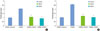
Statistically significant reductions in the proliferation indexes of allogenic and xenogenic PBMCs were found in mixed cell cultures including PDLSCs (Fig. 1). The reduced values of all experimental groups, i.e., mixed cell cultures with stem cells, were nearly the same as the values of the positive control. The degrees of reduction in the proliferation indexes of the PDLSC groups were similar to those of the BMSC groups in both allogenic (Fig. 1A) and xenogenic (Fig. 1B) mixed cell cultures.
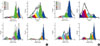
We also examined the cell division of PHA-stimulated PBMCs co-cultured with or without PDLSCs and BMSCs (Fig. 2). In the groups of PHA-stimulated canine and human PBMCs without stem cells, the peak generation phases of cell division were G3 and G7, respectively. However, in the presence of PDLSCs and BMSCs, cell division peaked at G0, i.e., in the parent cells. The histogram of CFSE assays in the PDLSC groups showed slightly different patterns when compared to the BMSC groups. The populations in the PDLSC groups shifted more to the left than the BMSC groups in both allogenic (Fig. 2A) and xenogenic (Fig. 2B) co-cultures.
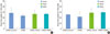
To measure the apoptosis of PBMCs after PHA stimulation, trypan blue uptake methods were used (Fig. 3). In all groups, no significant differences in the percentages of trypan blue-positive PBMCs were found.
DISCUSSION
Recently, the identification of MSC-like populations, such as PDLSCs, from dental tissues has indicated the possibility of applications to tissue engineering [9]. These techniques have the potential to develop into new methods for regenerative periodontal therapy. In previous studies [10,11], we found that canine PDLSCs are similar to human PDLSCs and other MSCs with respect to clonality, surface antigen profiles, and generation of multiple types of differentiated cells. In addition, it was demonstrated that transplantation of autologous canine PDLSCs and BMSCs enhanced alveolar bone regeneration in canine peri-implant defects [11].
The clinical challenges of stem cell-based periodontal therapy include immune rejection after administration, the oncogenic properties of stem cells, and the functional integration of transplanted tissues into the host [12]. High among these challenges is the nullification of the immune reaction. The use of autologous stem cells is regarded as a standard solution. However, allogenic and xenogenic PDLSCs have some advantages over autologous PDLSCs since their use is limited by the difficulties of patient age, tissue quality, and the disease status of the patient [13].
However, MSCs can modulate the actions of immune cells including antigen presenting cells (APCs), dendritic cells, T cells, B cells, and natural killer (NK) cells [14]. This MSC capacity has been shown to reduce the numbers of both allogenic and xenogenic immune cells [15,16]. PDLSCs have shown similar properties to those of canine [10] and human MSCs [13]. Therefore, we presumed that PDLSCs could also have immunomodulatory effects on allogenic and xenogenic immune cells.
In the present study, not only did PDLSCs inhibit the proliferation of allogenic and xenogenic PBMCs that had been stimulated by PHA, but this inhibition was similar to that of BMSCs. There was complete suppression of PBMC proliferation by colcemid (positive control), and the proliferation indexes of the groups that were co-cultured with stem cells were almost the same as that of the positive control. In addition, this inhibition was not related to the apoptosis of PBMCs because all groups showed 20-30% apoptosis rates that were not statistically different. Instead of apoptosis of PBMCs, the stem cells suppressed the proliferation of PBMCs by inhibiting the cell division of PBMCs in mixed cell cultures. Both PDLSCs and BMSCs arrested the division of PBMCs, and the most abundant generation of PBMCs after co-culture was that of the parent cell. These results are in accordance with those of previous studies [2,13,17]. In this study, the histogram of PDLSCs in CFSE assays showed a slight shift towards the left side in allogenic and xenogenic co-cultures, when compared to BMSCs. This finding demonstrates that PDLSCs show slightly less of an inhibitory effect on cell division of PBMCs than BMSCs. This result seems to be related to the statement that PDLSCs are more differentiated than BMSCs [18,19].
Most studies agree that soluble factors are involved in the immunomodulation of MSCs [1,20-22]. The suppressive factors are not constitutively secreted by MSCs; rather they require dynamic cross communication between MSCs and immune cells [14]. In contrast, the inhibitory effect of MSCs on NK-cells required cell-to-cell contact, suggesting different mechanisms for this MSC-mediated NK-cell suppression [22]. Several possible mechanisms have been proposed for MSC-mediated immunomodulation, including TGF-β1, HGF, cytokines (produced by MSCs), SDF-1, RANK-L, and PGE2, as well as osteoprotegerin (OPG) interactions and indoleamine 2,3-dioxygenase (IDO) induced by INF-γ [1,23-27]. The activated human PBMCs induced PDLSCs to secrete soluble factors to mediate, in part, the suppression of the PBMC proliferation. The resulting upregulated expressions of TGF-β1, HGF, and IDO (after stimulation with INF-γ) were seen in the mixed cell cultures of all mesenchymal stem cell types [13].
The results associated with MSC-induced inhibition of immune cells are somewhat contradictory and may include various suppressive mechanisms. Those varying results may be due to multifactorial issues including model systems, enriched cell populations, or unfractionated PBMCs, as well as the different species and sources of MSCs. Other laboratory parameters include isolation protocol, variable timings for measurements, and the lack of a clear-cut definition of MSCs [28].
In summary, canine PDLSCs have an immunomodulatory effect on allogenic and xenogenic PBMCs. This effect is not due to apoptosis of PBMCs but to inhibition of the cell division of PBMCs. These results suggest the potential application of allogenic and xenogenic PDLSCs for periodontal tissue regeneration. The present study is the first to study xenogenic immune cell inhibition by PDLSCs. Future studies are needed to investigate the mechanisms of immunomodulation of PDLSCs. Additional in vivo studies using animal models are also needed.
 XML Download