

 PDF
PDF ePub
ePub Citation
 Print
Print


INTRODUCTION
The incidence of unstable intertrochanteric fractures has grown substantially in the elderly along with an increasing life expectancy. Intertrochanteric fractures are more common in elderly patients and result in a high morbidity and a more difficult rehabilitation resulting from a deterioration of muscle strength and proprioceptive function12). For these reasons, stable bone healing is the most critical element after a fracture occurs. When selecting hip arthroplasty for the treatment of fractures, stable fixation of the greater trochanteric fragment is essential for complete bone union and functional recovery of the hip joint. A variety of internal fixation devices and fixation methods have been developed to achieve bone healing with stable fixation of fracture fragments, but the rate of sequelae, such as nonunion after early failure of fixation, has reportedly increased up to 50%3456). In particular, elderly patients with unstable intertrochanteric fractures of the femur associated with osteoporosis have difficulty performing early weight bearing, an outcome that can potentially prolong treatment time and result in an increased rate of systemic complications and poor functional recovery and clinical outcomes.
Because of unfavorable prognosis, instead of osteosynthesis, hip arthroplasty has become increasingly more popular as a treatment option. Stern and Goldstein7) have recommended prosthetic replacement to allow for early weight bearing in elderly patients with an unstable intertrochanteric fracture, and Cho et al.8) have demonstrated favorable results at short-term follow-up after performing cementless total hip arthroplasty for unstable intertrochanteric fractures.
After arthroplasty for unstable intertrochanteric fractures with the greater trochanteric fragment, nonunion of the greater trochanteric fragment may cause pain in the trochanteric region, functional gait abnormality and dislocation due to reduced hip abductor strength. Therefore, anatomical reduction and rigid fixation of the greater trochanter are important. Previously introduced fixation techniques or devices include: i) tension band wiring, ii) modified tension band wiring, iii) greater trochanteric reattachment device (GTRD), iv) claw plate, and v) others. Nam et al.9) have reported satisfactory results with cementless total hip arthroplasty and double tension band wiring in fixation of unstable intertrochanteric fractures with the greater trochanteric fragment. Moreover, Cho et al.10) and Kho et al.11) documented satisfactory results with hip hemiarthroplasty and cerclage wiring in fixation of unstable intertrochanteric fractures.
The authors of this study aimed to classify fracture patterns based on fracture type of the greater trochanter and report clinical and radiological results by applying fixation techniques that vary depending on the fracture pattern.
MATERIALS AND METHODS
For the purposes of this study, unstable intertrochanteric fractures were those classified as type 4 and type 5 (using Evan's criteria) and associated with a fracture of the medial cortex including the lesser trochanter. Of the 137 patients who underwent hip arthroplasty for an intertrochanteric fracture with osteoporosis (bone mineral density, <−2.5) between March 2014 and October 2015, 63 met our eligibility criteria and were included in this study. Patients were divided into three groups according to fracture pattern of the greater trochanter (as determined using three-dimensional computed tomography [3D CT] images) and different fixation methods were then applied by a single orthopaedic surgeon.
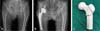
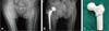
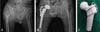
The mean age at time of surgery was 81.2 years (range, 71-95 years); 49 patients were female and 14 were male. The average follow-up period after the surgery was 11.6 months (range, 5-24 months). Operation time and volume of blood loss were evaluated based on anesthesia records. All operations were done in a lateral position. Using a modified Gibson approach, about a 15-cm skin incision was made and the fractured head and neck of the femur was removed. After temporary fixation using a 1.0-mm wire with double strands, the appropriate femoral component was inserted. To examine the potential benefits of differential fixation techniques, fracture patterns were divided into three groups (A, B, and C) based on 3D CT images. Patients in group A (n=18) presented with an oblique or spiral fracture with relatively large fragment (Fig. 1A). Group B (n=4) included the rarer cases of a transverse fracture of the greater trochanter (Fig. 1B). Group C (n=41) included those with comminuted fracture of the greater trochanter (Fig. 1C). For group A, fixation was accomplished using a 1.0-mm roll wire and a spiral band in a figure of 8 wiring the intertrochanteric area between the greater and lesser trochanter (Fig. 2). This conventional technique can prevent escape of the greater trochanteric fragment with resultant hip abductor weakness. For group B, we used tension band wiring using a 1.0 double-stranded wire after inserting two 1.8 K-wires from the tip of the greater trochanter to the distal part (Fig. 3). This method can help prevent bone loss of the greater trochanteric fragment by increasing compression across the transverse fracture of the greater trochanter. For group C, solid fixation of comminuted fractures was carried out using two 2.0 cables with the GTRD (Fig. 4). After splitting the abductors, comminuted fragments were reattached in place using cables by connecting a metal claw to the bone fragment12).
To evaluate postoperative function in patients, we measured: i) recovery of ambulation ability, ii) thigh and groin pain, and iii) the Harris hip score (HHS). For radiographic assessment, bone union was evaluated based on anteroposterior views on plain X-rays taken pre- and postoperatively and at final follow-up.
RESULTS
In the present study, the mean operative time, defined as the time from skin incision to the end of the skin closure, was 75 minutes and the mean intra-operative blood loss was 800 mL. Partial weight loading and standing were allowed in an average of 7 days after surgery (range, 4-10 days). Clinically, 50 out of 63 patients returned to normal daily activities without a decrease in walking distance, and we observed improvements in HHS from 74.8 to 85.7 points at the latest follow-up (considered relatively favorable scores; e.g., fair to good). Upon radiological assessment, bone union was observed in 62 (98.4%) cases, with the lone exception being a patient who experienced osteolysis, and the average time to radiographic union was 9 months (range, 8-12 months). The most common postoperative complications were irritation or impingement caused by wire or cable breakage (n=3), and in each case the metallic fragments were removed. A single case of traumatic dislocation was observed and managed with closed reduction without additional surgical intervention. There were no cases of deep vein thrombosis, pressure ulcer, pulmonary embolism or other medical complications.
DISCUSSION
Intertrochanteric fractures of the femur commonly occur in the elderly and anatomical reduction of this fracture type is challenging; the frequency of total hip arthroplasty has been increasing in recent years. Even though the fracture is reduced, stable fixation and maintenance of anatomical reduction are difficult. The incidence of complications (e.g., pressure ulcer, pneumonia, pulmonary embolism, atelectasis, urinary tract infection, and others) is relatively high as a result of patients being bedridden for prolonged periods of time13), and is considered a critical factor impacting mortality in the elderly population. The importance of early ambulation after surgery has been highlighted as it may reduce morbidity and increase the likelihood of favorable clinical results14).
Previous studies have demonstrated that the rates of sequelae range between 18% and 50% after open reduction and internal fixation for unstable intertrochanteric fractures345715). Laros and Moore5) have stated that the failure rate after internal fixation of unstable intertrochanteric fractures were 25%, and of these, 29% underwent secondary revision.
Chan and Gill16) and Haentjens et al.1718) compared the clinical results between bipolar arthroplasty and internal fixation for treatment of unstable intertrochanteric and subtrochanteric fractures. Rehabilitation was easier and faster and the incidence of pressure ulcers, pneumonia and atelectasis was dramatically lower in the bipolar arthroplasty group, an outcome that was found to be attributable to early ambulation. Furthermore, Stern and Angerman6) have suggested that hip arthroplasty can achieve more favorable clinical results (i.e., recovery of functional activities) than internal fixation for unstable intertrochanteric fracture of the femur in elderly patients. Kim et al.19) have recommended cemented bipolar hemiarthroplasty as a useful option for the treatment of unstable intertrochanteric fractures in elderly patients aged 65 years and have obtained good results in 88% of patients.
Since a comminuted fracture of the greater and lesser trochanters is commonly associated with an intertrochanteric femur fracture in arthroplasty, surgeons may face technical challenges during surgery in cases of non-rigid wire fixation or bone deformity in unwanted directions despite slight wire tension. Since the greater trochanter serves as the main attachment site for abductor muscles, anatomical reduction and maintenance of reduction depending on leg positions are uneasy in cases of comminution of the greater trochanter. Hamadouche et al.20) have achieved satisfactory outcomes with the use of the GTRD for greater trochanteric nonunion following revision total hip arthroplasty.
In this study, we thought more deeply about acquiring solid fixation in various fracture types and fragment patterns. We aimed to reduce the time and effort of fixation by applying different fixation methods to three types of fracture patterns (classified using 3D CT images). Recovery of preoperative ambulation status was obtained in 79% of patients, and we observed substantial improvements in HHS, both considered to be relatively favorable outcomes. The mean operative time was 75 minutes, confirming that fixation was completed in a relatively short period of time. This retrospective study was limited by the relatively short follow-up period and small sample size. Additional studies are warranted for further investigation.
CONCLUSION
Choosing an appropriate fixation method (e.g., cerclage wire fixation, tension band wiring and greater trochanteric reattachment) for distinct fracture patterns will help us to easily acquire proper reduction and fixation of greater trochanteric fractures and shorten bone union time when performing hip arthroplasty for unstable intertrochanteric fractures. It will also assist with early ambulation and rehabilitation, allowing early return to activities of daily living.

 XML Download